Case Reports
. 2013 May;125(5):753-69.
doi: 10.1007/s00401-013-1096-7. Epub 2013 Feb 12.

α-Synucleinopathy associated with G51D SNCA mutation: a link between Parkinson's disease and multiple system atrophy?

Affiliations
Case Reports

α-Synucleinopathy associated with G51D SNCA mutation: a link between Parkinson's disease and multiple system atrophy?

Aoife P Kiely et al. Acta Neuropathol. 2013 May.

Abstract

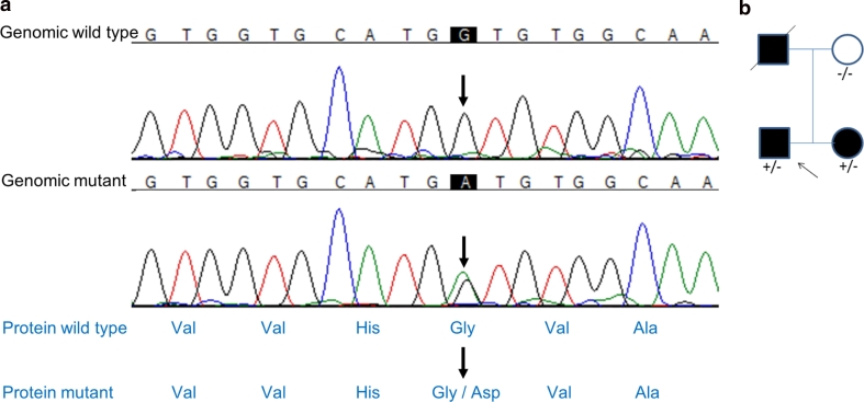
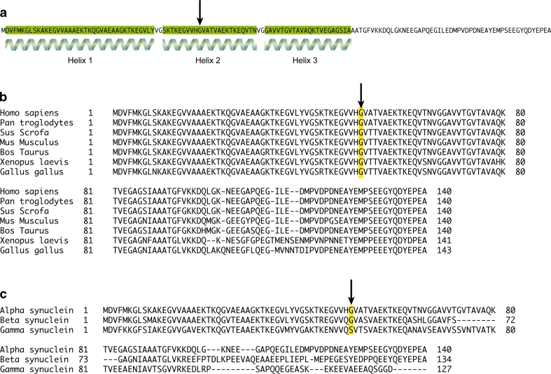
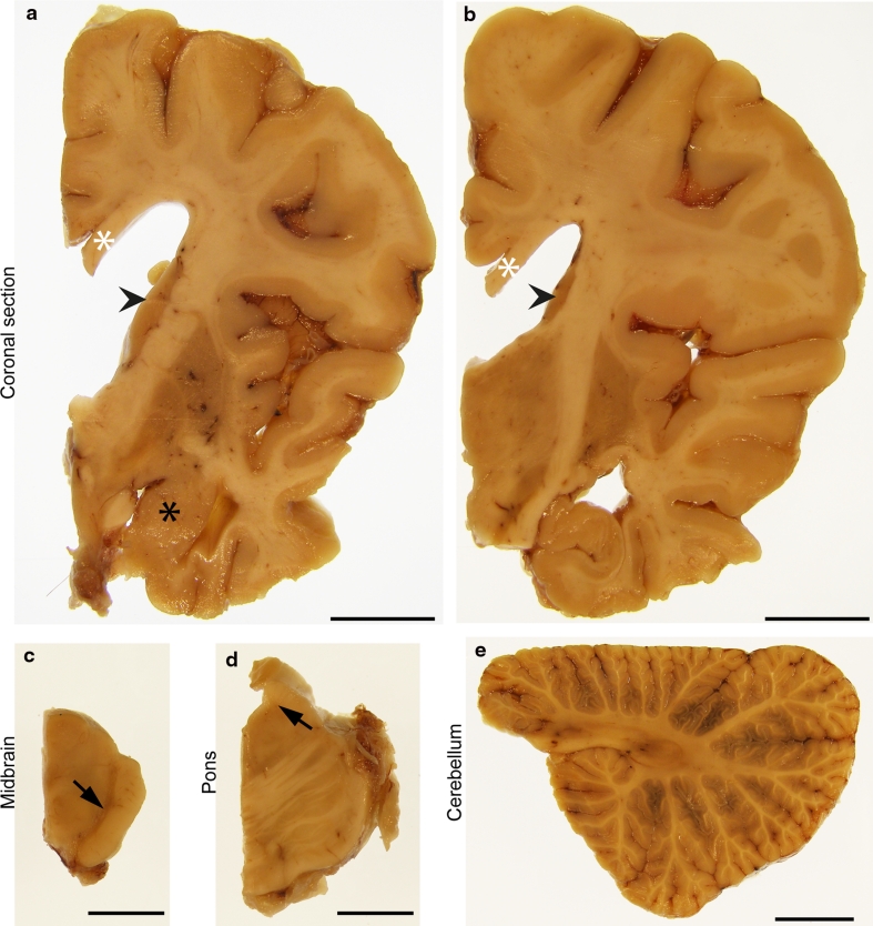
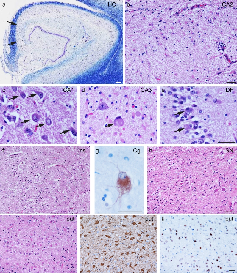
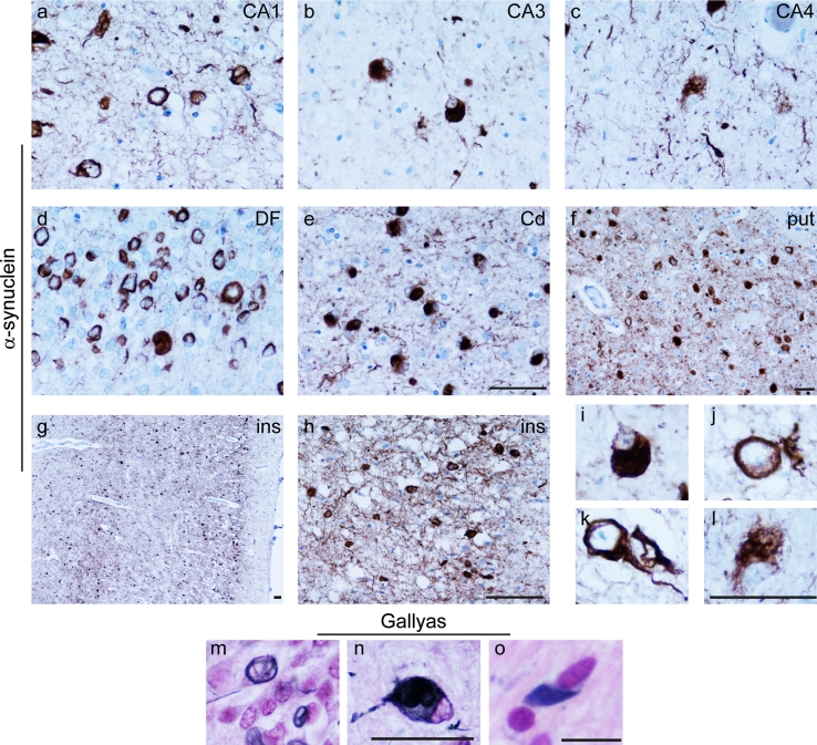
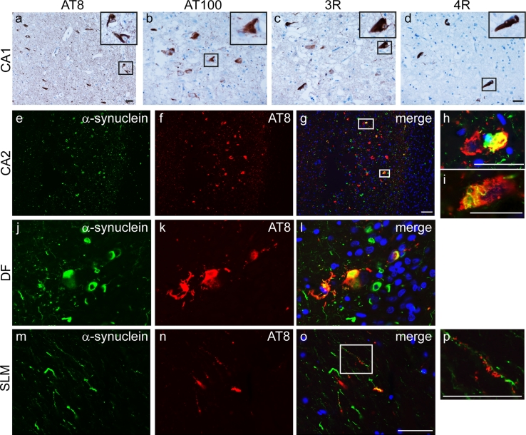
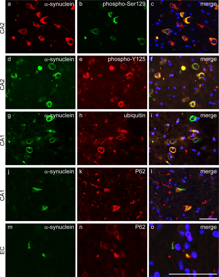
We report a British family with young-onset Parkinson's disease (PD) and a G51D SNCA mutation that segregates with the disease. Family history was consistent with autosomal dominant inheritance as both the father and sister of the proband developed levodopa-responsive parkinsonism with onset in their late thirties. Clinical features show similarity to those seen in families with SNCA triplication and to cases of A53T SNCA mutation. Post-mortem brain examination of the proband revealed atrophy affecting frontal and temporal lobes in addition to the caudate, putamen, globus pallidus and amygdala. There was severe loss of pigmentation in the substantia nigra and pallor of the locus coeruleus. Neuronal loss was most marked in frontal and temporal cortices, hippocampal CA2/3 subregions, substantia nigra, locus coeruleus and dorsal motor nucleus of the vagus. The cellular pathology included widespread and frequent neuronal α-synuclein immunoreactive inclusions of variable morphology and oligodendroglial inclusions similar to the glial cytoplasmic inclusions of multiple system atrophy (MSA). Both inclusion types were ubiquitin and p62 positive and were labelled with phosphorylation-dependent anti-α-synuclein antibodies In addition, TDP-43 immunoreactive inclusions were observed in limbic regions and in the striatum. Together the data show clinical and neuropathological similarities to both the A53T SNCA mutation and multiplication cases. The cellular neuropathological features of this case share some characteristics of both PD and MSA with additional unique striatal and neocortical pathology. Greater understanding of the disease mechanism underlying the G51D mutation could aid in understanding of α-synuclein biology and its impact on disease phenotype.


Figures

Fig. 1
Fig. 1
SNCA mutation G51D. Chromatogram (red thymine, blue cytosine, green adenine and black guanine). Sanger sequencing of the SNCA gene identified a c.G152A mutation (upper arrows) in exon 3 of this gene as shown. This created a glycine to aspartic acid amino acid change (lower arrows)
Fig. 2
Fig. 2
α-Synuclein protein structure and conservation as shown by amino acid sequence alignment. Amino acid sequence of human α-synuclein, showing regions of secondary structure (alpha helices highlighted in green). The G51 residue is indicated by an arrow, and sits in the middle of helix 2 (a). Sequence alignment of α-synuclein amino acid sequences from Homo sapiens, Pan troglodytes, Sus scrofa, Mus musculus, Bos taurus, Xenopus laevis and Gallus gallus. The G51 residue highlighted in yellow and indicated by an arrow is conserved throughout these organisms (b). Sequence alignment of human α-, β- and γ-synuclein amino acid sequences, with the G51 residue is highlighted in yellow and indicated by an arrow (c). This residue is conserved in α- and β-synuclein, but is replaced by a serine residue in γ-synuclein
Fig. 3
Fig. 3
Macroscopic features. Macroscopic images of formalin fixed right hemispheric brain slices (a, b), midbrain (c), pons (d) and cerebellum (e). There was severe atrophy of the temporal cortex with relative preservation of the superior temporal gyrus (a, b) and severe reduction in volume of white matter with thinning of the corpus callosum (a, b white asterisks). The caudate nucleus was reduced in volume (a, b arrow heads). There was reduction in size and grey discolouration of both the putamen and the globus pallidus (a, b). The amygdala was small and darkly coloured (a, black asterisk). The hippocampus was moderately reduced in volume (b). The midbrain showed marked depigmentation of the substantia nigra (c, arrow). The pons was well preserved with severe pallor of the locus coeruleus (d, arrow). The cerebellum was macroscopically normal and the white matter was well preserved (e). Scale bars represent 15 mm
Fig. 4
Fig. 4
Histological findings. Representative images from the hippocampus (ae), insular cortex (ins, f), cingulate gyrus (Cg, g), substantia nigra (SN, h) and putamen (put, ik). In the hippocampus, there was severe neuronal loss in the CA2 and CA3 (a, arrows). The CA2 showed few residual neurons (b). Neuronal inclusions (arrows) with varying morphology are illustrated in the CA1 (c), CA3 (d) and the dentate fascia (DF, e). Superficial laminae of the neocortex showed marked neuronal loss with microvacuolation illustrated in the insular cortex (ins, f). Ballooned neurons were most frequent in the cingulate gyrus (Cg, g). The substantia nigra (SN) shows severe loss of pigmented neurons accompanied by gliosis (h). Abundant eosinophilic reactive astrocytes are visualised in the putamen (put, i) and confirmed by GFAP immunohistochemistry (j). TDP-43 immunoreactive inclusions were also detected in the putamen (k). Luxol fast blue (a), haematoxylin and eosin (H&E) (bf, h, i), αB-crystallin (g), GFAP (j) TDP-43 (k). Scale bar 150 μm (a), scale bars in b, eh, k represent 50 μm (ce are at the same magnification, as are ik)
Fig. 5
Fig. 5
Characterisation of neuronal and glial α-synuclein inclusions. Thread-like α-synuclein immunoreactivity was observed to be widespread, shown here within the hippocampal regions CA1, CA3 and CA4 (ac), caudate nucleus (Cd, e), putamen (put, f) and insular cortex (ins, g, h). Neuronal cytoplasmic inclusions immunoreactive for α-synuclein had a number of different morphological appearances globular (i), annular (j), neurofibrillary tangle-like (k) and diffuse (l). Gallyas silver impregnation demonstrated the presence of fibrillar protein in neuronal inclusions (DF, m and CA4, n) and also in GCI-like inclusions (pontine base, o). Scale bars in eh, l represent 50 μm (an) (ae are at the same magnification as are il, m and n). Scale bar in o represents 10 μm
Fig. 6
Fig. 6
Glial inclusions. Representative double immunofluorescence images probed with α-synuclein (red) and the oligodendroglial marker, olig2 (green). Composite merged images show α-synuclein immunoreactive inclusions in oligodendrocytes resembling GCIs in the white matter of the frontal cortex (a, d), the pons (b) and the alveus of the hippocampus (c). Close proximity is observed between a subset of activated microglia, as detected by iba-1 (green) and α-synuclein (red) inclusion containing neurons, this occasionally involved encircling of α-synuclein-containing neurons by iba-1-positive processes (eg). A small proportion of these microglia contained α-synuclein immunoreactivity (g, inset arrowheads). A high level of reactive astrogliosis was detected throughout all brain regions examined, without evidence of α-synuclein expression within astrocytes or their processes (hj). DAPI nuclear stain (blue). Scale bar in j represents 50 μm (aj). Scale bar in g inset represents 25 μm
Fig. 7
Fig. 7
Tau expression and co-localisation with α-synuclein. Representative images from CA1 probed using immunohistochemistry with phospho-tau antibodies: AT8 (a) and AT100 (b). Inclusions contained a mixture of 3-repeat (c) and 4-repeat (d) tau isoforms. Double immunofluorescence images of CA2 (ei) and DF (jl) probed for α-synuclein (green) and AT8 (red) show strong co-localisation in a subset of neurons. AT8 immunoreactivity is detected on the dendritic processes of DF granule cells which express α-synuclein in the stratum lacunosum-moleculare (SLM, mp). DAPI nuclear stain (blue). Scale bars represent 50 μm (bd are at the same magnification as are eg, jo)
Fig. 8
Fig. 8
α-Synuclein is phosphorylated and inclusions contain ubiquitin and p62. Representative images of the CA2 region of the hippocampus probed by double immunofluorescence with total α-synuclein (red) and α-synuclein phospho-S129 (green) (ac), or α-synuclein (green) and α-synuclein phospho-Y125 (red) (df) show near complete co-localisation indicating that α-synuclein is phosphorylated at both epitopes. The majority of α-synuclein immunoreactivity (green) within neuronal inclusions co-localised with ubiquitin (red), shown in CA1 neurons (gi). Many α-synuclein-positive (green) neuronal inclusions also contained P62 (red) (jl) and a similar pattern was observed in GCI-like inclusions illustrated in the white matter underlying the entorhinal cortex (EC, mo). DAPI nuclear stain (blue). Scale bars represent 50 μm (al are at the same magnification as are mo)

References

    1. Ahmed Z, Asi YT, Sailer A, et al. The neuropathology, pathophysiology and genetics of multiple system atrophy. Neuropathol Appl Neurobiol. 2012;38:4–24. doi: 10.1111/j.1365-2990.2011.01234.x. - DOI - PubMed
    1. Al-Chalabi A, Durr A, Wood NW, et al. Genetic variants of the alpha-synuclein gene SNCA are associated with multiple system atrophy. PLoS One. 2009;4:e7114. doi: 10.1371/journal.pone.0007114. - DOI - PMC - PubMed
    1. Amador-Ortiz C, Lin W-L, Ahmed Z, et al. TDP-43 immunoreactivity in hippocampal sclerosis and Alzheimer’s disease. Ann Neurol. 2007;61:435–445. doi: 10.1002/ana.21154. - DOI - PMC - PubMed
    1. Armstrong RA, Cairns NJ, Lantos PL. Multiple system atrophy (MSA): topographic distribution of the alpha-synuclein-associated pathological changes. Parkinsonism Relat Disord. 2006;12:356–362. doi: 10.1016/j.parkreldis.2006.02.005. - DOI - PubMed
    1. Athanassiadou A, Voutsinas G, L Psiouri, et al. Genetic analysis of families with Parkinson disease that carry the Ala53Thr mutation in the gene encoding alpha-synuclein. Am J Hum Genet. 1999;65:555–558. doi: 10.1086/302486. - DOI - PMC - PubMed

Publication types