. 2013 Feb 12;4(1):e00638-12.
doi: 10.1128/mBio.00638-12.

Kdo hydrolase is required for Francisella tularensis virulence and evasion of TLR2-mediated innate immunity

Affiliations

Kdo hydrolase is required for Francisella tularensis virulence and evasion of TLR2-mediated innate immunity

Nihal A Okan et al. mBio. .

Abstract

The highly virulent Francisella tularensis subsp. tularensis has been classified as a category A bioterrorism agent. A live vaccine strain (LVS) has been developed but remains unlicensed in the United States because of an incomplete understanding of its attenuation. Lipopolysaccharide (LPS) modification is a common strategy employed by bacterial pathogens to avoid innate immunity. A novel modification enzyme has recently been identified in F. tularensis and Helicobacter pylori. This enzyme, a two-component Kdo (3-deoxy-d-manno-octulosonic acid) hydrolase, catalyzes the removal of a side chain Kdo sugar from LPS precursors. The biological significance of this modification has not yet been studied. To address the role of the two-component Kdo hydrolase KdhAB in F. tularensis pathogenesis, a ΔkdhAB deletion mutant was constructed from the LVS strain. In intranasal infection of mice, the ΔkdhAB mutant strain had a 50% lethal dose (LD(50)) 2 log(10) units higher than that of the parental LVS strain. The levels of the proinflammatory cytokines tumor necrosis factor alpha (TNF-α) and interleukin-1β (IL-1β) in bronchoalveolar lavage fluid were significantly higher (2-fold) in mice infected with the ΔkdhAB mutant than in mice infected with LVS. In vitro stimulation of bone marrow-derived macrophages with the ΔkdhAB mutant induced higher levels of TNF-α and IL-1β in a TLR2-dependent manner. In addition, TLR2(-/-) mice were more susceptible than wild-type mice to ΔkdhAB bacterial infection. Finally, immunization of mice with ΔkdhAB bacteria elicited a high level of protection against the highly virulent F. tularensis subsp. tularensis strain Schu S4. These findings suggest an important role for the Francisella Kdo hydrolase system in virulence and offer a novel mutant as a candidate vaccine.

Importance: The first line of defense against a bacterial pathogen is innate immunity, which slows the progress of infection and allows time for adaptive immunity to develop. Some bacterial pathogens, such as Francisella tularensis, suppress the early innate immune response, killing the host before adaptive immunity can mature. To avoid an innate immune response, F. tularensis enzymatically modifies its lipopolysaccharide (LPS). A novel LPS modification-Kdo (3-deoxy-d-manno-octulosonic acid) saccharide removal--has recently been reported in F. tularensis. We found that the kdhAB mutant was significantly attenuated in mice. Additionally, the mutant strain induced an early innate immune response in mice both in vitro and in vivo. Immunization of mice with this mutant provided protection against the highly virulent F. tularensis strain Schu S4. Thus, our study has identified a novel LPS modification important for microbial virulence. A mutant lacking this modification may be used as a live attenuated vaccine against tularemia.


Figures

FIG 1
FIG 1
LPSs from F. tularensis LVS and ΔkdhAB mutant strain differ from each other by a side chain Kdo. The structure of core oligosaccharide synthesized by LVS (55) and the proposed structure of core oligosaccharide synthesized by the ΔkdhAB mutant, as determined by HPAEC (14), are shown.
FIG 2
FIG 2
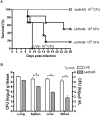
The virulence of the F. tularensis LVS ΔkdhAB mutant strain is attenuated in mice. (A) Survival of mice after F. tularensis intranasal challenge. BALB/cByJ mice (n = 6) were challenged intranasally with LVS (103 CFU) or ΔkdhAB bacteria (103, 104, or 105 CFU). (B) Bacterial burden in lung, spleen, liver, and blood of mice 6 days after infection. BALB/cByJ mice (n = 3) were challenged intranasally with LVS bacteria (103 CFU) or ΔkdhAB bacteria (103 CFU).
FIG 3
FIG 3
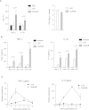
Enhanced proinflammatory cytokine response to the F. tularensis LVS ΔkdhAB mutant strain. (A) BALB/cByJ mice (n = 3) were challenged intranasally with LVS bacteria (103 CFU), ΔkdhAB bacteria (103 CFU), or PBS (control). BAL fluid was collected 24 h after infection, and the levels of TNF-α and IL-1β were measured by ELISA (left panel). The bacterial burden in lung was measured 24 h after infection (right panel). (B) Levels of TNF-α and IL-1β released by ex vivo BMMs after stimulation with LVS or ΔkdhAB bacteria (MOI of 10, 20, or 50). Culture supernatants were analyzed by ELISA 24 h after infection. (C) TNF-α and IL-1β gene expression in BMMs infected with LVS or ΔkdhAB bacteria (MOI of 10). Gene expression was measured by real-time PCR, normalized with RP II gene expression, and reported as relative expression compared with that in uninfected macrophages. All data are presented as mean plus SD (error bars) values (n = 3).
FIG 4
FIG 4
Bacteria of the F. tularensis LVS ΔkdhAB mutant strain activate NF-κB through the TLR2 signaling pathway. (A) Macrophage-like cell lines were stimulated with LVS or ΔkdhAB bacteria for 2 h (MOI = 100). The IL-1β level in cell lysates was assessed by ELISA. Measurements were expressed as fold induction in infected versus uninfected cells (mean plus SD; n = 3). (B) HEK293 cells stably expressing TLR2 or TLR4/MD2 were transiently transfected with an NF-κB luciferase reporter plasmid and stimulated for 24 h with LVS or ΔkdhAB bacteria (MOI = 20) or with Pam3CSK (1 µg/ml) or E. coli LPS (100 ng/ml) as positive controls. Purified LPSs from LVS or ΔkdhAB bacteria (5 µg/ml) were tested in a mixture with soluble CD14 recombinant protein (5 µg/ml). The data are reported as relative luciferase units and represent fold induction of luciferase activity in the cell lysate compared with that in unstimulated cells (mean plus SD; n = 3).
FIG 5
FIG 5
Mutation in kdhAB leads to enhanced accessibility of the bacterial surface. (A) Investigation of the leaky-membrane phenotype. Mid-logarithmic-phase cultures were spread onto CHAH plates, and a 10-µl volume of SDS (20, 10, and 5%) or EDTA (500, 250 and 125 mM) was spotted onto filter discs. Images were captured after 48 h of bacterial growth. Black arrows indicate zone of inhibition around 6-mm filter discs. (B) F. tularensis LVS and ∆kdhAB and ∆wbtA mutant strains were tested for binding of mouse anti-FopA antibodies to their surface FopA proteins in an antigen accessibility assay. Bacteria—along with surface-bound FopA antibodies—were lysed, and proteins were resolved by 5 to 20% SDS-PAGE. Bound FopA antibodies were detected as IgG heavy chain (HC) and IgG light chain (LC). Total FopA and bacterioferritin (Bfr) protein levels are shown as protein loading controls.
FIG 6
FIG 6
The F. tularensis ΔkdhAB mutant strain is not altered in the biosynthesis of O antigen or lipid A. (A) SDS-PAGE analysis of F. tularensis LVS and ΔkdhAB mutant outer membranes. LPS was visualized by zinc staining. (B) Negative-ion MALDI-TOF analysis of lipid A from LVS and ΔkdhAB bacteria.
FIG 7
FIG 7
Bacteria of the F. tularensis LVS ΔkdhAB mutant strain elicit protective immunity against F. tularensis LVS and type A strain Schu S4. (A and B) Groups of BALB/cByJ mice (10 mice in each group) were immunized intranasally with 3 doses of LVS or ∆kdhAB bacteria; 28 days after the last dose, the mice were challenged intranasally with either 104 CFU of LVS (10 × LD50) (A) or 10 CFU of strain Schu S4 (10 × LD50) (B). (C) Hematoxylin and eosin (H&E) staining of liver tissue on day 5 after Schu S4 challenge. The black arrowhead indicates extensive tissue necrosis (10×). (D) Bacterial burden in lung, spleen, and liver of mice 5 days after Schu S4 challenge (mean plus SD; n = 3). (E) Titers of serum antibody to F. tularensis LPS were determined by ELISA. Sera from LVS-immunized, ΔkdhAB mutant-immunized, and unimmunized control mice were collected on the day before challenge. ELISA was performed as described in Materials and Methods.

Similar articles

Cited by

References

    1. Bosio CM, Bielefeldt-Ohmann H, Belisle JT. 2007. Active suppression of the pulmonary immune response by Francisella tularensis Schu4. J. Immunol. 178:4538–4547 - PubMed
    1. Hall JD, Woolard MD, Gunn BM, Craven RR, Taft-Benz S, Frelinger JA, Kawula TH. 2008. Infected-host-cell repertoire and cellular response in the lung following inhalation of Francisella tularensis Schu S4, LVS, or U112. Infect. Immun. 76:5843–5852 - PMC - PubMed
    1. Malik M, Bakshi CS, Sahay B, Shah A, Lotz SA, Sellati TJ. 2006. Toll-like receptor 2 is required for control of pulmonary infection with Francisella tularensis. Infect. Immun. 74:3657–3662 - PMC - PubMed
    1. Mares CA, Ojeda SS, Morris EG, Li Q, Teale JM. 2008. Initial delay in the immune response to Francisella tularensis is followed by hypercytokinemia characteristic of severe sepsis and correlating with upregulation and release of damage-associated molecular patterns. Infect. Immun. 76:3001–3010 - PMC - PubMed
    1. Moreland JG, Hook JS, Bailey G, Ulland T, Nauseef WM. 2009. Francisella tularensis directly interacts with the endothelium and recruits neutrophils with a blunted inflammatory phenotype. Am. J. Physiol. Lung Cell. Mol. Physiol. 296:L1076–L1084 - PMC - PubMed

Publication types

MeSH terms

LinkOut - more resources