Review
. 2013 May;91(5):603-22.
doi: 10.1002/jnr.23197. Epub 2013 Feb 13.

Organization and maintenance of molecular domains in myelinated axons

Affiliations
Review

Organization and maintenance of molecular domains in myelinated axons

Elizabeth D Buttermore et al. J Neurosci Res. 2013 May.

Abstract

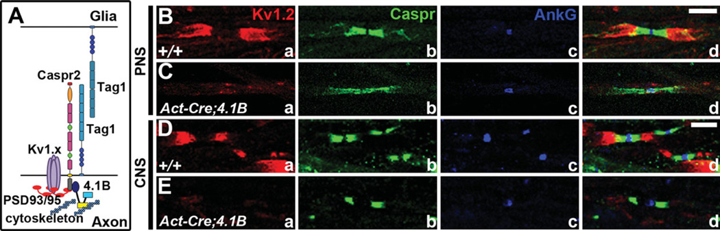
Over a century ago, Ramon y Cajal first proposed the idea of a directionality involved in nerve conduction and neuronal communication. Decades later, it was discovered that myelin, produced by glial cells, insulated axons with periodic breaks where nodes of Ranvier (nodes) form to allow for saltatory conduction. In the peripheral nervous system (PNS), Schwann cells are the glia that can either individually myelinate the axon from one neuron or ensheath axons of many neurons. In the central nervous system (CNS), oligodendrocytes are the glia that myelinate axons from different neurons. Review of more recent studies revealed that this myelination created polarized domains adjacent to the nodes. However, the molecular mechanisms responsible for the organization of axonal domains are only now beginning to be elucidated. The molecular domains in myelinated axons include the axon initial segment (AIS), where various ion channels are clustered and action potentials are initiated; the node, where sodium channels are clustered and action potentials are propagated; the paranode, where myelin loops contact with the axolemma; the juxtaparanode (JXP), where delayed-rectifier potassium channels are clustered; and the internode, where myelin is compactly wrapped. Each domain contains a unique subset of proteins critical for the domain's function. However, the roles of these proteins in axonal domain organization are not fully understood. In this review, we highlight recent advances on the molecular nature and functions of some of the components of each axonal domain and their roles in axonal domain organization and maintenance for proper neuronal communication.


Figures

Fig. 1
Fig. 1
Axonal domains in central and peripheral myelinated axons. A: Wild-type cerebellar section immunostained against calbindin (Calb, red), myelin basic protein (MBP, green), and Caspr (blue) highlights the segregation of axonal domains in Purkinje neuron (P) myelinated axons. B: Representative drawing of a Purkinje neuron (P) showing the localization of molecular domains in myelinated axons, including the axon initial segment (AIS), node of Ranvier (node), paranode, and myelinated internode. C: Teased wild-type sciatic nerve fiber immunostained against potassium channels (KV1.2, red), Nfasc186 (NF186, green), and Caspr (blue) highlights the distinct axonal domains flanking the node. D: Representative drawing of the axonal domains flanking the node, which reflects the distinct segregation of each of these domains. Scale bars = 10 µm.
Fig. 2
Fig. 2
Molecular organization of the Purkinje neuron AIS. A: Diagram of molecular components at the AIS. B: Wild-type Purkinje neuron immunostained against Calb (green) and Nfasc (red). C: AnkG−/− Purkinje neuron immunostained against Calb (green) and Nfasc (red) highlights the diffusion of Nfasc distally from the AIS in the absence of AnkG (Ango et al., 2004). D,E: Cultured hippocampal neurons with AnkG shRNA (green cell) or without AnkG shRNA (cell with star) reveal that Nfasc (PAN NF, D) and NaV channels (PAN NaV; E) are absent from the AIS when AnkG is knocked down (Hedstrom et al., 2007). F–I: Cerebellar sections from P10 (F,G) and P20 (H,I) wild-type (F,H) and Pcp2-Cre; NfascFlox/Flox(G,I) mice immunostained against NaV1.6 (a, green), AnkG (b, red), and Calb (c, blue, merged) show that the mature isoform of NaV channels, NaV1.6, clusters to the distal part of the AIS at P10 and fails to localize at the AIS proper at P20 in the absence of Nfasc. Purkinje cell protein 2-Cre (Pcp2-Cre) is expressed specifically in Purkinje neurons in the cerebellum. J–L: Cerebellar sections from wild-type (J), Pcp2-Cre;NfascFlox/Flox(K), and Parv-Cre;NfascFlox/Flox(L) mice immunostained against Calb (red) and KV1.2 (green) reveal severely disorganized pinceau surrounding the Nfasc-deficient Purkinje AIS (Buttermore et al., 2012). Parv-Cre is expressed in parvalbumin-expressing GABAergic neurons throughout the nervous system. Scale bars = 10 µm. (B,C: Reprinted from Cell 2004; 119:257–272, with permission from Elsevier; D,E: © Hedstrom KL, Xu X, Ogawa Y, Frischknecht R, Seidenbecher CI, Shrager P, Rasband MN, Neurofascin assembles a specialized extracellular matrix at the axon initial segment; originally published in J Cell Biol 2007; 178:875–886; J–L: Reprinted from J Neurosci 2012; 32:4724–4742, with permission from The Journal of Neuroscience.)
Fig. 3
Fig. 3
Organization of the node and formation of a molecular barrier against invading paranodes. A: Diagram of molecular components of the PNS node. B–E: Sciatic nerve (B,C) and spinal cord (D,E) fibers from wild-type (B,D) and Nefl-Cre;NfascFlox (C,E) mice immuno-stained against Caspr (a, green), pan-NaV channels (b, red), and NfascNF186 (c, blue) reveal loss of NaV channel clustering at the node in the absence of NfascNF186 (Thaxton et al., 2011). Nefl-Cre allows neuron-specific expression of Cre under the neurofilament light chain promoter. F–I: Electron micrographs of sciatic nerve (F,G) and spinal cord (H,I) fibers from wild-type (F,H) and Nefl-Cre;NfascFlox (G,I) mice reveal the aberrant overlapping of adjacent paranodal loops when the node has been disrupted by loss of NfascNF186 (Thaxton et al., 2011). Scale bars 5 0.5 µm. (B–I: Reprinted from Neuron 2011; 69:244–257, with permission from Elsevier.)
Fig. 4
Fig. 4
Organization and stabilization of the paranodal domain. A: Diagram of the molecular components of the paranode. B,C: Sciatic nerve fibers from wild-type (B) and Caspr−/− (C) mice immuno-stained against potassium channels (KV1.1, red), Caspr (blue), and sodium channels (NaCh, green) reveal the role of the paranodal AGSJs to segregate potassium channels in the JXP from sodium channels in the node (Bhat et al., 2001). D,E: Electron micrographs of wild-type (D) and Caspr−/− (E) axons showing parallel arrays of axonal cytoskeleton in the wild-type axon and disorganization of the axonal cytoskel-eton in the Caspr−/− (Garcia-Fresco et al., 2006). F,G: Sciatic nerves from wild-type (F) and Act-Cre;4.1BFlox (G) mice immunostained against Nfasc (a, red), Caspr (b, green), AnkG (c, blue), and merged (d) reveal the disruption of paranodes in P30 4.1B mutant PNS axons. Act-Cre allows ubiquitous expression of Cre under β-actin promoter. H,I: Spinal cord sections from wild-type (H) and Act-Cre;4.1BFlox (I) mice immunostained against 4.1B (a, red), Caspr (b, green), AnkG (c, blue), and merged (d) reveal disruption of the paranode in 4-month-old 4.1B mutant CNS axons. J,M: Electron micrographs of sciatic nerve fibers (J,K) and spinal cord fibers (L,M) from wild-type (J,L) and Act-Cre;4.1BFlox (K,M) mice, revealing disrupted AGSJ compaction in the PNS (K) and loss of AGSJs in the CNS (M; Buttermore et al., 2011). Scale bars = 20 µm in C (applies to B,C); 10 µm in F (applies to F,G); 5 µm in H (applies to H,I); 0.2 µm in J–M. (B,C: Reprinted from Neuron 2001; 30:369–383, with permission from Elsevier; D,E: © Garcia-Fresco GP, Sousa AD, Pillai AM, Moy SS, Crawley JN, Tessarollo L, Dupree JL, Bhat MA. Disruption of axo–glial junctions causes cytoskeletal disorganization and degeneration of Purkinje neuron axons. Originally published in Proc Natl Acad Sci USA 2006; 103:5137–5142; F–M: Reprinted from J Neurosci 2011; 31:8013–8024, with permission from The Journal of Neuroscience.)
Fig. 5
Fig. 5
Organization and stabilization of the juxtaparanode. A: Diagram of molecular components of the JXP. B–E: Sciatic nerve (B,C) and spinal cord (D,E) myelinated fibers from wild-type (B,D) and Act-Cre;4.1BFlox (C,E) mice immunostained against potassium channels (a, KV1.2, red), Caspr (b, green), AnkG (c, blue), and merged (d) reveal the importance of the cytoskeletal adaptor protein 4.1B in the organization of the JXP, because loss of 4.1B results is disruption of the JXP in the PNS (C) and CNS (E; Buttermore et al., 2011). Scale bars 5 10 µm in B (applies to B,C); 5 µm in D (applies to D,E). (B–E: Reprinted from J Neurosci 2011; 31:8013–8024, with permission from The Journal of Neuroscience.)

References

    1. Ango F, di Cristo G, Higashiyama H, Bennett V, Wu P, Huang ZJ. Ankyrin-based subcellular gradient of neurofascin, an immunoglobulin family protein, directs GABAergic innervation at purkinje axon initial segment. Cell. 2004;119:257–272. - PubMed
    1. Arroyo EJ, Xu T, Poliak S, Watson M, Peles E, Scherer SS. Internodal specializations of myelinated axons in the central nervous system. Cell Tissue Res. 2001;305:53–66. - PubMed
    1. Arroyo EJ, Xu T, Grinspan J, Lambert S, Levinson SR, Brophy PJ, Peles E, Schere SS. Genetic dysmyelination alters the molecular architecture of the nodal region. J Neurosci. 2002;22:1726–1737. - PMC - PubMed
    1. Baba H, Akita H, Ishibashi T, Inoue Y, Nakahira K, Ikenaka K. Completion of myelin compaction, but not the attachment of oligodendroglial processes triggers K+ channel clustering. J Neurosci Res. 1999;58:752–764. - PubMed
    1. Banerjee S, Sousa AD, Bhat MA. Organization and function of septate junctions: an evolutionary perspective. Cell Biochem Biophys. 2006;46:65–77. - PubMed

Publication types

LinkOut - more resources