. 2013 Mar 29;288(13):9447-56.
doi: 10.1074/jbc.M112.438010. Epub 2013 Feb 12.

A pathophysiologic role for epidermal growth factor receptor in pemphigus acantholysis

Affiliations

A pathophysiologic role for epidermal growth factor receptor in pemphigus acantholysis

Meryem Bektas et al. J Biol Chem. .

Abstract

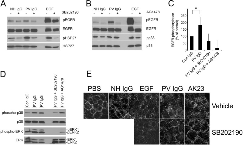
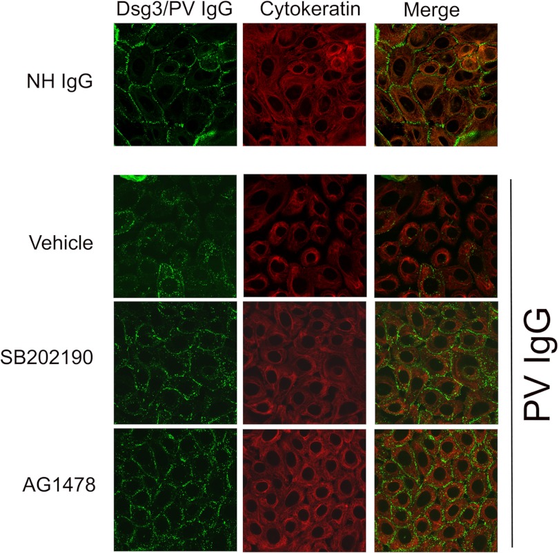
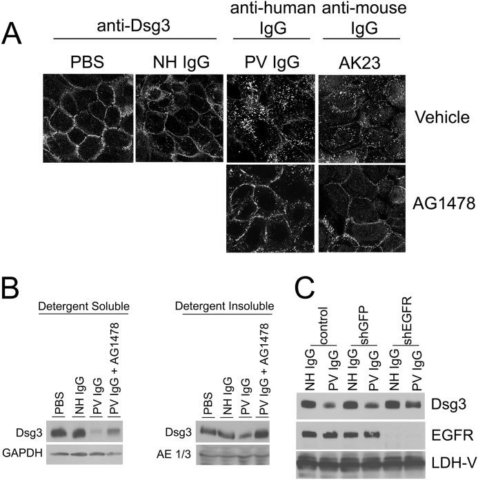
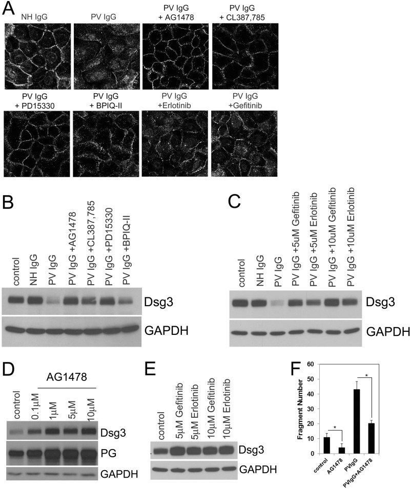
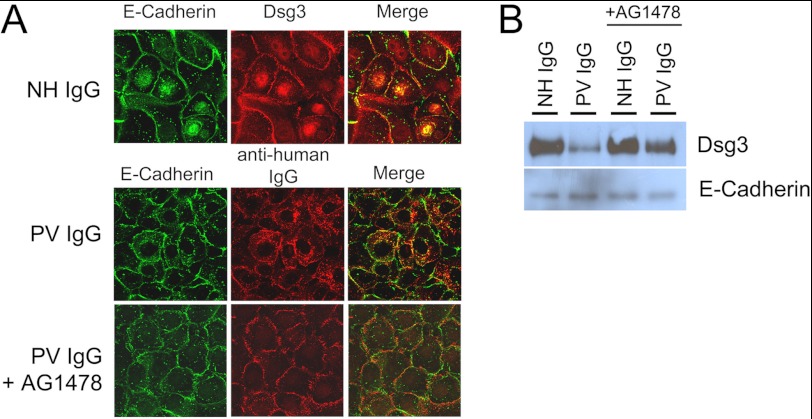
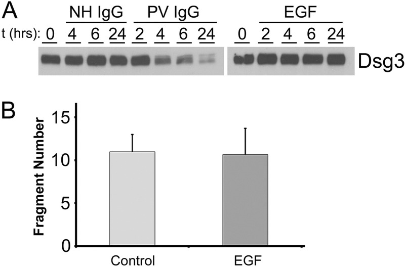
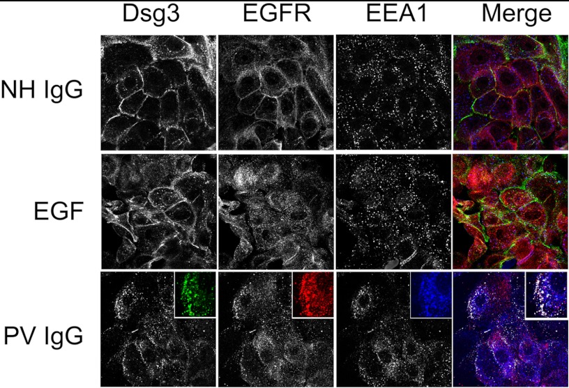
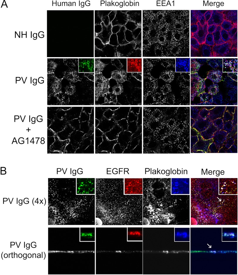
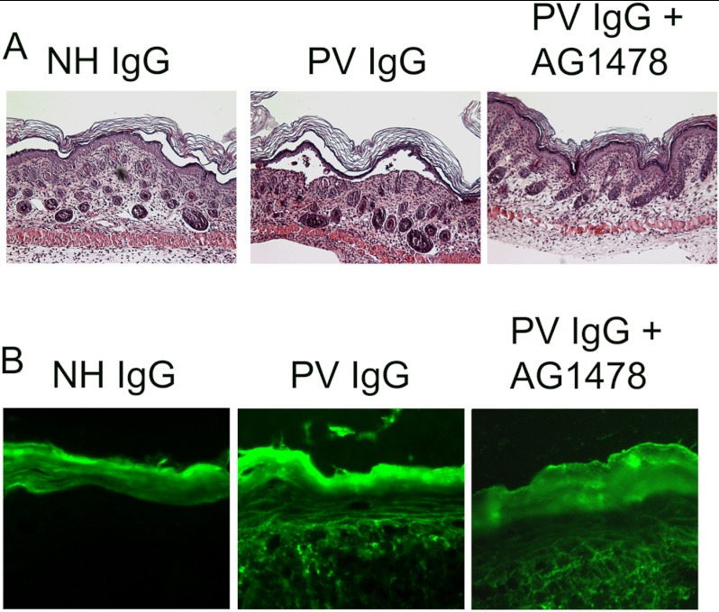
The pemphigus family of autoimmune bullous disorders is characterized by autoantibody binding to desmoglein 1 and/or 3 (dsg1/dsg3). In this study we show that EGF receptor (EGFR) is activated following pemphigus vulgaris (PV) IgG treatment of primary human keratinocytes and that EGFR activation is downstream of p38 mitogen-activated protein kinase (p38). Inhibition of EGFR blocked PV IgG-triggered dsg3 endocytosis, keratin intermediate filament retraction, and loss of cell-cell adhesion in vitro. Significantly, inhibiting EGFR prevented PV IgG-induced blister formation in the passive transfer mouse model of pemphigus. These data demonstrate cross-talk between dsg3 and EGFR, that this cross-talk is regulated by p38, and that EGFR is a potential therapeutic target for pemphigus. Small-molecule inhibitors and monoclonal antibodies directed against EGFR are currently used to treat several types of solid tumors. This study provides the experimental rationale for investigating the use of EGFR inhibitors in pemphigus.


Figures

FIGURE 1.
FIGURE 1.
p38 is upstream of EGFR activation by PV IgG. A and B, immunoblot analysis of keratinocyte lysates probed with antibodies to phospho-EGFR (pEGFR), total EGFR (EGFR), phospho-HSP27 (pHSP27), total HSP27 (HSP27), phospho-p38 (pp38), and total p38 (p38). Unlike NH IgG, PV IgG stimulated EGFR, p38, and HSP27 phosphorylation. The p38 inhibitor SB202190 blocked PV IgG-induced phosphorylation of EGFR (A) as well as HSP27, previously identified to be downstream of PV IgG-induced p38 activity. In contrast, the EGFR inhibitor AG1478 blocked PV IgG-induced EGFR phosphorylation but not PV IgG-induced p38 phosphorylation (B), indicating that p38 acts upstream of EGFR in PV IgG-treated keratinocytes. C, quantitation of PV IgG-stimulated EGFR phosphorylation in the presence and absence of SB202190 and AG1478. PV IgG stimulated EGFR phosphorylation 1.8 ± 0.6-fold compared with the control (Con). *, p = 0.031. Primary human keratinocytes were grown to confluence, serum-starved overnight, preincubated for 3 h with 0.5 mm calcium and for 1 h in vehicle (dimethyl sulfoxide), the EGFR inhibitor AG1478 (10 μm) (A), or the p38 inhibitor SB202190 (10 μm) (B) and then treated with NH IgG or PV IgG for 30 min or EGF (100 ng/ml, positive control) for 5 min. Cell lysates were prepared, and immunoblot analysis was performed. In addition to EGFR, each blot was also probed for HSP27 (A) or p38 (B) as loading controls. Results shown are representative of three independent experiments. D, PV IgG induced ERK phosphorylation. Keratinocyte monolayers were treated with normal human control IgG (Con IgG, 2 mg/ml) or PV IgG (2 mg/ml) for 30 min. Alternatively, keratinocytes were pretreated for 1 h with either SB202190 (10 μm) or AG1478 (10 μm) and then treated with PV IgG for 30 min. Lysates were prepared, separated by SDS-PAGE, and probed by immunoblot analysis with antibodies to phosphorylated p38 (phospho-p38), total p38 (p38), phosphorylated ERK (phospho-ERK), and ERK1 and ERK2. E, confocal immunofluorescent images of EGFR-stained keratinocytes. Keratinocytes were grown as above on coverslips; treated with PBS, EGF (100 ng/ml), NH IgG (2 mg/ml), PV IgG (2 mg/ml), or AK23 (2 mg/ml) for 4 h, and then fixed and stained with Cy-2 (1:100)-conjugated anti-EGFR secondary antibodies. In contrast to buffer (PBS)- and NH IgG-treated controls, EGF, PV IgG and AK23 induced EGFR endocytosis. SB202190 blocked both PV IgG- and AK23-induced EGFR endocytosis but not EGF-triggered EGFR endocytosis. Results shown are representative of at least three independent experiments.
FIGURE 2.
FIGURE 2.
The EGFR inhibitor blocks PV IgG-induced keratin intermediate filament retraction. Confocal immunofluorescent images of keratinocytes treated with NH or PV IgG ± the p38 inhibitor SB202190 or the EGFR inhibitor AG1478. Primary human keratinocytes were grown as described above; pretreated for 1 h with either vehicle (dimethyl sulfoxide), SB202190 (10 μm), or AG1478 (10 μm) as indicated; and then treated with NH IgG or PV IgG for 4 h. To study the effects on dsg3 and cytokeratin, Cy2-conjugated (1:100) and Cy3-conjugated (1:75) secondary antibodies were used to stain for PV IgG/Dsg3 (green) and keratin 5 (red), respectively. As expected, when compared with NH IgG (top panel), PV IgG caused dsg3 endocytosis and keratin intermediate filament retraction. In contrast, both SB202190 and AG1478 blocked PV IgG-induced dsg3 endocytosis and keratin retraction, indicating that both p38 and EGFR activation occur upstream of these acantholysis-related events.
FIGURE 3.
FIGURE 3.
The EGFR inhibitor AG1478 blocks PV IgG- and AK23-triggered dsg3 internalization. A, no dsg3 endocytosis was observed in PBS or NH IgG controls. Both PV IgG- and AK23-stimulated internalization was blocked by AG1478. Keratinocytes were preincubated with AG1478 or vehicle (as described in Fig. 2) and treated with PBS, NH IgG, PV IgG, or AK23, as indicated, for 4 h. B, the EGFR inhibitor AG1478 antagonized PV IgG-induced dsg3 depletion from the detergent-soluble and detergent-insoluble cell fractions. Primary human keratinocytes were grown as described above; preincubated with either vehicle control (dimethyl sulfoxide) or AG1478 (10 μm); and then treated with PBS, NH IgG, or PV IgG. After 18 h, detergent-soluble and detergent-insoluble lysates were prepared, and Western blot analysis was performed. In detergent-soluble fractions, PV IgG treatment caused a marked depletion of dsg3 that was blocked by AG1478. Similarly, PV IgG-induced dsg3 depletion from the detergent-insoluble fraction was blocked by AG1478. C, down-regulating the EGFR prevents dsg3 depletion by PV IgG. Primary keratinocytes were transfected with shRNA against GFP or EGFR. After 3 days, cells were treated with PBS, NH IgG, or PV IgG. After 4 h, cell lysates were prepared, and Western blot analysis was performed with antibodies to dsg3, EGFR, or lactate dehydrogenase V (LDH-V) as a loading control. Results shown are representative of three independent experiments.
FIGURE 4.
FIGURE 4.
EGFR inhibitors block dsg3 depletion. A, keratinocytes grown on coverslips were serum-starved overnight and preincubated with 0.5 mm calcium and vehicle ± 10 μm AG1478; CL387,785; PD15330; BPIQ-II; erlotinib; or gefitinib; treated as indicated for 4 h; and then fixed and stained with Cy-2-conjugated (1:100) secondary antibodies against dsg3 in NH IgG-treated cells or Cy-2-conjugated anti-human IgG for PV IgG-treated cells. In contrast to NH IgG-treated controls, PV IgG caused the endocytosis of dsg3. EGFR inhibitors blocked PV IgG-induced dsg3 internalization. B and C, EGFR inhibitors blocked PV IgG-induced dsg3 depletion. Keratinocytes were grown and preincubated with 0.5 mm calcium and the EGFR inhibitor as indicated. NH IgG or PV IgG were added 2 h later. After 18 h, cell lysates were prepared and subjected to immunoblot analysis with antibodies to dsg3 and GAPDH (loading control). D and E, EGFR inhibitors increase basal dsg3 levels. Keratinocytes were cultured as above and incubated in the presence of AG1478, gefitinib, or erlotinib at the indicated concentrations for 18 h. Immunoblot analysis of cell lysates with antibodies to dsg3, plakoglobin (PG), and GAPDH show increased levels of dsg3 and PG with EGFR inhibition. F, EGFR inhibition stabilizes cell-cell adhesion and antagonizes the acantholytic effects of PV IgG in vitro. Cells were grown to confluence and pretreated with 0.5 mm calcium and 10 μm AG1478. Two hours later, 2 mg/ml PVIgG was added. 24 h later, a dispase assay was performed. Data are expressed as the number of fragments/well + S.D. n = 3. *, p < 0.05.
FIGURE 5.
FIGURE 5.
The effect of PV IgG and EGFR inhibition is dsg3-specific. PV IgG-induced cell surface depletion of dsg3 was blocked by the EGFR inhibitor AG1478. In contrast, PV IgG did not induce E-cadherin depletion. A, keratinocytes were treated with NH IgG, PV IgG, or PV IgG and the EGFR inhibitor AG1478 and then examined by confocal immunofluorescent microscopy. PV IgG stimulated the endocytosis of dsg3 (Cy2, 1:100) but not E-cadherin (Cy3, 1:75). PV IgG-stimulated dsg3 endocytosis was blocked with AG1478. B, cell surface biotinylation studies. Keratinocytes were treated with NH IgG or PV IgG ± AG1478 and labeled with a membrane-impermeable biotin cell surface label. Biotin-labeled proteins were purified from lysates on NeutraAvidin-agarose beads and subjected to immunoblot analysis with antibodies to dsg3 or E-cadherin.
FIGURE 6.
FIGURE 6.
In contrast to PV IgG, EGF does not induce dsg3 depletion or loss of cell-cell adhesion. A, keratinocyte monolayers were treated with NH IgG (2 mg/ml), PV IgG (2 mg/ml), or EGF (100 ng/ml) for the indicated times. Lysates were prepared and probed by immunoblot analysis with antibodies to dsg3. B, no additional cell fragments were observed by the dispase assay in EGF-treated versus control keratinocyte monolayers. Cells were grown to confluence and incubated for 24 h in the presence (EGF) or absence (Control) of 100 ng/ml EGF, after which the dispase assay was performed. The data are expressed as number of fragments/well + S.D. (n = 6).
FIGURE 7.
FIGURE 7.
EGFR and dsg3 colocalize in early endosomes following PV IgG treatment. Confocal images of control NH IgG, EGF, and PV IgG-treated keratinocytes labeled with antibody specific for dsg3 (green), EGFR (red), and the early endosome marker EEA1 (blue). Primary human keratinocytes were treated for 4 h with NH IgG, EGF, or PV IgG and probed for PV IgG (dsg3-Cy2, 1:100), EGFR (Cy3, 1:75), and EEA1 (Cy5, 1:50). In NH IgG-treated cells, both dsg3 and EGFR remained at the cell surface and did not colocalize with EEA1. In EGF-treated cells, EGFR, but not dsg3, was internalized. EGFR and EEA1 appeared to partially colocalize in early endosomes. In PV IgG-treated cells, dsg3 and EGFR undergo endocytosis and partially colocalize with EEA1 in early endosomes.
FIGURE 8.
FIGURE 8.
Plakoglobin colocalizes with dsg3 and EGFR in early endosomes following PV IgG treatment. Primary human keratinocytes were treated for 4 h with NH IgG, PV IgG, or PV IgG and the EGFR inhibitor AG1478 and probed for PV IgG (dsg3-Cy2, 1:100), plakoglobin (Cy3, 1:75), and EEA1 (Cy5, 1:50). In PV IgG-treated cells, dsg3 and plakoglobin partially colocalized in early endosomes (A). In addition, following PV IgG treatment, dsg3, EGFR, and plakoglobin also partially colocalized (B). Conventional (A) and orthogonal (B) views are shown.
FIGURE 9.
FIGURE 9.
The EGFR inhibitor AG1478 blocks blister formation in vivo. Neonatal mice were preinjected with either vehicle or AG1478 for 2 h and then treated by intradermal injection with either NH IgG or PV IgG (n = 3 per treatment group). After 18 h, the mice were sacrificed, and skin from the injection site was harvested and stained by H&E (A) or examined by direct immunofluorescence using anti-human IgG to detect bound PV IgG (B). PV IgG-treated but not NH IgG-treated mice demonstrated characteristic suprabasilar acantholysis. PV IgG-induced blistering was blocked in mice pretreated with AG1478. Mice were examined for the presence of human anti-dsg3 PV IgG by direct immunofluorescence using a mouse anti-human Cy-2-conjugated monoclonal antibody. A honeycomb pattern of staining in the epidermis is seen in both PV IgG-treated and PV IgG + AG1478-treated mice, demonstrating that the EGFR inhibitor does not prevent binding of PV autoantibodies to the keratinocyte cell surface. No bound human IgG was detected in skin biopsies of NH IgG-treated mice. The statistical significance between the PV IgG-treated and PV IgG+AG1478-treated groups was determined using the chi square test. p = 0.014.

Similar articles

Cited by

References

    1. Stanley J. R., Koulu L., Klaus-Kovtun V., Steinberg M. S. (1986) A monoclonal antibody to the desmosomal glycoprotein desmoglein I binds the same polypeptide as human autoantibodies in pemphigus foliaceus. J. Immunol. 136, 1227–1230 - PubMed
    1. Koulu L., Kusumi A., Steinberg M. S., Klaus-Kovtun V., Stanley J. R. (1984) Human autoantibodies against a desmosomal core protein in pemphigus foliaceus. J. Exp. Med. 160, 1509–1518 - PMC - PubMed
    1. Amagai M., Klaus-Kovtun V., Stanley J. R. (1991) Autoantibodies against a novel epithelial cadherin in pemphigus vulgaris, a disease of cell adhesion. Cell 67, 869–877 - PubMed
    1. Eyre R. W., Stanley J. R. (1988) Identification of pemphigus vulgaris antigen extracted from normal human epidermis and comparison with pemphigus foliaceus antigen. J. Clin. Invest. 81, 807–812 - PMC - PubMed
    1. Seishima M., Iwasaki-Bessho Y., Itoh Y., Nozawa Y., Amagai M., Kitajima Y. (1999) Phosphatidylcholine-specific phospholipase C, but not phospholipase D, is involved in pemphigus IgG-induced signal transduction. Arch. Dermatol. Res. 291, 606–613 - PubMed

Publication types

MeSH terms