. 2013 Mar 22;288(12):8176-8186.
doi: 10.1074/jbc.M112.439299. Epub 2013 Feb 12.

The propeptides of VEGF-D determine heparin binding, receptor heterodimerization, and effects on tumor biology

Affiliations

The propeptides of VEGF-D determine heparin binding, receptor heterodimerization, and effects on tumor biology

Nicole C Harris et al. J Biol Chem. .

Abstract

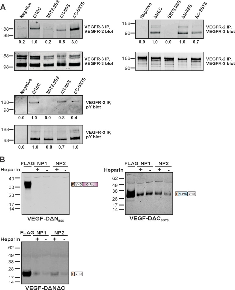
VEGF-D is an angiogenic and lymphangiogenic glycoprotein that can be proteolytically processed generating various forms differing in subunit composition due to the presence or absence of N- and C-terminal propeptides. These propeptides flank the central VEGF homology domain, that contains the binding sites for VEGF receptors (VEGFRs), but their biological functions were unclear. Characterization of propeptide function will be important to clarify which forms of VEGF-D are biologically active and therefore clinically relevant. Here we use VEGF-D mutants deficient in either propeptide, and in the capacity to process the remaining propeptide, to monitor the functions of these domains. We report for the first time that VEGF-D binds heparin, and that the C-terminal propeptide significantly enhances this interaction (removal of this propeptide from full-length VEGF-D completely prevents heparin binding). We also show that removal of either the N- or C-terminal propeptide is required for VEGF-D to drive formation of VEGFR-2/VEGFR-3 heterodimers which have recently been shown to positively regulate angiogenic sprouting. The mature form of VEGF-D, lacking both propeptides, can also promote formation of these receptor heterodimers. In a mouse tumor model, removal of only the C-terminal propeptide from full-length VEGF-D was sufficient to enhance angiogenesis and tumor growth. In contrast, removal of both propeptides is required for high rates of lymph node metastasis. The findings reported here show that the propeptides profoundly influence molecular interactions of VEGF-D with VEGF receptors, co-receptors, and heparin, and its effects on tumor biology.


Figures

FIGURE 1.
FIGURE 1.
Characterization of VEGF-DΔNIISS and VEGF-DΔCSSTS, and of receptor binding and activation. A, schematic representation of VEGF-DΔNIISS and VEGF-DΔCSSTS, asterisks denote arginine residues in the proteolytic cleavage sites that have been mutated to serine, N-pro and C-pro denote the propeptides, F denotes FLAG peptide and numbers denote positions in the primary structure of VEGF-D. The processing site mutated in VEGF-DΔCSSTS is the one at which the N-terminal propeptide has been reported to be most commonly cleaved from the VHD (defined previously as the “major” site) (28). B, immunoprecipitation and Western blotting (IP, Blot) and Coomassie Blue staining (Coom). Conditioned media from transiently transfected 293F cells expressing VEGF-DΔNIISS (ΔN-IISS) or VEGF-DΔCSSTS (ΔC-SSTS) were subjected to immunoprecipitation, reducing SDS-PAGE and Western blotting. The immunoprecipitation and Western blots were conducted with M2 anti-FLAG antibody for VEGF-DΔNIISS, and with anti-VHD antibodies for VEGF-DΔCSSTS. For Coomassie staining, VEGF-DΔNIISS and VEGF-DΔCSSTS stably expressed in 293EBNA cells were purified by anti-FLAG affinity chromatography and subjected to reducing SDS-PAGE. Sizes of molecular weight markers (kDa) are shown to the left and schematics of VEGF-D species to the right. C, results from biosensor analysis of purified VEGF-DΔNIISS and VEGF-DΔCSSTS binding to the extracellular domains of VEGFR-2 and VEGFR-3. Previously published results for purified VEGF-DSSTS.IISS and VEGF-DΔNΔC (29) are shown for comparison. Representative sensorgrams for the interactions of VEGF-DΔNIISS and VEGF-DΔCSSTS with receptor constructs are shown in supplemental Fig. S1. D, receptor activation. HMVECs were stimulated for 10 min with the indicated concentrations of purified VEGF-D variants and used in VEGFR-2 (left panel) or VEGFR-3 (right panel) phosphorylated and total ELISAs. Receptor activation is measured by the ratio of phosphorylated to total VEGF receptor. Error bars indicate S.E., and asterisks denote p < 0.05 (Student's t test).
FIGURE 2.
FIGURE 2.
Induction of VEGFR-2/VEGFR-3 heterodimers and neuropilin interactions. A, induction of receptor heterodimers. HMVECs were stimulated for 10 min with purified VEGF-DΔNIISS (ΔN-IISS), VEGF-DΔCSSTS (ΔC-SSTS), VEGF-DSSTS.IISS (SSTS.IISS) (all at 500 ng/ml), or VEGF-DΔNΔC (200 ng/ml), or left unstimulated (Negative). Lysates were immunoprecipitated with an antibody against VEGFR-3 and analyzed by reducing SDS-PAGE and Western blot with an antibody against VEGFR-2 to assess heterodimers, or with an antibody against VEGFR-3 to confirm the presence of this receptor (upper, left panels). Alternatively, lysates were immunoprecipitated with an antibody against VEGFR-2 and analyzed by SDS-PAGE/Western blot with an antibody against VEGFR-3 to assess heterodimers, or with an antibody against VEGFR-2 to confirm the presence of this receptor (upper right panels). Activation of receptors was assessed by immunoprecipitation with VEGFR-2 or VEGFR-3 antibodies and SDS-PAGE/Western blot with an antibody against phosphotyrosine (bottom panels). VEGFR-2 migrates predominantly at ∼230 kDa, whereas VEGFR-3 migrates as three bands, a ∼125 kDa cleaved form and two uncleaved forms of ∼175 kDa and ∼195 kDa that differ in the degree of glycosylation (56). Sizes of molecular weight markers (kDa) are shown to the left. Numbers below panels represent the relative intensities of the major bands, with the values for the major ΔNΔC band in each blot defined as 1.0. Black lines within Western blots indicate removal of irrelevant tracks from the images, all lanes in a blot come from the same exposure of the same membrane. B, neuropilin interactions. Conditioned media from 293EBNA-1 cells expressing VEGF-D variants were precipitated with NP1- or NP2-Ig fusion proteins, in the presence or absence of 10 μg/ml heparin, and analyzed by reducing SDS-PAGE and Western blotting for VEGF-D with antibodies recognizing the FLAG tag. Tracks labeled FLAG indicate controls in which VEGF-D variants were precipitated with anti-FLAG beads. Sizes of molecular weight markers (kDa) are shown to the left and schematics of VEGF-D variants to the right.
FIGURE 3.
FIGURE 3.
Analysis of heparin binding. Binding of VEGF-D variants, and VEGF-A165, to heparin was analyzed by affinity chromatography. Purified proteins were loaded onto heparin-Sepharose columns and flowthrough (FT), containing unbound protein, was collected. Bound proteins were eluted from columns using buffers of increasing NaCl concentration (M), as indicated above the blots. Fractions were analyzed by reducing SDS-PAGE and Western blotting with antibodies targeting FLAG, to detect VEGF-D variants, or with an anti-VEGF-A antibody. Purified protein samples (S) that had not been subjected to heparin Sepharose affinity chromatography are shown for comparison. Sizes of molecular weight markers (kDa) are shown to the left.
FIGURE 4.
FIGURE 4.
Growth and morphology of tumors expressing VEGF-D variants, and analysis of angiogenesis. 293EBNA-1 cells stably expressing various forms of VEGF-D were injected subcutaneously into the flanks of SCID/NOD mice to generate tumors (n = 10 per study group). A, VEGF-D expression in tumors. VEGF-D proteins in lysates from tumors expressing VEGF-DΔNΔC (ΔNΔC), VEGF-DΔNIISS (ΔN-IISS) or VEGF-DΔCSSTS (ΔC-SSTS), or from Vector Control tumors, were immunoprecipitated and analyzed by reducing SDS-PAGE and Western blotting using antibodies targeting the VHD. Sizes of molecular weight markers (in kDa) are shown to the left. The black line within the Western blot indicates removal of irrelevant tracks from the image, all lanes in the blot come from the same exposure of the same membrane. The blot compares one tumor from each study group, and the results are representative of a series of immunoprecipitations/Western blots comparing five tumors from each group. Protein concentrations of all lysates were measured by spectrophotometry to ensure that all lysate samples used for immunoprecipitations were matched for total protein content. B, growth rates of subcutaneous tumors. Asterisks indicate days on which VEGF-DΔNIISS tumors were significantly smaller than both VEGF-DΔNΔC and VEGF-DΔCSSTS tumors (p < 0.05; Student's t test). Error bars represent S.E. C, macroscopic appearance of tumors upon reaching a volume of ∼2,500 mm3. Data generated from VEGF-DΔNΔC tumors has been reported previously (34) and is shown for comparison. D, analysis of tumor blood vessels. Tissue sections from tumors expressing different forms of VEGF-D were analyzed by immunohistochemistry with antibodies to PECAM-1 (brown staining) for blood vessels (left panels). Graph shows quantitation of blood vessel endothelium in tumor sections as assessed by the number of pixels positively stained for PECAM-1. Asterisks indicate statistically significant differences in the abundance of blood vessel endothelium (p < 0.01; Student's t test). Error bars represent standard error of the mean. The experiments described in this figure were conducted twice with similar results in each case, the results of one experiment are shown here.
FIGURE 5.
FIGURE 5.
Impact of N- and C-terminal propeptides on lymph node metastasis and lymphangiogenesis. A, effects on lymph node metastasis. Representative images of lymph node tissue sections, stained with hematoxylin and eosin, that were taken from mice with primary tumors ∼2,500 mm3 in size. Top panel shows lymph node from a mouse with a metastatic VEGF-DΔNΔC tumor; bottom panel shows a lymph node from a mouse with a non-metastatic Vector Control tumor. Arrowheads indicate clusters of tumor cells. B, occurrence of lymph node metastasis in tumor xenografts. Mice were scored positive for metastasis if tumor cells were observed in the axillary lymph nodes. Numbers indicate the proportion of mice with primary tumors that scored positive for lymph node metastasis. Asterisks indicate those groups with significantly lower rates of metastasis than the VEGF-DΔNΔC study group, as determined by the Kruskal-Wallis test (p < 0.05). Data generated from VEGF-DΔNΔC tumors has been reported previously (34) and is shown for comparison. The data from two independent experiments is combined in the table. C, analysis of tumor lymphatic vessels. Tissue sections were analyzed by immunohistochemistry with antibodies to LYVE-1 (brown staining) for lymphatic vessels (left panels). Graph shows quantitation of lymphatic endothelium in tumor sections as assessed by the number of pixels positively stained for LYVE-1. ΔN-IISS denotes VEGF-DΔNIISS and ΔC-SSTS denotes VEGF-DΔCSSTS. Asterisks indicate statistically significant differences in abundance of lymphatic endothelium (p < 0.01: Student's t test). Error bars represent S.E. The experiments described in this figure were conducted twice with similar results in each case, the results of one experiment are shown here in panels A and C.

Similar articles

Cited by

References

    1. Tammela T., Alitalo K. (2010) Lymphangiogenesis: Molecular mechanisms and future promise. Cell 140, 460–476 - PubMed
    1. Achen M. G., McColl B. K., Stacker S. A. (2005) Focus on lymphangiogenesis in tumor metastasis. Cancer Cell 7, 121–127 - PubMed
    1. Potente M., Gerhardt H., Carmeliet P. (2011) Basic and therapeutic aspects of angiogenesis. Cell 146, 873–887 - PubMed
    1. Seyama K., Kumasaka T., Kurihara M., Mitani K., Sato T. (2010) Lymphangioleiomyomatosis: a disease involving the lymphatic system. Lymphat. Res. Biol. 8, 21–31 - PubMed
    1. Achen M. G., Jeltsch M., Kukk E., Mäkinen T., Vitali A., Wilks A. F., Alitalo K., Stacker S. A. (1998) Vascular endothelial growth factor D (VEGF-D) is a ligand for the tyrosine kinases VEGF receptor 2 (Flk1) and VEGF receptor 3 (Flt4). Proc. Natl. Acad. Sci. U.S.A. 95, 548–553 - PMC - PubMed

Publication types

MeSH terms

Substances