. 2013 Mar 12;110(11):E986-95.
doi: 10.1073/pnas.1214313110. Epub 2013 Feb 12.

Direct activation of full-length proapoptotic BAK

Affiliations

Direct activation of full-length proapoptotic BAK

Elizaveta S Leshchiner et al. Proc Natl Acad Sci U S A. .

Abstract

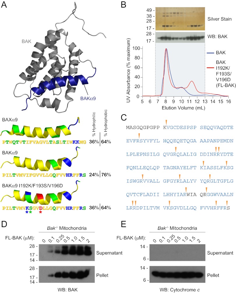
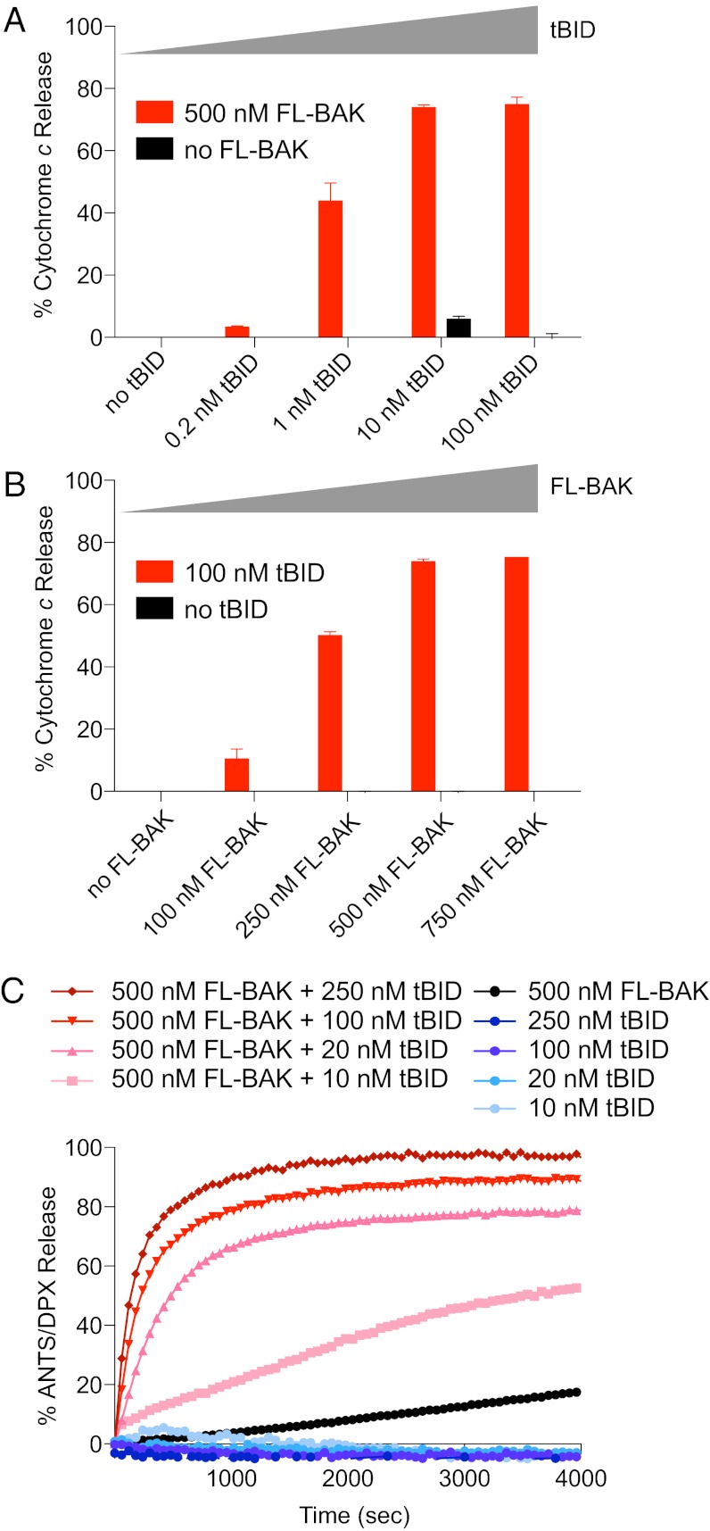
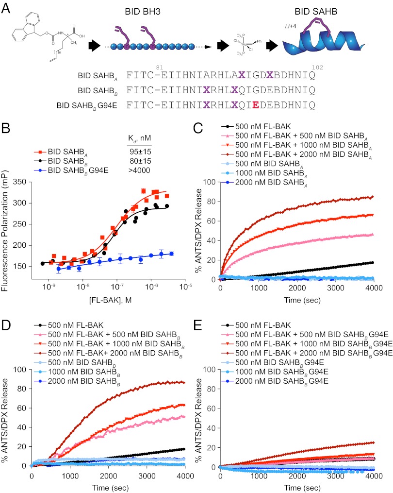
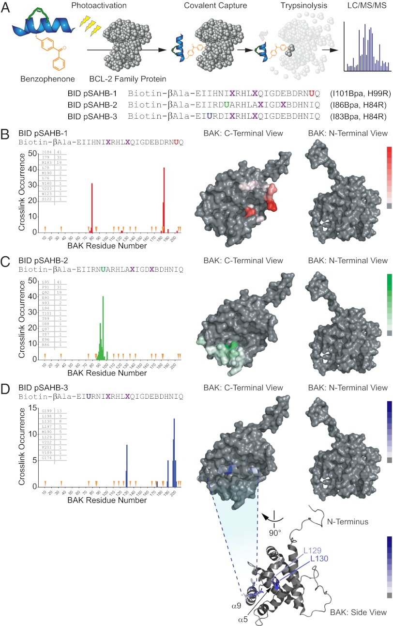
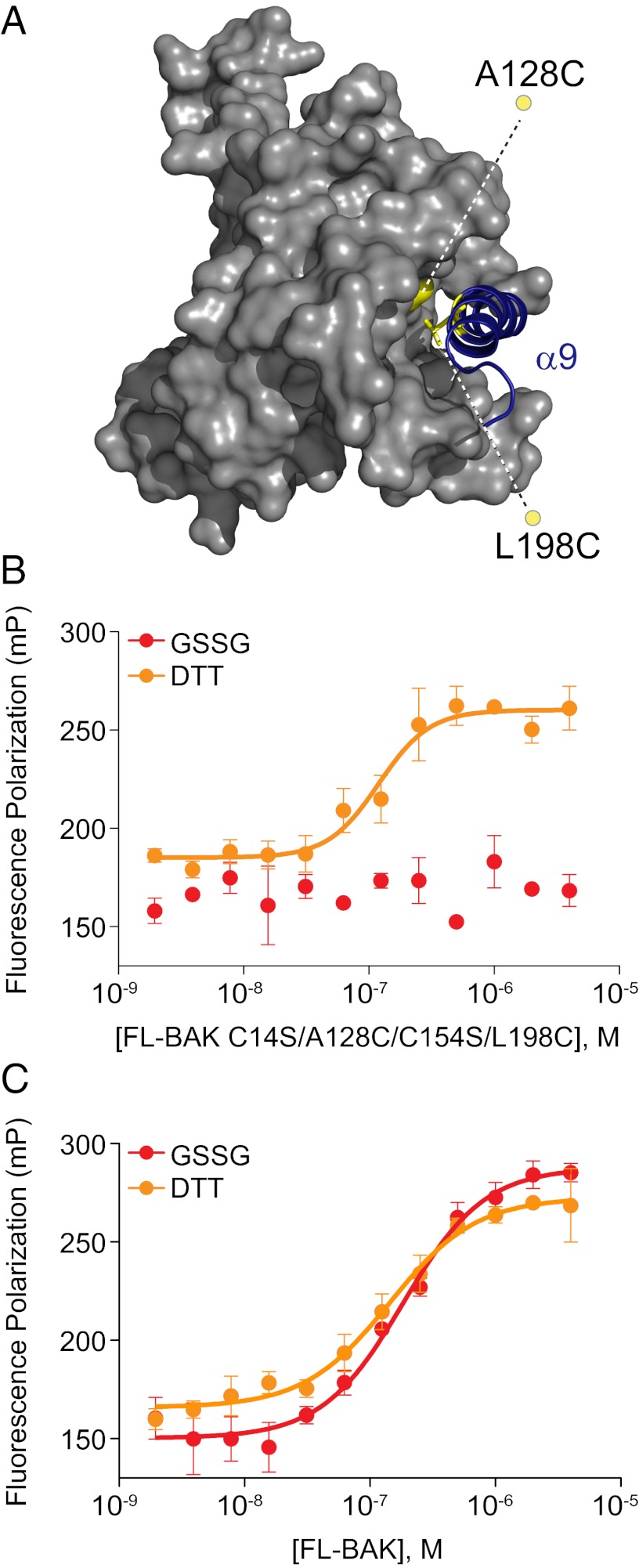
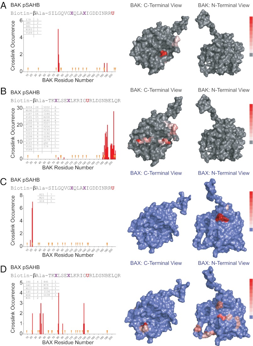
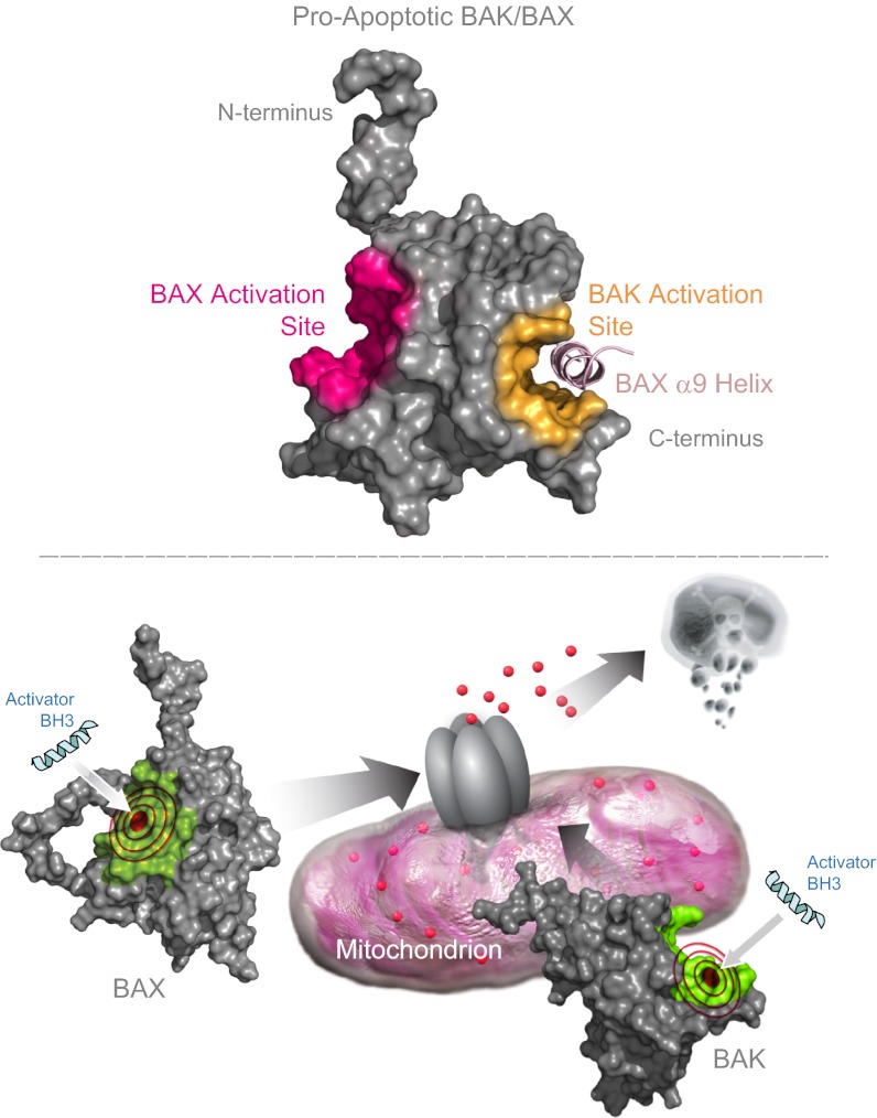
Proapoptotic B-cell lymphoma 2 (BCL-2) antagonist/killer (BAK) and BCL-2-associated X (BAX) form toxic mitochondrial pores in response to cellular stress. Whereas BAX resides predominantly in the cytosol, BAK is constitutively localized to the outer mitochondrial membrane. Select BCL-2 homology domain 3 (BH3) helices activate BAX directly by engaging an α1/α6 trigger site. The inability to express full-length BAK has hampered full dissection of its activation mechanism. Here, we report the production of full-length, monomeric BAK by mutagenesis-based solubilization of its C-terminal α-helical surface. Recombinant BAK autotranslocates to mitochondria but only releases cytochrome c upon BH3 triggering. A direct activation mechanism was explicitly demonstrated using a liposomal system that recapitulates BAK-mediated release upon addition of BH3 ligands. Photoreactive BH3 helices mapped both triggering and autointeractions to the canonical BH3-binding pocket of BAK, whereas the same ligands crosslinked to the α1/α6 site of BAX. Thus, activation of both BAK and BAX is initiated by direct BH3-interaction but at distinct trigger sites. These structural and biochemical insights provide opportunities for developing proapoptotic agents that activate the death pathway through direct but differential engagement of BAK and BAX.


Conflict of interest statement

Conflict of interest statement: L.D.W. is a scientific advisory board member and consultant for Aileron Therapeutics.

Figures

Fig. 1.
Fig. 1.
Expression and purification of recombinant, full-length, and monomeric BAK. (A) The α9 helix of BAK was subjected to triple mutagenesis (I192K/F193S/V196D) to increase its hydrophilicity and thereby better match that of soluble, full-length BAX. The pictured model structure of BAK was calculated based on the solution structure of BAX using Modeller software (51), with BAK α9 colored blue. The hydrophobic, hydrophilic, positively charged, and negatively charged residues of the BAK and BAX α9 helices are colored yellow, green, blue, and red, respectively. (B) A comparison of the SEC elution profiles of BAK and FL-BAK demonstrates that mutagenesis enabled the isolation of a monomeric species (11- to 12-mL fractions). Silver staining and anti-BAK Western analysis of the electrophoresed FL-BAK fractions documented the isolation of monomeric BAK protein (∼23 kDa). (C) The identity of the isolated monomeric protein was confirmed to be FL-BAK by MS analysis. Tryptic sites are indicated by the arrowheads, and the FL-BAK sequences identified by LC-MS/MS are colored blue. (D) Upon exposure to isolated Bak−/− mouse liver mitochondria, FL-BAK dose-responsively translocated to the mitochondria-containing pellet fraction, as detected by anti-BAK Western analysis. (E) Autotranslocation of FL-BAK to the mitochondrial fraction did not induce cytochrome c release from the pellet to the supernatant, as measured by anti-cytochrome c Western analysis.
Fig. 2.
Fig. 2.
The BH3-only protein tBID triggers FL-BAK–dependent mitochondrial and liposomal release. (A and B) tBID dose-responsively triggered FL-BAK–mediated cytochrome c release from isolated Bak−/− mitochondria, as measured by cytochrome c ELISA. Likewise, FL-BAK dose-responsively induced cytochrome c release upon exposure to fixed-dose tBID. No release was observed upon exposure of the mitochondria to tBID or FL-BAK alone. Experiments were performed in duplicate and were repeated at least three times using independent FL-BAK preparations with similar results. Data are mean ± SD. (C) tBID dose-responsively induced FL-BAK–mediated liposomal release of entrapped fluorophore, whereas tBID or FL-BAK alone had little to no effect. Data are representative of at least three independent experiments with similar results.
Fig. 3.
Fig. 3.
BID SAHBs bind to FL-BAK in a sequence-dependent manner and directly trigger FL-BAK activation. (A) SAHBs corresponding to the BH3 motif of the BH3-only protein BID were generated by substituting nonnatural amino acids bearing olefin tethers at i, i+4 positions followed by ruthenium-catalyzed olefin metathesis. G94E point mutagenesis of the hydrophobic binding interface yielded a negative control SAHB for biochemical studies. X, stapling amino acid; B, norleucine (substituted for methionine to avoid thioether-based interference with ruthenium catalysis). (B) BID SAHBs A and B bound to FL-BAK with nanomolar affinity, whereas G94E point mutagenesis abrogated the interaction, as measured by FP assay. Experiments were performed at least in duplicate and were repeated three times with independent preparations of FL-BAK. Data are mean ± SEM. (CE) BID SAHBs A and B dose-responsively induced FL-BAK–mediated liposomal release of entrapped fluorophore, whereas BID SAHBB G94E, or BID SAHBs or FL-BAK alone, had little to no effect. Data are representative of at least three independent experiments with similar results.
Fig. 4.
Fig. 4.
Photoreactive BID pSAHBs localize the BH3 trigger site on BAK to its C-terminal BH3-binding pocket. (A) pSAHBs modeled after the BH3 domain of BID were generated for protein capture and binding-site identification by replacing select native residues with 4-benzoyl-L-phenylalanine (Bpa), followed by ring-closing metathesis of olefinic nonnatural amino acids installed at i, i+4 positions. To facilitate tryptic digestion of pSAHBs into shorter and more identifiable fragments by MS, single arginine substitutions were made as indicated, taking advantage of natural sites of homology between the human and mouse BID BH3 domains (i.e., H84R, H99R). X, stapling amino acid; B, norleucine; U, Bpa. (BD) BID pSAHBs 1–3 were incubated individually with FL-BAK, and the mixtures were subjected to UV irradiation, electrophoresis, excision of the crosslinked protein, trypsin proteolysis, and LC-MS/MS analysis. The plots depict the frequency of crosslinked sites identified across the FL-BAK polypeptide sequence. Crosslinked residues are mapped onto a calculated model structure of FL-BAK (based on sequence homology to BAX) and colored according to the frequency of occurrence for each pSAHB (1, red; 2, green; 3, blue). For BID pSAHBs 1 and 2, which do not crosslink to residues of the C-terminal helix of FL-BAK, α9 has been removed from the structure to better visualize the crosslinked residues at the surface of the canonical BH3-binding pocket. For BID pSAHB-3, a side view of FL-BAK is also shown to demonstrate the localization of crosslinked residues at both the surface of the canonical BH3-binding pocket and the inner surface of α9. The latter dataset shows that, after α9 displacement, previously unexposed and inward-facing α9 residues become available for interaction with BID pSAHB-3.
Fig. 5.
Fig. 5.
The binding interaction between BID SAHB and FL-BAK requires access to the canonical BH3-binding pocket. (A) To determine if the BID SAHB/FL-BAK interaction is explicitly dependent on displacement of α9 (blue) and exposure of the canonical pocket, FL-BAK residues A128 of α5 and L198 of α9 were mutated to cysteines (yellow), and native cysteines C14 and C154 were mutated to serines, yielding an α9-tethered FL-BAK construct in which the C-terminal helix is reversibly locked into its binding pocket by redox conditions. (B) Oxidized FL-BAK (2 mM GSSG) showed no interaction with FITC-BID SAHBB, whereas binding activity was completely restored upon addition of reducing agent (10 mM DTT), as assessed by FP assay. Experiments were performed at least in duplicate and were repeated at least two times with independent preparations of FL-BAK. Data are mean ± SEM. (C) In contrast, redox conditions have no effect on the binding interaction between FITC-BID SAHBB and FL-BAK. Experiments were performed in at least duplicate and repeated at least twice with independent preparations of FL-BAK. Data are mean ± SEM.
Fig. 6.
Fig. 6.
The BAK/BAX BH3 helices likewise engage distinct trigger surfaces on FL-BAK and BAX. (A and B) BAK and BAX pSAHBs were incubated individually with FL-BAK, and the mixtures were subjected to UV irradiation, electrophoresis, excision of the crosslinked protein, trypsin proteolysis, and LC-MS/MS analysis. The plots depict the frequency of crosslinked sites identified across the FL-BAK polypeptide sequence. Crosslinked residues are mapped onto the calculated model structure of FL-BAK (gray) and colored according to the frequency of occurrence as in Fig. 4. Because BAK pSAHB crosslinks to FL-BAK α9 were not identified, the C-terminal helix was removed from the calculated FL-BAK structure to better visualize the crosslinked residues at the surface of the canonical BH3-binding pocket. (C and D) When exposed to full-length BAX, the identical BAK and BAX pSAHBs crosslinked to a series of residues at the α1/α6 trigger site, with no canonical site crosslinks observed for BAK pSAHB and a select few identified for BAX pSAHB. The plots depict the frequency of crosslinked sites identified across the BAX polypeptide sequence. Crosslinked residues are mapped onto the structure of BAX (blue) and colored according to the frequency of occurrence as in Fig. 4. Because pSAHB crosslinks to BAX α9 were not evident, the C-terminal helix was removed from the BAX structure to better visualize the BAX pSAHB-crosslinked residues at the surface of the canonical BH3-binding pocket. X, stapling amino acid; B, norleucine; U, Bpa.
Fig. P1.
Fig. P1.
BH3 interaction at geographically distinct trigger sites initiates the direct activation of full-length BAK and BAX. (Upper) The expression and purification of recombinant, monomeric, and full-length BAK enabled a comparison of the direct activation mechanisms for BAK and BAX, the two essential executioner proteins of the mitochondrial apoptosis pathway. The initiating site for BH3-triggered direct BAX activation lies at the N-terminal side of BAX, whereas the BH3 interaction site for direct BAK activation localizes to the canonical BH3-binding site at the C-terminal face of BAK. (Lower) Our biochemical and structural analyses suggest that activating BH3 interactions at the canonical BH3-binding pocket in the context of the mitochondrial outer membrane may reflect a common mechanism for propelling the activation and oligomerization of BAK and BAX, with the N-terminal triggering mechanism of cytosolic BAX representing a unique afferent step required to initiate its activation and mitochondrial translocation.

References

    1. Wei MC, et al. tBID, a membrane-targeted death ligand, oligomerizes BAK to release cytochrome c. Genes Dev. 2000;14(16):2060–2071. - PMC - PubMed
    1. Wei MC, et al. Proapoptotic BAX and BAK: A requisite gateway to mitochondrial dysfunction and death. Science. 2001;292(5517):727–730. - PMC - PubMed
    1. Jürgensmeier JM, et al. Bax directly induces release of cytochrome c from isolated mitochondria. Proc Natl Acad Sci USA. 1998;95(9):4997–5002. - PMC - PubMed
    1. Yang J, et al. Prevention of apoptosis by Bcl-2: Release of cytochrome c from mitochondria blocked. Science. 1997;275(5303):1129–1132. - PubMed
    1. Du C, Fang M, Li Y, Li L, Wang X. Smac, a mitochondrial protein that promotes cytochrome c-dependent caspase activation by eliminating IAP inhibition. Cell. 2000;102(1):33–42. - PubMed

Publication types

MeSH terms