Review
. 2013 Sep;9(3):285-306.
doi: 10.1007/s11302-013-9357-4. Epub 2013 Feb 13.

Purinergic signaling in inflammatory cells: P2 receptor expression, functional effects, and modulation of inflammatory responses

Affiliations
Review

Purinergic signaling in inflammatory cells: P2 receptor expression, functional effects, and modulation of inflammatory responses

Fenila Jacob et al. Purinergic Signal. 2013 Sep.

Abstract

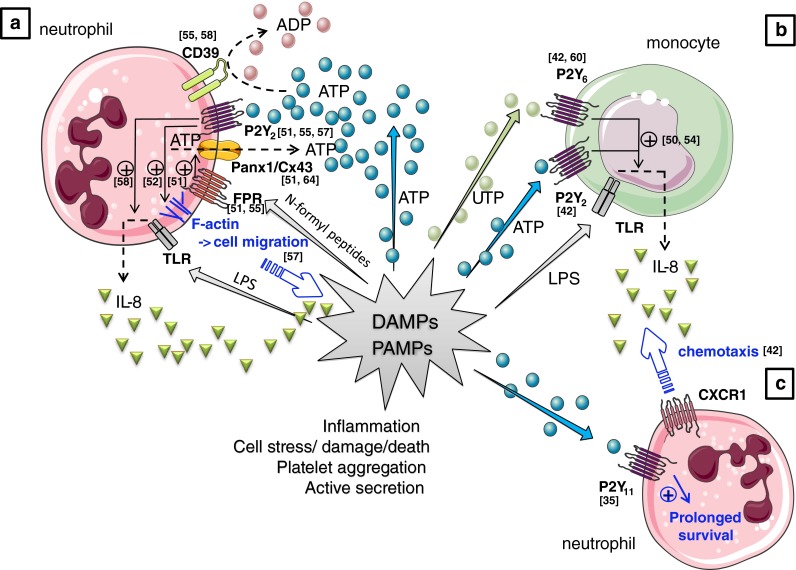
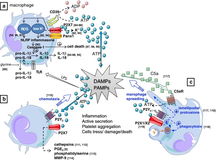
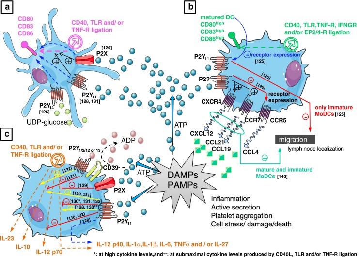
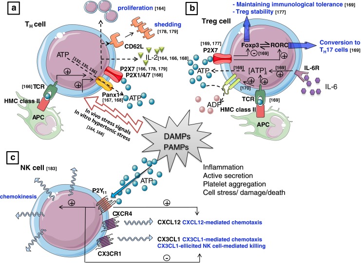
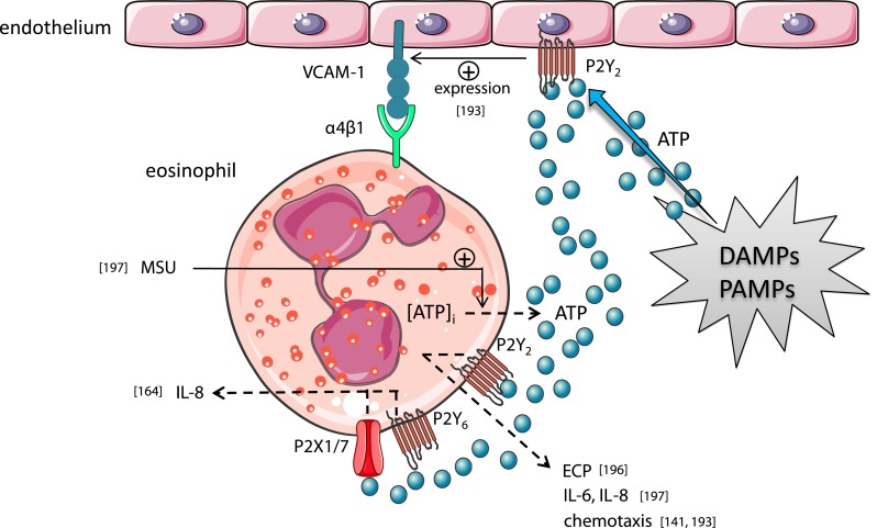
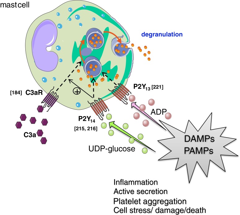
Extracellular ATP and related nucleotides promote a wide range of pathophysiological responses via activation of cell surface purinergic P2 receptors. Almost every cell type expresses P2 receptors and/or exhibit regulated release of ATP. In this review, we focus on the purinergic receptor distribution in inflammatory cells and their implication in diverse immune responses by providing an overview of the current knowledge in the literature related to purinergic signaling in neutrophils, macrophages, dendritic cells, lymphocytes, eosinophils, and mast cells. The pathophysiological role of purinergic signaling in these cells include among others calcium mobilization, actin polymerization, chemotaxis, release of mediators, cell maturation, cytotoxicity, and cell death. We finally discuss the therapeutic potential of P2 receptor subtype selective drugs in inflammatory conditions.


Figures

Fig. 1
Fig. 1
Purinergic mechanisms in neutrophil biology. a Activation of the formyl-peptide receptor (FPR) by N-formyl peptides produced by the degradation of either bacterial or host cells (which constitutes together with many other molecules, including ATP, the group of PAMPs and DAMPs) stimulates localized ATP release from neutrophils [51, 55] which occurs through pannexin-1 (panx1) and/or connexin 43 (Cx43) hemichannels [51, 64] resulting in the activation of nearby P2Y2 receptors on the neutrophils [51, 55, 57] This autocrine P2Y2 receptor activation will subsequently amplify gradient sensing of chemotactic signals (e.g., N-formyl peptides [51, 55] and interleukin (IL)-8 [57] by stimulating F-actin to the leading edge [51, 52, 55]. P2Y2 receptor ligation is also implicated in the potentiation of IL-8 production by human neutrophils in response to the bacterial endotoxin LPS [58], a mechanism under tight control by the ectonucleotidase “Ecto-nucleoside triphosphate diphosphohydrolase 1” (E-NTPDase1 or CD39) expressed on the surface of neutrophils [55, 58] by breaking down ATP to ADP. b Next to neutrophilic P2Y2 receptors, extracellular nucleotides (UTP and ATP) were also reported to regulate neutrophil migration by controlling TLR-induced IL-8 release from monocytes via P2Y2 and/or P2Y6 receptors expressed on the latter cell types [42, 60]. c ATP, via P2Y11 might also cause a long-lasting delay in constitutive neutrophil apoptosis [35]. DAMP damage-associated molecular patterns, LPS lipopolysaccharide, PAMP pathogen-associated molecular patterns, TLR toll-like receptor. Values between brackets in the figure represent reference numbers
Fig. 2
Fig. 2
a Challenging macrophages with PAMPs such as LPS stimulates synthesis of pro-IL-1β and pro-IL-18. Secretion of the active cytokines requires a secondary signal that is provided by ATP through P2X7 ligation [–88], yielding the conversion of pro-IL-1β into active IL-1β by NLRP3 inflammasome activated caspase-1 enzymatic cleavage [90]. The activation process of caspase-1 is initiated via P2X7-mediated cytosolic K+ outflow [82, 94], or alternatively via the activation of ROS [94, 96, 97]. Also the pro-IL-1β and pro-IL-18 can be released via a distinct caspase-1 independent pathway which is blocked by glycine [89]. Prolonged stimulation with ATP also leads to caspase-1 dependent, but IL-1β and IL-18 cytokine processing independent cell death in macrophages [87, 98, 99]. b P2X7 receptor activation has also been shown to signal caspase-1 and IL-1β/IL-18 independent release of cathepsins [111, 112], PGE2 [8], phosphatidylserine [113], and MMP-9 [114], all of which are implicated in cellular processes that play a defined role in inflammation. ATP is also reported to act as a long-range “find me” signal (chemoattractant) to recruit monocytes and macrophages [115]. c The role of ATP as “real” direct chemoattractant for macrophages is however controversial; the purinergic system can also act as an indirect chemoattractant that navigates macrophages in a gradient of the “real” chemoattractant C5a via autocrine release of ATP induced by the C5a receptor (C5aR), yielding an amplification in the gradient sensing via an orientated generation of lamellipodial membrane protrusions involving P2Y2 and P2Y12 receptors to furthermore induce an indirect effect of chemotaxis and the promotion of phagocytosis [117, 118]. Enhanced phagocytic effect in macrophages by purinergic stimuli can also involve the ligation of P2X1 or P2X3 receptors [119]. DAMP damage-associated molecular patterns, LPS lipopolysaccharide, MMP-9 matrix metalloproteinase-9, PAMP pathogen-associated molecular patterns, TLR toll-like receptor, PGE2 prostaglandin E2. Values between brackets in the figurerepresent reference numbers
Fig. 3
Fig. 3
Purinergic responses in monocyte-derived dendritic cells (MoDCs). a MoDC maturation: chronic stimulation of DCs with noncytotoxic doses of ATP increases membrane expression of CD80, CD83, and CD86, Moreover, ATP also enhanced soluble CD40 ligand (sCD40L), LPS, and/or TNF-α-induced maturation of moDCs via P2X receptor activation [129] These processes might also involve P2Y11 [128] and/or P2Y14 [126] receptor activation. b MoDC migration: ATP provides a signal for enhanced lymph node localization of DCs; ATP increased migration of immature and mature DCs to CXCL12, CCL19, and CCL21, whereas responses to CCL4 were reduced [140]. In contrast, exposure of MoDCs to gradients of ATP inhibited their migratory capacity via P2Y11 receptors [125]. During maturation (by sCD40L, LPS, TNF-α, INFγ, and/or PGE2), MoDCs downregulated P2Y11 receptor expression which yield mature DCs that are less sensitive to ATP-mediated inhibition of migration [125]. c MoDC cytokine release—equivocal data in the literature report either a potentiating or inhibitory effect on sCD40L LPS and/or TNF-α-induced expression of inflammatory cytokines. CCL19 chemokine (C–C motif) ligand 19, CCR7 C–C chemokine receptor type 7, CXCL12 chemokine (C–X–C motif) ligand 12, CXCR4 C–X–C chemokine receptor type 4, DAMP damage-associated molecular patterns, INFγ interferon gamma, INFGR INFγ receptor, LPS lipopolysaccharide, PAMP pathogen-associated molecular patterns, PGE2 prostaglandin E2, EP2/4-R PGE2 receptor 2 and/or 4, TLR toll-like receptor, TNF-α tumor necrosis factor alpha, TNFR TNF receptor. Values between brackets in figurerepresent reference numbers
Fig. 4
Fig. 4
a T helper (TH) cells: hypertonic saline promotes T cell proliferation and enhances IL-2 production of stimulated T cells by inducing the release of cellular ATP and by the autocrine activation of stimulatory P2X receptors [164]. Also, T cell receptor (TCR) stimulation itself triggers the release of ATP from the T cells which - via an autocrine feedback mechanism through P2X7 receptors [166] and/or P2X1 and P2X4 [168]—is required for the effective activation of T cells. This ATP release occurs through pannexin-1 hemichannels [167, 168]. ATP also triggers P2X7 receptor-dependent CD62L shedding in lymphocytes [178, 179] which is associated with T cell activation. b T regulatory (Treg) cells: CD4+ helper T cells upon stimulation of the TCR and potentiated by the inflammatory cytokine IL-6, increases the synthesis and release of ATP [169] which subsequently induces an autocrine P2X7-mediated signaling in Tregs [169] that inhibits the suppressive potential and stability of Tregs [177], and promotes their conversion to IL-17-secreting T Th17 effector cells [169]. However, TCR stimulation also enhances the expression of CD39 [170] which degrades ATP to ADP and AMP. Consequently, activated Treg cells are able to abrogate the P2X7-mediated suppression of Treg function and stability in an autologous way [169]. c Natural killer (NK) cells: NK cells stimulated with ATP increases chemokinesis and chemotaxis to CXCL12 via P2Y11 receptor activation, while chemotaxis and NK cell-mediated killing in response to CX3CL1 chemokine is inhibited [183]. APC antigen presenting cell, CX3CL1 chemokine (C–X3–C motif) ligand 1, CX3CR1 CX3C chemokine receptor 1, CXCL12 chemokine (C–X–C motif) ligand 12, CXCR4 C–X–C chemokine receptor type 4, DAMP damage-associated molecular patterns, Foxp3 forkhead box P3, HMC class II major histocompatibility complex class II, IL-6R interleukin-6 receptor, PAMP pathogen-associated molecular patterns, RORC retinoic acid receptor-related orphan receptor C. Values between brackets in figurerepresent reference numbers
Fig. 5
Fig. 5
Human eosinophils release eosinophil cationic protein (ECP) [196] IL-6 and IL-8 [197] by P2Y2 receptor stimulation, while P2X1, P2X7, and P2Y6 receptors might also be implicated in the release of IL-8 [196]. The P2Y2 receptor subtype expressed on eosinophils is also suggested to be implicated in eosinophil chemotaxis [141, 193]. Also endothelial P2Y2 receptors can affect eosinophil migration by regulating the expression of endothelial VCAM-1 which mediates migration and adhesion of eosinophils via the α4β1 integrin [193]. Monosodium urate (MSU) crystals induce the release of autocrine ATP from eosinophils which provides a pivotal positive feedback signal via P2 receptors on eosinophils [197]. DAMP damage-associated molecular patterns, PAMP pathogen-associated molecular patterns, VCAM-1 vascular cell adhesion molecule 1. Values between brackets in the figurerepresent reference numbers
Fig. 6
Fig. 6
P2Y13 and P2Y14 receptor-activation directly induces mast cell degranulation [215, 221] while P2Y14 receptors also potentiate C3a-induced degranulation [216]. DAMP damage-associated molecular patterns, PAMP pathogen-associated molecular patterns, C3a complement 3a, C3aR complement 3a receptor. Values between brackets in the figurerepresent reference numbers

Similar articles

Cited by

References

    1. Lotze MT, Zeh HJ, Rubartelli A, Sparvero LJ, Amoscato AA, et al. The grateful dead: damage-associated molecular pattern molecules and reduction/oxidation regulate immunity. Immunol Rev. 2007;220:60–81. doi: 10.1111/j.1600-065X.2007.00579.x. - DOI - PubMed
    1. Rubartelli A, Lotze MT. Inside, outside, upside down: damage-associated molecular-pattern molecules (DAMPs) and redox. Trends Immunol. 2007;28(10):429–436. doi: 10.1016/j.it.2007.08.004. - DOI - PubMed
    1. Khakh BS, North RA. P2X receptors as cell-surface ATP sensors in health and disease. Nature. 2006;442(7102):527–532. doi: 10.1038/nature04886. - DOI - PubMed
    1. Myrtek D, Idzko M. Chemotactic activity of extracellular nucleotideson human immune cells. Purinergic Signal. 2007;3(1–2):5–11. doi: 10.1007/s11302-006-9032-0. - DOI - PMC - PubMed
    1. Vitiello L, Gorini S, Rosano G, la Sala A (2012) Immunoregulation through extracellular nucleotides. Blood 120(3):511–518 - PubMed

Substances