. 2013 Apr 15;145(4):357-70.
doi: 10.1530/REP-12-0369. Print 2013 Apr.

Telocytes: ultrastructural, immunohistochemical and electrophysiological characteristics in human myometrium

Affiliations

Telocytes: ultrastructural, immunohistochemical and electrophysiological characteristics in human myometrium

Sanda M Cretoiu et al. Reproduction. .

Abstract

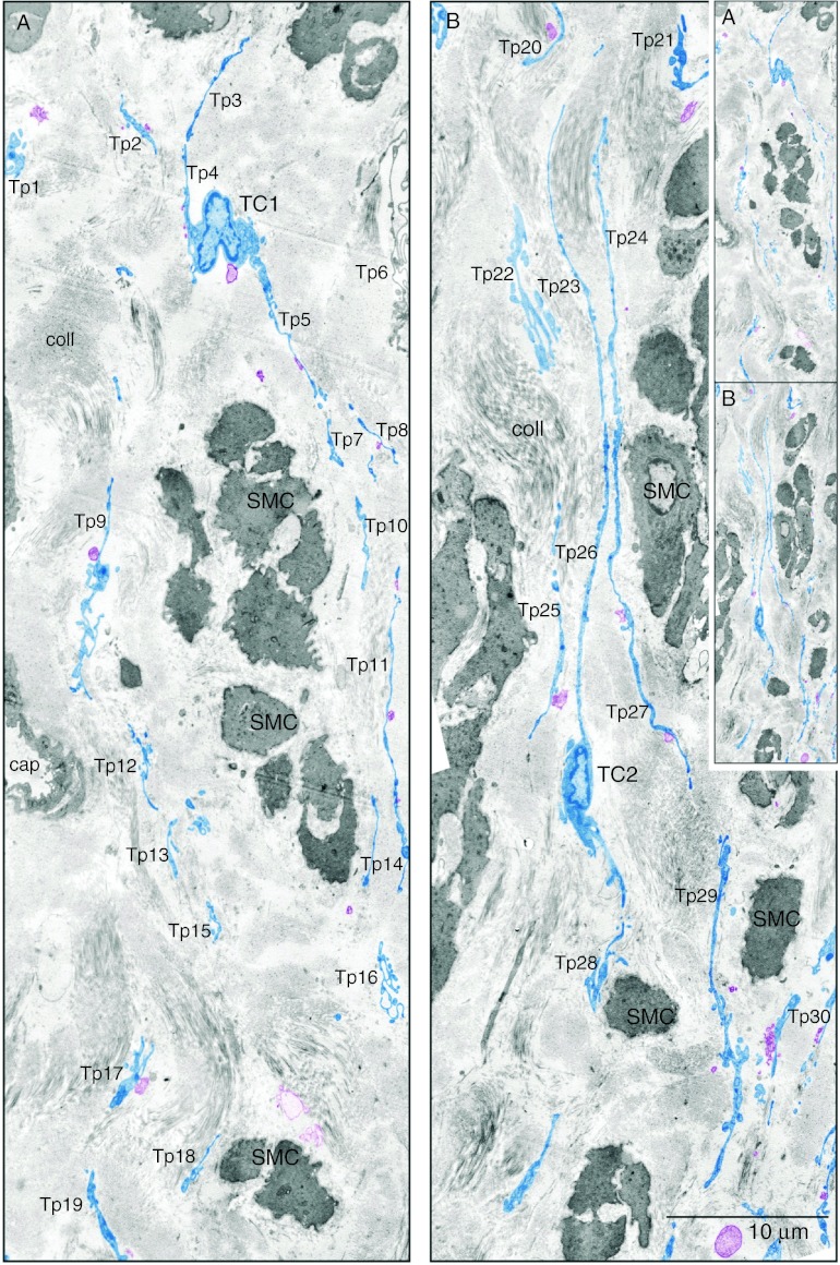
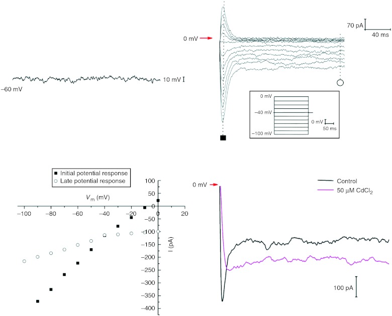
Telocytes (TCs) have been described in various organs and species (www.telocytes.com) as cells with telopodes (Tps) - very long cellular extensions with an alternation of thin segments (podomers) and dilated portions (podoms). We examined TCs using electron microscopy (EM), immunohistochemistry (IHC), immunofluorescence (IF), time-lapse videomicroscopy and whole-cell patch voltage clamp. EM showed a three-dimensional network of dichotomous-branching Tps, a labyrinthine system with homocellular and heterocellular junctions. Tps release extracellular vesicles (mean diameter of 160.6±6.9 nm in non-pregnant myometrium and 171.6±4.6 nm in pregnant myometrium), sending macromolecular signals to neighbouring cells. Comparative measurements (non-pregnant and pregnant myometrium) of podomer thickness revealed values of 81.94±1.77 vs 75.53±1.81 nm, while the podoms' diameters were 268.6±8.27 vs 316.38±17.56 nm. IHC as well as IF revealed double c-kit and CD34 positive results. Time-lapse videomicroscopy of cell culture showed dynamic interactions between Tps and myocytes. In non-pregnant myometrium, patch-clamp recordings of TCs revealed a hyperpolarisation-activated chloride inward current with calcium dependence and the absence of L-type calcium channels. TCs seem to have no excitable properties similar to the surrounding smooth muscle cells (SMCs). In conclusion, this study shows the presence of TCs as a distinct cell type in human non-pregnant and pregnant myometrium and describes morphometric differences between the two physiological states. In addition, we provide a preliminary in vitro electrophysiological evaluation of the non-pregnant state, suggesting that TCs could influence timing of the contractile activity of SMCs.


Figures

Figure 1
Figure 1
Human pregnant myometrium. Semi-thin sections (∼1 μm thick) of Epon-embedded samples, stained with toluidine blue. Typical TCs (red dashed lines) are seen embracing the cross-sectioned SMC. Note the triangular morphology of the TC body (*). Original magnification 100×, oil immersion.
Figure 2
Figure 2
Human pregnant myometrium. Phenotypic profile assessments of paraffin-embedded sections: (A) two spindle-shaped TCs are marked by c-kit staining and can be easily distinguished from a mast cell which can be recognised by its granular content. (B) An elongated TC is detected between myocytes by CD34 staining. Nuclei are counterstained with Mayer's hemalun. Original magnification 100×, oil immersion.
Figure 3
Figure 3
TCs in human non-pregnant myometrium. Two-dimensional sequenced concatenation of six electron micrographs portraying TCs and their Tps digitally coloured in blue. At least 30 Tps can be counted in the two-dimensional reconstruction. The inset demonstrates the continuity of figures (A) and (B). (A) One can observe a triangular TC1 cell body in the interstitial space between smooth muscle cells (SMC). (B) A spindle-shaped TC2 from which are detached very long Tps. In both TCs note the heterochromatin mostly confined to the periphery of the nucleus but also dispersed throughout. Tps of different TCs can be seen across the space between SMC. Tps are highlighted in blue revealing a network disposition. Most times they are disposed in parallel (B) and surround collagen bundles of different orientations. All Tps and have dilated regions – podoms – and thin segments – podomers. Released vesicles (coloured purple) can be seen emerging or in close proximity of Tps. Vesicles can be formed either by TC membrane blebbing (SMVs) or by TC secretion (exosomes). Sometimes, Tps are seen forming a labyrinthine system by three-dimensional convolution and overlapping – Tp9, Tp16 and Tp21 – and are communicating through junctions. The inset shows that images (A) and (B) are continuous and represent a two-dimensional concatenation of eight sequenced micrographs.
Figure 4
Figure 4
High-magnification micrographs of ultrathin sections. TC–TC junctions between Tps in human non-pregnant (A and B) and pregnant myometrium (C). (A) Three overlapping Tps (Tp1, Tp2 and Tp3) are connected by nanocontacts. In the encircled dotted area the two plasma membranes overlap over a length of 1.1 μm and are spaced at an average distance of 13 nm. The rectangle dotted area depicts an intermediate phase before the nanocontacts are formed that we called ante-nanocontacts where overlapping membranes (over a distance of 2.1 μm) are at a mean distance of 30 nm. Note the mitochondria (m) which are frequently seen in the podoms. SMVs (arrowheads) are often captured and suggest a transfer of information between Tp4 and Tp5. (B) Numerous Tps (Tp1–Tp7) in a region of close interrelations. Tp1 came in close contact with a cross-cut (*) Tp2 at distances between 7 and 12 nm within a 0.2-μm distance. In the middle upper part of the image one can observe a nanocontact between Tp3 and a podom rich in mitochondria (m) caveolae (arrowhead) and ER. Intermembrane distance is 13 nm and the planar contact is 0.30 μm long. The encircled dotted area reveals different junctions between three Tps: a gap junction – intermembrane space 4 nm, 0.26 long, and two close contacts – both of 8 nm wide, 0.3 and 0.2 μm long respectively. The white dotted rectangle shows details of the gap junction: the intercellular space in this region which is crossed by a central densification. (C) Maximum magnification micrographs in human pregnant myometrium expose an electron-dense nanostructure – a puncta adherentia minima junction between the Tp3 and Tp4 ends. Scale bars: (A) 2 μm, (B) 0.5 μm and (C) 0.5 μm.
Figure 5
Figure 5
Tps width in human pregnant and non-pregnant myometrium. Third-order polynomial (poly.) trend line that has the best fit to a series of data points showed the distribution of podomers. The Tps widths are more evenly distributed (flatter trend line) in pregnant myometrium compared with non-pregnant myometrium. Most frequent Tps are in the range of 51–60 nm diameter for pregnant myometrium and 61–70 nm diameter for non-pregnant myometrium.
Figure 6
Figure 6
Extracellular vesicles released by Tps into the microenvironment. Boxplots displaying the extremes, the upper and lower quartiles and the median of the maximum difference between the two physiological conditions: pregnant and non-pregnant myometrium (abscissa). The boundary of the box closest to zero indicates the 25th percentile, a green triangle within the box marks the median, and the boundary of the box farthest from zero indicates the 75th percentile. The measured diameter of membranous extracellular organelles which include SMVs (100–1000 nm) and exosomes (30–90 nm) was expressed in nanometres on the ordinate.
Figure 7
Figure 7
Human pregnant myometrium. TCs in primary semi-confluent cultures (day 4) are vitally stained with Janus green B. The vital staining reveals mitochondria at the level of the cell body and of the podoms (arrowheads). Original magnification 20×. The very long Tps (dashed lines) have a ‘bead on a string’ appearance – given by the dilated podoms (which accommodate mitochondria) – and pass through or establish contacts with the myocytes.
Figure 8
Figure 8
Human myometrium cells in culture (the second passage): (A) control phase-contrast microscopy and (B) IF for c-kit and CD34 (red and green respectively) of the same microscopic fields. One can observe that TCs co-express c-kit and CD34 in the same area (yellow). Original magnification 60×, nuclear counterstaining with Hoechst 33342 (blue).
Figure 9
Figure 9
Human non-pregnant myometrium. Cell culture, the first passage. (A, B, C, D, E, F, G and H) Successive snapshots chosen from a time-lapse videomicroscopy recording. (A and B) A TC is moving towards the upper part of the microscopic field leaving behind a very long telopode. Immediately after, an SMC can be seen approaching the telopode (C) and establishing connections with it (D, arrowheads). Soon the SMC will twirl around the Tp (using it as guiding wire) (E) and then will detach from the dish (F, asterisk), divide (G, double asterisk) and then reattach (H). Objective, 20×. Scale bar 10 μm.
Figure 10
Figure 10
Patch-clamp recordings on TCs from human non-pregnant myometrium. (A) A zero current-clamp recording of the resting potential from a TC. (B) Hyperpolarisation-activated inward currents generated by 200 ms duration pulses from −100 to 0 mV, starting from a holding potential of −40 mV. (C) Current–voltage plot for the current records presented in (B) showing a slight inward rectification. (D) Calcium dependence of the hyperpolarisation-activated inward currents tested by extracellular cadmium (50 μM) application.

Similar articles

Cited by

References

    1. Allix S, Reyes-Gomez E, Aubin-Houzelstein G, Noël D, Tiret L, Panthier JJ, Bernex F. Uterine contractions depend on KIT-positive interstitial cells in the mouse: genetic and pharmacological evidence. Biology of Reproduction. 2008;79:510–517. doi: 10.1095/biolreprod.107.066373. - DOI - PubMed
    1. Ardeleanu C, Bussolati G. Telocytes are the common cell of origin of both PEComas and GISTs: an evidence-supported hypothesis. Journal of Cellular and Molecular Medicine. 2011;15:2569–2574. doi: 10.1111/j.1582-4934.2011.01461.x. - DOI - PMC - PubMed
    1. Bani D, Formigli L, Gherghiceanu M, Faussone-Pellegrini MS. Telocytes as supporting cells for myocardial tissue organization in developing and adult heart. Journal of Cellular and Molecular Medicine. 2010;14:2531–2538. doi: 10.1111/j.1582-4934.2010.01119.x. - DOI - PMC - PubMed
    1. Barile L, Lionetti V. Prometheus's heart: what lies beneath. Journal of Cellular and Molecular Medicine. 2012;16:228–236. doi: 10.1111/j.1582-4934.2011.01487.x. - DOI - PMC - PubMed
    1. Bigsby RM. Control of growth and differentiation of the endometrium: the role of tissue interactions. Annals of the New York Academy of Sciences. 2002;955:110–117. doi: 10.1111/j.1749-6632.2002.tb02771.x. - DOI - PubMed

Publication types