. 2013 May 1;98(2):277-85.
doi: 10.1093/cvr/cvt029. Epub 2013 Feb 11.

Cardiomyocyte ryanodine receptor degradation by chaperone-mediated autophagy

Affiliations

Cardiomyocyte ryanodine receptor degradation by chaperone-mediated autophagy

Zully Pedrozo et al. Cardiovasc Res. .

Abstract

Aims: Chaperone-mediated autophagy (CMA) is a selective mechanism for the degradation of soluble cytosolic proteins bearing the sequence KFERQ. These proteins are targeted by chaperones and delivered to lysosomes where they are translocated into the lysosomal lumen and degraded via the lysosome-associated membrane protein type 2A (LAMP-2A). Mutations in LAMP2 that inhibit autophagy result in Danon disease characterized by hypertrophic cardiomyopathy. The ryanodine receptor type 2 (RyR2) plays a key role in cardiomyocyte excitation-contraction and its dysfunction can lead to cardiac failure. Whether RyR2 is degraded by CMA is unknown.

Methods and results: To induce CMA, cultured neonatal rat cardiomyocytes were treated with geldanamycin (GA) to promote protein degradation through this pathway. GA increased LAMP-2A levels together with its redistribution and colocalization with Hsc70 in the perinuclear region, changes indicative of CMA activation. The inhibition of lysosomes but not proteasomes prevented the loss of RyR2. The recovery of RyR2 content after incubation with GA by siRNA targeting LAMP-2A suggests that RyR2 is degraded via CMA. In silico analysis also revealed that the RyR2 sequence harbours six KFERQ motifs which are required for the recognition Hsc70 and its degradation via CMA. Our data suggest that presenilins are involved in RyR2 degradation by CMA.

Conclusion: These findings are consistent with a model in which oxidative damage of the RyR2 targets it for turnover by presenilins and CMA, which could lead to removal of damaged or leaky RyR2 channels.


Figures
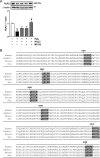
Figure 1
Figure 1
(A) The degradation of RyR2 is stimulated by hydrogen peroxide (H2O2). Representative western blot against RyR2 or β-actin in cultured cardiomyocytes treated with H2O2 in the presence or absence of NH4Cl, MG132, or NH4Cl and MG132. The bar graph shows the ratio of RyR2/β-actin. (B) Motifs biochemically related to the pentapeptide KFERQ (box) in the RyR2 aminoacidic sequence from Rattus novergicus (GenBank ID: NP_114467.1), human (GenBank ID: NP_001026.2), rabbit (GenBank ID: NP_001076226.1), and mouse (GenBank ID: NP_076357.2). Values are mean ± SEM (n = 5–6). *P < 0.05 H2O2 vs. control and #P < 0.05 H2O2 vs. H2O2 plus NH4Cl and MG132.
Figure 2
Figure 2
Geldanamycin (GA) stimulates chaperone-mediated autophagy and RyR2 degradation in cultured cardiomyocytes. (A) Representative western blot obtained with anti-LAMP-2A or anti-β-actin in total cell lysates. The bar graph shows the ratio LAMP–2A/β-actin calculated from densitometric analysis of western blot. (B) Colocalization of LAMP-2A and Hsc70 in cells stimulated with GA. Bar graph indicated Mander's coefficients of the overlap. The scale bar is 10 µm. (C) Percent of cell death measured by LDH release is indicated in the bar graph. (D) Representative western blot obtained with anti-RyR2 or anti-β-actin in total cell lysates. The bar graph shows the ratio RyR2/β-actin calculated from densitometric analysis of western blot. Values are mean ± SEM (n = 9 independent experiments). *P < 0.05 GA vs. control.
Figure 3
Figure 3
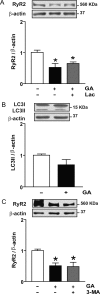
Effect of proteasome or macroautophagy inhibitors on RyR2 content in cardiomyocytes treated with geldanamycin (GA). (A) Representative western blot obtained with anti-RyR2 or anti-β-actin in total cell lysates obtained from controls or after incubation with GA or GA plus the proteasome inhibitor clasto-lactacystin-β-lactone (Lac). The bar graph shows the ratio RyR2/β-actin calculated from densitometric analysis of western blot. (B) Effect of GA on LC3-II content after incubation with GA. Representative western blot obtained with anti-LC3B or anti-β-actin. The bar graph shows the ratio LC3–II/β-actin calculated from densitometric analysis. (C) Effect of 3-methyladenine (3-MA) on RyR2 content after incubation with GA. Representative western blot obtained with anti-RyR2 or anti-β-actin in total cell lysates obtained from controls or after incubation with GA or GA and 3-MA. Bars show the ratio RyR2/β-actin calculated from densitometric analysis of western blot. Values are mean ± SEM (n = 3–9). *P < 0.05 GA, GA plus Lac, or GA plus 3-MA vs. control.
Figure 4
Figure 4
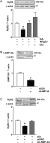
Chaperone-mediated autophagy degrades RyR2 in cardiomyocytes stimulated with geldanamycin (GA). (A) Representative western blot obtained with anti-RyR2 or anti-β-actin from controls, GA, GA plus ammonium chloride (NH4Cl), or chloroquine (Clo) to inhibit lysosomes. Bar graph shows the ratio RyR2/β-actin. (B) Representative western blot against LAMP-2A or β-actin in cardiomyocytes transfected with siRNA for LAMP-2A (siLAMP-2A) or scrambled siRNA (siSCR). Bar graph shows the ratio LAMP-2A/β-actin. (C) Western blot against RyR2 or β-actin obtained in the same cells. Bars show the ratio RyR2/β-actin. Values are mean ± SEM (n = 3–5). *P < 0.05 GA vs. control; siLAMP-2A vs. siSCR; or GA plus siSCR vs. siSCR.
Figure 5
Figure 5
ROS generation and CMA stimulation are two independent effects of geldanamycin (GA). (A) ROS were measured by flow cytometry after 6 h with GA in the presence or absence of N-acetylcysteine (NAC). (B) Representative western blot obtained with anti-RyR2 or anti-β-actin in total cell lysates obtained from controls or after incubation with GA or GA plus NAC. Bar graph shows the ratio RyR2/β-actin. (C) Representative western blot obtained with anti-LAMP-2A or anti-β-actin in total cell lysates. The bar graph shows the ratio LAMP-2A/β-actin calculated from densitometric analysis of western blot. Values are mean ± SEM (n = 5–7). *P < 0.05 GA vs. control or GA plus NAC vs. control.
Figure 6
Figure 6
Presenilins are implicated in RyR2 degradation in cardiomyocytes treated with geldanamycin (GA). (A) Western blot representative of α-spectrin proteolysis. Asterisk indicates α-spectrin total (top) or calpain-induced proteolytic fragments (150 and 145 KDa) of α-spectrin. Ionomycin was used as positive control (Io, 0.5+ and 1++ µM). Bar graph shows the ratio within proteolytic fragments 145 KDa and total α-spectrin. (B) Representative western blot obtained with anti-RyR2 or anti-β-actin in total cell lysates from controls or after incubation with GA in the presence or absence of compound E (CE). Bar graph shows the ratio within RyR2/β-actin. Values are mean ± SEM (n = 4). *P < 0.05 GA or Io vs. control.

References

    1. Bers DM. Cardiac excitation-contraction coupling. Nature. 2001;415:198–205. doi:10.1038/415198a. - DOI - PubMed
    1. Walsh LG, Tormey JMcD. Subcellular electrolyte shifts during in vitro myocardial ischemia and reperfusion. Am J Physiol. 1998;255:H917–H928. - PubMed
    1. Chohan PK, Singh RB, Dhalla NS, Netticadan T. L-arginine administration recovers sarcoplasmic reticulum function in ischemic reperfused hearts by preventing calpain activation. Cardiovasc Res. 2006;69:152–163. doi:10.1016/j.cardiores.2005.07.016. - DOI - PubMed
    1. Singh RB, Chohan PK, Dhalla NS, Netticadan T. The sarcoplasmic reticulum proteins are targets for calpain action in the ischemic-reperfused heart. J Mol Cell Cardiol. 2004;37:101–110. doi:10.1016/j.yjmcc.2004.04.009. - DOI - PubMed
    1. Takeda S, Mochizuki S, Saini HK, Elimban V, Dhalla NS. Modification of alterations in cardiac function and sarcoplasmic reticulum by vanadate in ischemic-reperfused rat hearts. J Appl Physiol. 2005;99:999–1005. doi:10.1152/japplphysiol.00234.2005. - DOI - PubMed

Publication types

MeSH terms