. 2013;8(2):e55031.
doi: 10.1371/journal.pone.0055031. Epub 2013 Feb 6.

Binding of the radioligand SIL23 to α-synuclein fibrils in Parkinson disease brain tissue establishes feasibility and screening approaches for developing a Parkinson disease imaging agent

Affiliations

Binding of the radioligand SIL23 to α-synuclein fibrils in Parkinson disease brain tissue establishes feasibility and screening approaches for developing a Parkinson disease imaging agent

Devika P Bagchi et al. PLoS One. 2013.

Abstract

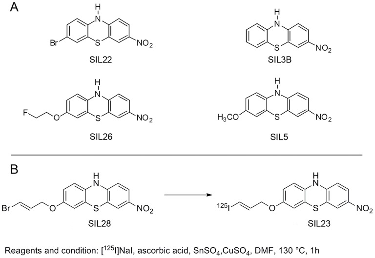
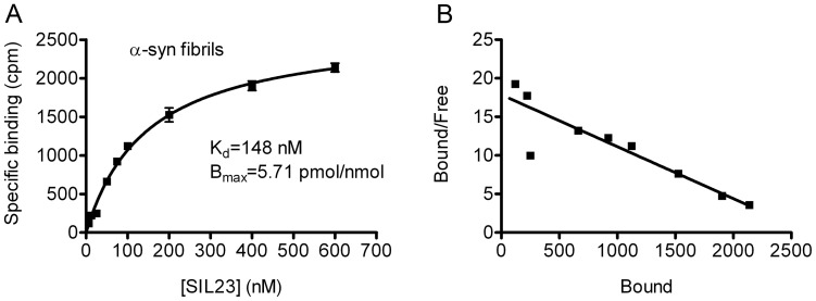
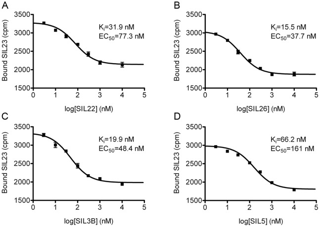
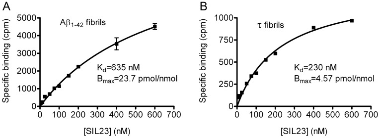
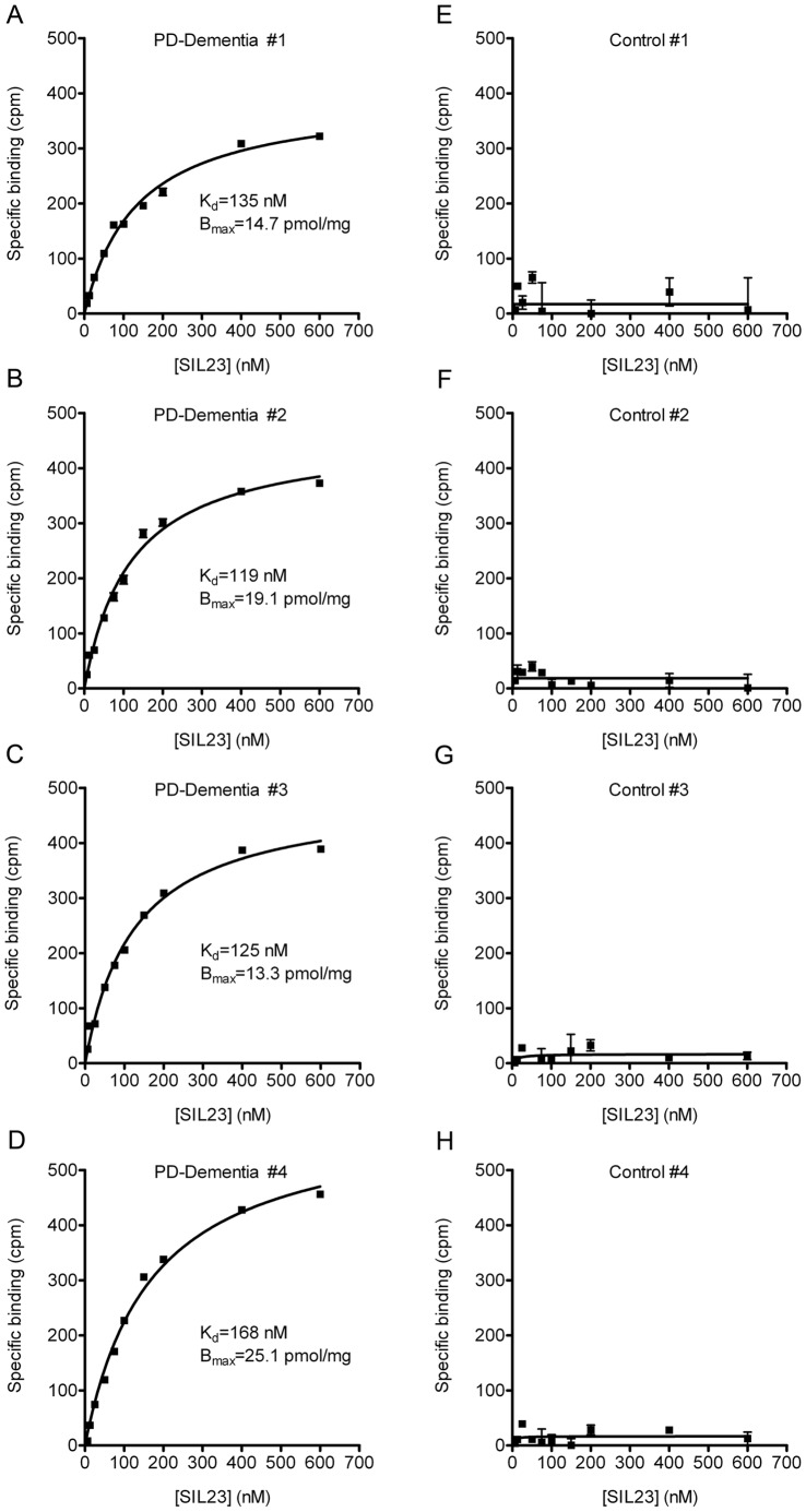
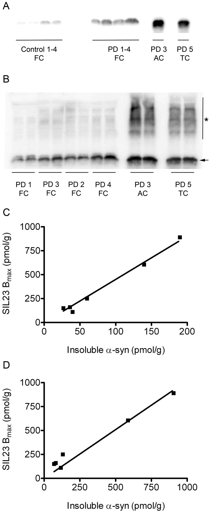
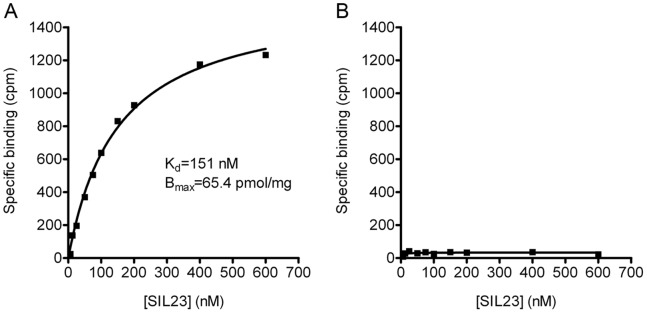
Accumulation of α-synuclein (α-syn) fibrils in Lewy bodies and Lewy neurites is the pathological hallmark of Parkinson disease (PD). Ligands that bind α-syn fibrils could be utilized as imaging agents to improve the diagnosis of PD and to monitor disease progression. However, ligands for α-syn fibrils in PD brain tissue have not been previously identified and the feasibility of quantifying α-syn fibrils in brain tissue is unknown. We report the identification of the (125)I-labeled α-syn radioligand SIL23. [(125)I]SIL23 binds α-syn fibrils in postmortem brain tissue from PD patients as well as an α-syn transgenic mouse model for PD. The density of SIL23 binding sites correlates with the level of fibrillar α-syn in PD brain tissue, and [(125)I]SIL23 binding site densities in brain tissue are sufficiently high to enable in vivo imaging with high affinity ligands. These results identify a SIL23 binding site on α-syn fibrils that is a feasible target for development of an α-syn imaging agent. The affinity of SIL23 for α-syn and its selectivity for α-syn versus Aβ and tau fibrils is not optimal for imaging fibrillar α-syn in vivo, but we show that SIL23 competitive binding assays can be used to screen additional ligands for suitable affinity and selectivity, which will accelerate the development of an α-syn imaging agent for PD.


Conflict of interest statement

Competing Interests: Authors LY, RHM, PTK and ZT are listed as inventors on a patent application covering the phenothiazine compounds described in this manuscript. The patent application number is 61/642,025 and it was filed on May 3, 2012. This does not alter the authors' adherence to all the PLOS ONE policies on sharing data and materials.

Figures

Figure 1
Figure 1. Phenothiazine structures and radiosynthesis of [125I]SIL23.
Structures of SIL23 analogues are shown in A. The radiosynthesis of [125I]SIL23 is shown in B.
Figure 2
Figure 2. The radioligand [125I]SIL23 binds recombinant α-syn fibrils with a Kd of 148 nM.
Fibrils prepared from recombinant α-syn were incubated with increasing concentrations of [125I]SIL23. Nonspecific binding was determined in parallel reactions utilizing 50 µM ThioT as competitor. A representative plot of specific binding versus [125I]SIL23 concentration is shown in A. Data points represent mean +/− s.d. (n = 3). The data was analyzed by curve fitting to a one-site binding model using nonlinear regression. The Kd value was determined by fitting the data to the equation Y = Bmax*X/(X+Kd). Scatchard analysis of binding is shown in B. Similar results were obtained in more than three independent experiments.
Figure 3
Figure 3. SIL23 competitive binding assays demonstrate higher affinity binding to recombinant α-syn fibrils for additional phenothiazine analogues, indicating that the structure of this compound class can be optimized to improve binding affinity using SIL23 assays.
α-Syn fibrils were incubated with 200 nM [125I]SIL23 and increasing concentrations of SIL22 (A), SIL26 (B), SIL3B (C), and SIL5 (D). The amount of bound radioligand is plotted as a function of the concentration of unlabeled competitor ligand in the incubation mixture. Data points represent mean +/− s.d. (n = 3). EC50 values were determined by fitting the data to the equation Y = bottom+(top-bottom)/(1+10(x-logEC50)). Similar results were obtained in two independent experiments.
Figure 4
Figure 4. Radioligand binding studies demonstrate that [125I]SIL23 has selectivity for binding recombinant α-syn fibrils compared to synthetic Aβ1–42 fibrils or recombinant tau fibrils.
Binding affinities of [125I]SIL23 to Aβ (A) and tau (B) fibrils were determined in saturation binding studies. The determined Kd values of Aβ and tau fibrils were 635 nM and 230 nM, respectively. Data points represent mean +/− s.d. (n = 3). Similar results were obtained in more than three independent experiments for saturation binding studies.
Figure 5
Figure 5. [125I]SIL23 exhibits specific binding to insoluble protein from human PD brain samples but not control human brain samples.
Homogenized insoluble fractions from human brain samples (n = 8) were incubated with increasing concentrations of [125I]SIL23. Nonspecific binding was determined in parallel reactions utilizing 50 µM ThioT as competitor. Representative plots of specific binding versus [125I]SIL23 concentration are shown. A–D show four different PD cases and E–H show four control cases. The data was analyzed by curve fitting to a one-site binding model using nonlinear regression. Kd values for binding to PD-dementia brain samples range from 119.1 nM to 168.3 nM and Bmax values range from 13–25 pmol/mg insoluble protein. No significant binding of [125I]SIL23 to control samples was observed. Results were verified with at least two independent experiments.
Figure 6
Figure 6. Binding of [125I]SIL23 is correlated with levels of insoluble α-syn present in PD brain.
Levels of α-syn were measured in insoluble fractions from human brain samples (n = 10) by sequential extraction and western blot. A representative syn1 western blot with SDS extracts from PD and control cases is shown in A. A quantitative syn303 western blot with SDS extracts from PD cases is shown in B, in which higher molecular weight α-syn species (asterisk) are observed in combination with monomeric α-syn (arrow). The correlation of Bmax values for [125I]SIL23 binding to levels of total insoluble α-syn quantified from the monomer band on western blot is shown in C. The correlation of Bmax values for [125I]SIL23 binding to levels of total insoluble α-syn quantified from monomer plus high molecular weight species on western blot is shown in D. Results were verified with more than three independent experiments.
Figure 7
Figure 7. [125I]SIL23 binding sites are present at high density in a transgenic A53T α-syn mouse model for PD.
[125I]SIL23 binding was determined in mouse brain samples obtained from (A) M83 mice with transgenic expression of human A53T α-syn, and (B) M7 mice with transgenic expression of human WT α-syn. Representative plots of specific binding versus [125I]SIL23 concentration are shown. The data was analyzed by curve fitting to a one-site binding model using nonlinear regression. [125I]SIL23 binding was detected in M83 brain samples with a Kd of 150.3 nM and a Bmax of 65 pmol/mg insoluble protein. No significant [125I]SIL23 binding was detected in M7 brain. The results were verified with two independent experiments.

References

    1. Hely MA, Reid WG, Adena MA, Halliday GM, Morris JG (2008) The Sydney multicenter study of Parkinson’s disease: the inevitability of dementia at 20 years. Mov Disord 23: 837–844. - PubMed
    1. Compta Y, Parkkinen L, O’Sullivan SS, Vandrovcova J, Holton JL, et al. (2011) Lewy- and Alzheimer-type pathologies in Parkinson’s disease dementia: which is more important? Brain 134: 1493–1505. - PMC - PubMed
    1. Hurtig HI, Trojanowski JQ, Galvin J, Ewbank D, Schmidt ML, et al. (2000) Alpha-synuclein cortical Lewy bodies correlate with dementia in Parkinson’s disease. Neurology 54: 1916–1921. - PubMed
    1. Braak H, Ghebremedhin E, Rub U, Bratzke H, Del Tredici K (2004) Stages in the development of Parkinson’s disease-related pathology. Cell Tissue Res 318: 121–134. - PubMed
    1. Clayton DF, George JM (1998) The synucleins: a family of proteins involved in synaptic function, plasticity, neurodegeneration and disease. Trends Neurosci 21: 249–254. - PubMed

Publication types