. 2013;8(2):e55379.
doi: 10.1371/journal.pone.0055379. Epub 2013 Feb 6.

TGF-β and TGF-β/Smad signaling in the interactions between Echinococcus multilocularis and its hosts

Affiliations

TGF-β and TGF-β/Smad signaling in the interactions between Echinococcus multilocularis and its hosts

Junhua Wang et al. PLoS One. 2013.

Abstract

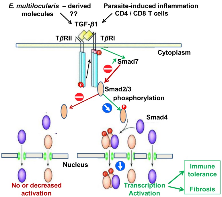
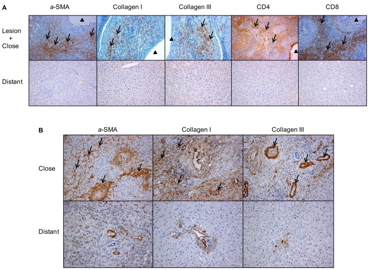
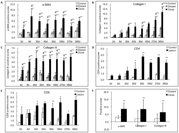
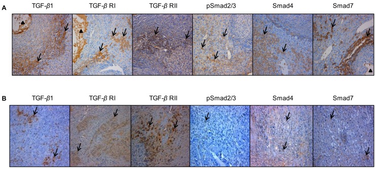
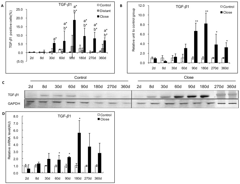
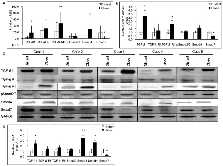
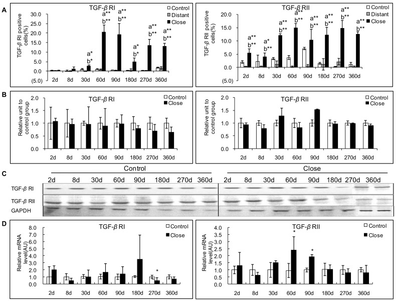
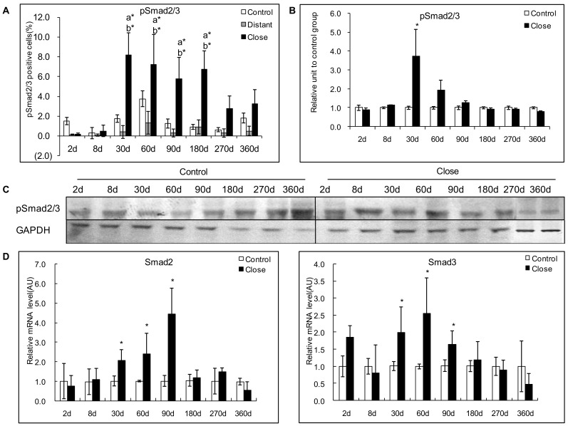
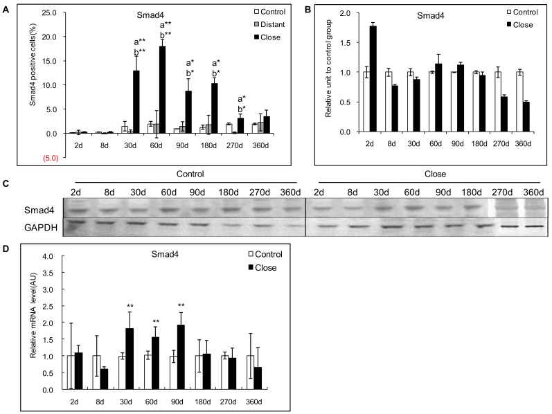
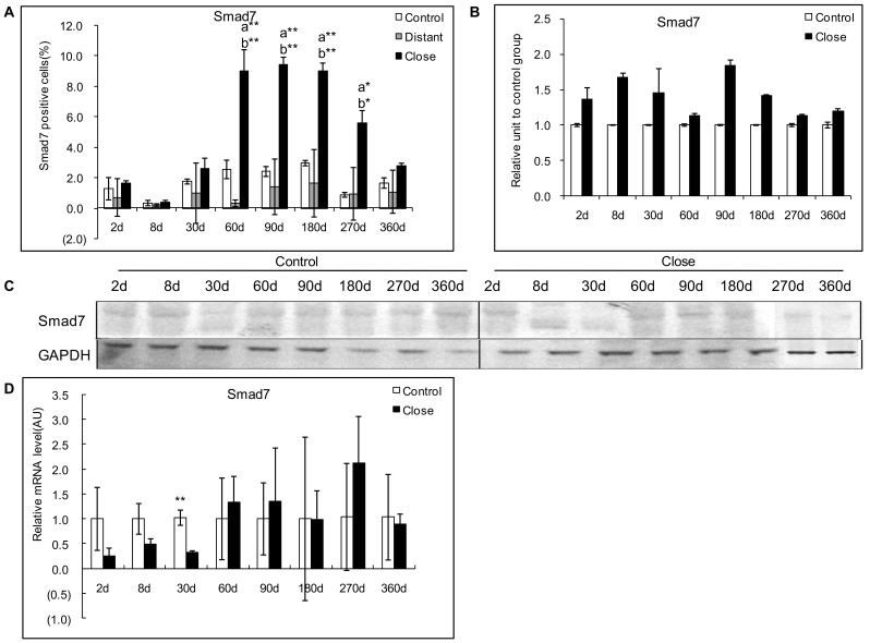
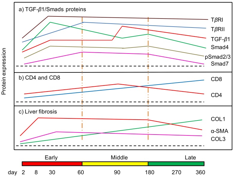
Alveolar echinococcosis (AE) is characterized by the development of irreversible fibrosis and of immune tolerance towards Echinococcus multilocularis (E. multilocularis). Very little is known on the presence of transforming growth factor-β (TGF-β) and other components of TGF-β/Smad pathway in the liver, and on their possible influence on fibrosis, over the various stages of infection. Using Western Blot, qRT-PCR and immunohistochemistry, we measured the levels of TGF-β1, TGF-β receptors, and down-stream Smads activation, as well as fibrosis marker expression in both a murine AE model from day 2 to 360 post-infection (p.i.) and in AE patients. TGF-β1, its receptors, and down-stream Smads were markedly expressed in the periparasitic infiltrate and also in the hepatocytes, close to and distant from AE lesions. Fibrosis was significant at 180 days p.i. in the periparasitic infiltrate and was also present in the liver parenchyma, even distant from the lesions. Over the time course after infection TGF-β1 expression was correlated with CD4/CD8 T-cell ratio long described as a hallmark of AE severity. The time course of the various actors of the TGF-β/Smad system in the in vivo mouse model as well as down-regulation of Smad7 in liver areas close to the lesions in human cases highly suggest that TGF-β plays an important role in AE both in immune tolerance against the parasite and in liver fibrosis.


Conflict of interest statement

Competing Interests: The authors have declared that no competing interests exist.

Figures

Figure 1
Figure 1. The TGF-β/Smad pathway; hypothesis for its involvement in the host-parasite relationship in E. multilocularis infection.
Figure 2
Figure 2. Immuno-histochemical expression of fibrosis markers in E. multilocularis-infected livers of experimental mice and AE patients, and of the periparasitic infiltration by CD4+ T and CD8+ T lymphocytes in the liver of experimental mice (arrow).
A: In experimental mice. α-SMA: expression at day 8, in the cytoplasm of smooth muscle cells, hepatic stellate cells and myofibroblasts in the liver parenchyma; collagen I: expression at day 360, in the peri-parasitic granuloma as concentric bundles extending from the laminated layer of the parasitic vesicles to the border of the normal liver; collagen III: expression at day 360, in the peri-parasitic granuloma as concentric bundles extending from the laminated layer of the parasitic vesicles to the border of the normal liver, also present as dotted lines between the cells at the outer part of the granulomatous infiltrate and, occasionally, in the cytoplasm of round cells in the sinusoids of the surrounding liver; CD4+ T cells: expression at day 90, in the periparasitic infiltrate surrounding the metacestode; CD8+ T cells: expression at day 180, in the periparasitic infiltrate surrounding the metacestode. B: In AE patients. α-SMA: expressed in the extracellular matrix; collagen I expressed both in the extracellular matrix and hepatocytes; collagen III expressed in the extracellular matrix and hepatocytes. The arrowheads indicate the parasitic lesions in the liver of infected mice and human patients. Final magnification: 200×. ‘Lesion’: E. multilocularis metacestode and surrounding immune infiltrate; ‘Close’: liver parenchyma close to E. multilocularis lesion; ‘Distant’: liver parenchyma distant from E. multilocularis lesion.
Figure 3
Figure 3. Semiquantitative expression of fibrosis markers in E. multilocularis-infected liver in experimental mice and in AE patients.
Score for each marker expression was calculated from quantitative analysis of the histo-immunostaining using both staining intensity and the percentage of cells stained at a specific range of intensities (arrow) (see Materials and Methods section). A: Course of α-SMA expression in E. multilocularis-infected mice. B: Course of collagen I expression in E. multilocularis-infected mice; C: Course of collagen III expression in E. multilocularis-infected mice; D: Course of CD4+ T cell infiltration in E. multilocularis-infected mice; E: Course of CD8+ T cell infiltration in E. multilocularis-infected mice; F: Expression of fibrosis markers in the liver of AE patients. a: close versus control; b: close versus distant. *P<0.05; **P<0.01. ‘Control’, non-infected mice; ‘Lesion’: E. multilocularis metacestode and surrounding immune infiltrate; ‘Close’: liver parenchyma close to E. multilocularis lesion; ‘Distant’: liver parenchyma distant from E. multilocularis lesion.
Figure 4
Figure 4. Immunohistochemical expression of the various components of the TGF-β/Smad pathway in the E. multilocularis-infected liver in experimental mice and in AE patients.
A: In experimental mice. Expression of the various components of the TGF-β/Smad pathway at their peak of expression in the liver. TGF-β1: expression at day 90, in most of the immune cells in most of areas with inflammatory granulomas, in the cytoplasm of hepatocytes, endothelial cells of the hepatic sinusoids and fibroblasts; TGF-β RI and RII: expression at day 60, in the cytoplasm of lymphocytes and macrophages in the periparasitic infiltrate and in most of the hepatocytes, fibroblasts, and endothelial cells close to the periparasitic infiltrate; pSmad2/3: expression at day 30, in both the cytoplasm and nuclear of the hepatocytes; Smad4: expression at day 60, in both the cytoplasm and nuclear of the hepatocytes; Smad7: expression at day 90, in the cytoplasm of the hepatocytes. B: In AE patients. Specimen ‘Close’ was taken close to the parasitic lesions (0.5 cm from the macroscopic changes due to the metacestode/granuloma lesion), and Specimen ‘Distant’ was taken in the liver distant from the lesions (the non-diseased lobe of the liver whenever possible, or at least at 10 cm from the lesion). TGF-β1: expressed in most of the immune cells in most of areas with inflammatory granulomas, in the cytoplasm of hepatocytes, endothelial cells of the hepatic sinusoids and fibroblasts; TGF-β RI and RII: expressed in the cytoplasm of lymphocytes and macrophages in the periparasitic infiltrate and in most of the hepatocytes, fibroblasts, and endothelial cells close to the periparasitic infiltrate; pSmad2/3: expressed in both the cytoplasm and nuclear of the hepatocytes; Smad4: expressed in both the cytoplasm and nuclear of the hepatocytes; Smad7: expressed in the cytoplasm of the hepatocytes. The arrowheads indicate the parasitic lesions in the liver of infected mice and human patients. Final magnification: 200×.
Figure 5
Figure 5. Course of TGF-β1 expression in the liver of experimental mice during E. multilocularis infection.
A: Course of TGF-β1 expression observed by immune-staining in the liver from E. multilocularis infected mice, calculated as the percent of positive cells to the total number of counted cells (see Materials and Methods section). B: Relative amount of TGF-β1 calculated from semi-quantitative analysis of the Western Blot using densitometry. C: Representative example of the course of TGF-β1 protein measured by Western Blot. D: Course of TGF-β1 mRNA expression measured by real time RT-PCR. a: ‘close’ versus ‘control’; b: ‘close’ versus ‘distant’. *P<0.05; **P<0.01. ‘Control’, non-infected mice; ‘Lesion’: E. multilocularis metacestode and surrounding immune infiltrate; ‘Close’: liver parenchyma close to E. multilocularis lesion; ‘Distant’: liver parenchyma distant from E. multilocularis lesion. AU: arbitrary units; GAPDH: glyceraldehyde-3-phosphate dehydrogenase.
Figure 6
Figure 6. Expression of the various components of the TGF-β1/Smad pathway in the liver of AE patients.
A: Expression of TGF-β1/Smads calculated as the percent of positive cells to the total number of counted cells after immunostaining (see Materials and Methods section). B: Relative amount of TGF-β1/Smads calculated from semi-quantitative analysis of the Western Blot using densitometry. C: Representative examples of Western Blot analyses performed on lysates from liver samples with antibodies that recognize TGF-β1, TGF-β RI, TGF-β RII, phosphorylated (p-) Smad2/3, Smad4 and Smad7. D: TGF-β1/Smads mRNA expression measured by real time RT-PCR. *P<0.05 versus control, **P<0.01 versus control. ‘Distant’: distant from lesion; ‘Close’: close to lesion; AU: arbitrary units; GAPH: glyceraldehyde-3-phosphate dehydrogenase.
Figure 7
Figure 7. Course of TGF-β1 receptors (TGF-β RI and TGF-β RII) expression in the liver of mice during E. multilocularis infection in experimental mice.
A: Course of TGF-β RI and RII expression observed by immune-staining in the liver from E. multilocularis infected mice compared to control mice, calculated as the percent of positive cells to the total number of counted cells (see Materials and Methods section). B: Relative amount of TGF-β RI and RII calculated from semi-quantitative analysis of the Western Blot using densitometry. C: Representative example of the course of TGF-β RI and RII protein measured by Western Blot in experimental mice. D: Course of TGF-β RI and RII mRNA expression measured by real time RT-PCR in experimental mice. a: ‘close’ versus ‘control’; b: ‘close’ versus ‘distant’. *P<0.05; **P<0.01. ‘Control’, non-infected mice; ‘Lesion’: E. multilocularis metacestode and surrounding immune infiltrate; ‘Close’: liver parenchyma close to E. multilocularis lesion; ‘Distant’: liver parenchyma distant from E. multilocularis lesion. AU: arbitrary units; GAPDH: glyceraldehyde-3-phosphate dehydrogenase.
Figure 8
Figure 8. Course of pSmad2/3 expression in the liver of mice during E. multilocularis infection in experimental mice.
A: Course of pSmad2/3 expression observed by immune-staining in the liver from E. multilocularis infected mice compared to control mice, calculated as the percent of positive cells to the total number of counted cells (see Materials and Methods section). B: Relative amount of pSmad2/3 calculated from semi-quantitative analysis of the Western blot using densitometry. C: Representative example of the course of pSmad2/3 protein measured by Western Blot in experimental mice. D: Course of Smad2 and Smad3 mRNA expression measured by real time RT-PCR in experimental mice. a: ‘close’ versus ‘control’; b: ‘close’ versus ‘distant’. *P<0.05; **P<0.01. ‘Control’, non-infected mice; ‘Lesion’: E. multilocularis metacestode and surrounding immune infiltrate; ‘Close’: liver parenchyma close to E. multilocularis lesion; ‘Distant’: liver parenchyma distant from E. multilocularis lesion. AU: arbitrary units; GAPDH: glyceraldehyde-3-phosphate dehydrogenase.
Figure 9
Figure 9. Course of Smad4 expression in the liver of mice during E. multilocularis infection in experimental mice.
A: Course of Smad4 expression observed by immune-staining in the liver from E. multilocularis infected mice compared to control mice, calculated as the percent of positive cells to the total number of counted cells (see Materials and Methods section). B: Relative amount of Smad4 calculated from semi-quantitative analysis of the Western blot using densitometry. C: Representative example of the course of Smad4 protein measured by Western Blot in experimental mice. D: Course of Smad4 mRNA expression measured by real time RT-PCR in experimental mice. a: ‘close’ versus ‘control’; b: ‘close’ versus ‘distant’. *P<0.05; **P<0.01. ‘Control’, non-infected mice; ‘Lesion’: E. multilocularis metacestode and surrounding immune infiltrate; ‘Close’: liver parenchyma close to E. multilocularis lesion; ‘Distant’: liver parenchyma distant from E. multilocularis lesion.AU: arbitrary units; GAPDH: glyceraldehyde-3-phosphate dehydrogenase.
Figure 10
Figure 10. Course of Smad7 expression in the liver of mice during E. multilocularis infection in experimental mice.
A: Course of Smad7 expression observed by immune-staining in the liver from E. multilocularis infected mice compared to control mice, calculated as the percent of positive cells to the total number of counted cells (see Materials and Methods section). B: Relative amount of Smad7 calculated from semi-quantitative analysis of the Western blot using densitometry. C: Representative example of the course of Smad7 protein measured by Western Blot in experimental mice. D: Course of Smad7 mRNA expression measured by real time RT-PCR in experimental mice. a: ‘close’ versus ‘control’; b: ‘close’ versus ‘distant’. *P<0.05; **P<0.01. ‘Control’, non-infected mice; ‘Lesion’: E. multilocularis metacestode and surrounding immune infiltrate; ‘Close’: liver parenchyma close to E. multilocularis lesion; ‘Distant’: liver parenchyma distant from E. multilocularis lesion. AU: arbitrary units; GAPDH: glyceraldehyde-3-phosphate dehydrogenase.
Figure 11
Figure 11. Course of the changes in the protein expression of TGF-β1/Smads (a), T cell subpopulation CDs (b) and liver fibrosis markers (c) during the process of E. multilocularis-induced liver injury in mice.

Similar articles

Cited by

References

    1. Vuitton DA, Zhou H, Bresson-Hadni S, Wang Q, Piarroux M, et al. (2003) Epidemiology of alveolar echinococcosis with particular reference to China and Europe. Parasitology 127 Suppl: S87–107 - PubMed
    1. Vuitton DA, Zhang SL, Yang Y, Godot V, Beurton I, et al. (2006) Survival strategy of Echinococcus multilocularis in the human host. Parasitol Int 55 Suppl: S51–55 - PubMed
    1. Grenard P, Bresson-Hadni S, El Alaoui S, Chevallier M, Vuitton DA, et al. (2001) Transglutaminase-mediated cross-linking is involved in the stabilization of extracellular matrix in human liver fibrosis. J Hepatol 35: 367–375. - PubMed
    1. Guerret S, Vuitton DA, Liance M, Pater C, Carbillet JP (1998) Echinococcus multilocularis: relationship between susceptibility/resistance and liver fibrogenesis in experimental mice. Parasitol Res 84: 657–667. - PubMed
    1. Ricard-Blum S, Bresson-Hadni S, Guerret S, Grenard P, Volle PJ, et al. (1996) Mechanism of collagen network stabilization in human irreversible granulomatous liver fibrosis. Gastroenterology 111: 172–182. - PubMed

Publication types

MeSH terms

Supplementary concepts