. 2013;8(2):e55559.
doi: 10.1371/journal.pone.0055559. Epub 2013 Feb 6.

De novo synthesized estradiol protects against methylmercury-induced neurotoxicity in cultured rat hippocampal slices

Affiliations

De novo synthesized estradiol protects against methylmercury-induced neurotoxicity in cultured rat hippocampal slices

Takeshi Yamazaki et al. PLoS One. 2013.

Erratum in

  • PLoS One. 2013;8(4). doi:10.1371/annotation/52376e1c-1a2d-44af-a129-849345da78a0

Abstract

Background: Estrogen, a class of female sex steroids, is neuroprotective. Estrogen is synthesized in specific areas of the brain. There is a possibility that the de novo synthesized estrogen exerts protective effect in brain, although direct evidence for the neuroprotective function of brain-synthesized estrogen has not been clearly demonstrated. Methylmercury (MeHg) is a neurotoxin that induces neuronal degeneration in the central nervous system. The neurotoxicity of MeHg is region-specific, and the molecular mechanisms for the selective neurotoxicity are not well defined. In this study, the protective effect of de novo synthesized 17β-estradiol on MeHg-induced neurotoxicity in rat hippocampus was examined.

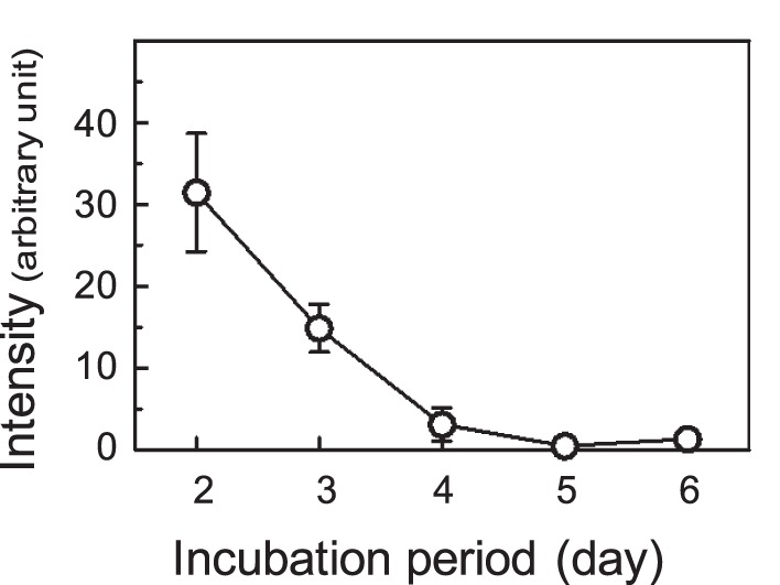
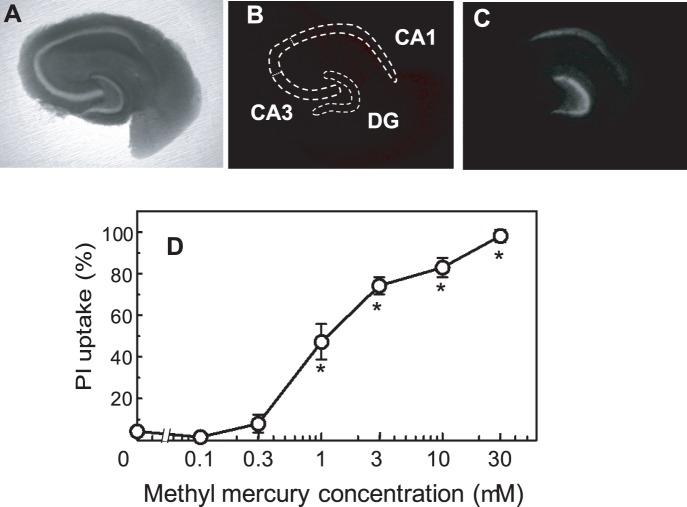
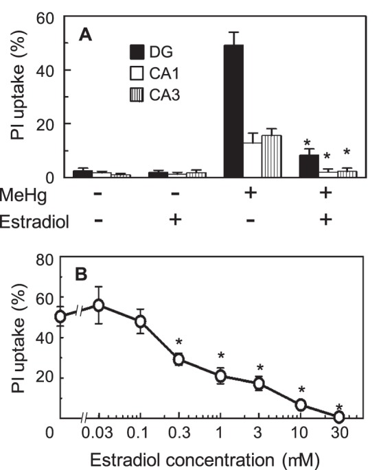
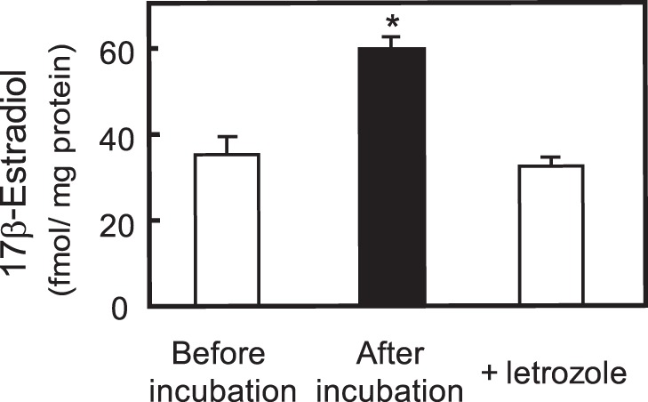
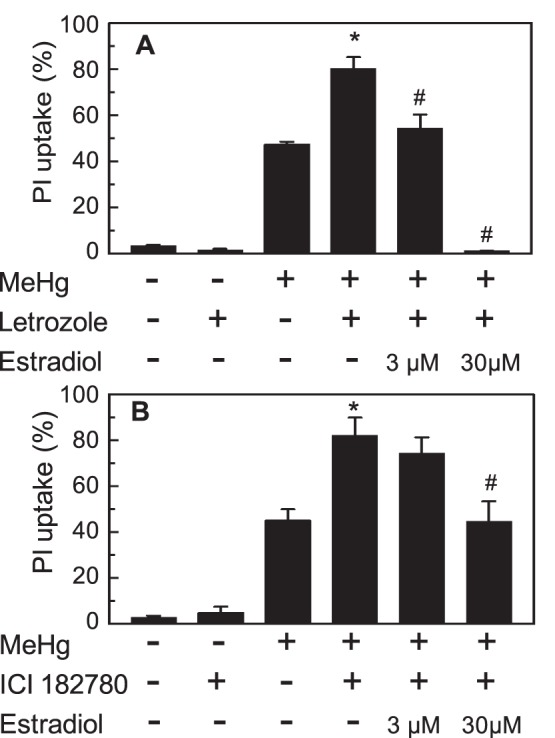
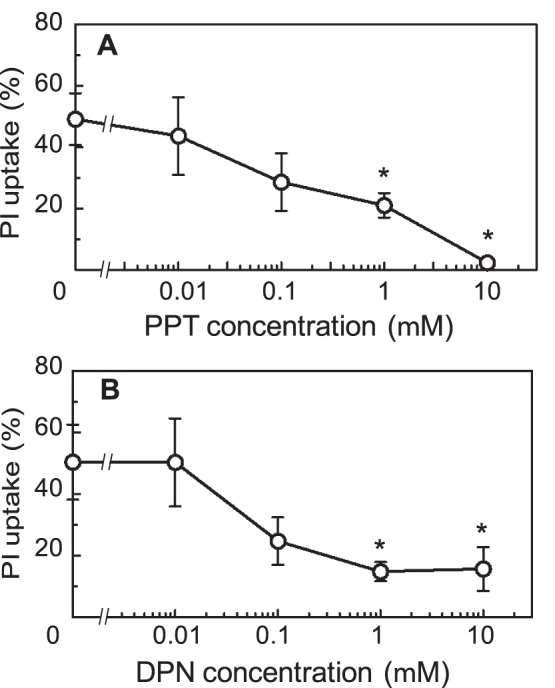
Methodology/principal findings: Neurotoxic effect of MeHg on hippocampal organotypic slice culture was quantified by propidium iodide fluorescence imaging. Twenty-four-hour treatment of the slices with MeHg caused cell death in a dose-dependent manner. The toxicity of MeHg was attenuated by pre-treatment with exogenously added estradiol. The slices de novo synthesized estradiol. The estradiol synthesis was not affected by treatment with 1 µM MeHg. The toxicity of MeHg was enhanced by inhibition of de novo estradiol synthesis, and the enhancement of toxicity was recovered by the addition of exogenous estradiol. The neuroprotective effect of estradiol was inhibited by an estrogen receptor (ER) antagonist, and mimicked by pre-treatment of the slices with agonists for ERα and ERβ, indicating the neuroprotective effect was mediated by ERs.

Conclusions/significance: Hippocampus de novo synthesized estradiol protected hippocampal cells from MeHg-induced neurotoxicity via ERα- and ERβ-mediated pathways. The self-protective function of de novo synthesized estradiol might be one of the possible mechanisms for the selective sensitivity of the brain to MeHg toxicity.


Conflict of interest statement

Competing Interests: The authors have declared that no competing interests exist.

Figures

Figure 1
Figure 1. The PI fluorescence intensity of cultured slices.
The hippocampal slices were cultured for 6 days with serum-containing medium. The intensity of PI fluorescence of whole slices is indicated. The values shown are the means of 9 slices in three different cultures, and the error bars indicate the standard error (SE).
Figure 2
Figure 2. MeHg-induced cell death as assessed by PI uptake.
Bright field (A) and PI fluorescence (B) images after 5 days of pre-culture and PI fluorescence after subsequent incubation with 1 µM MeHg for 24 h (C) of the cultured hippocampal slices. Neuronal cell dense regions of dentate gyrus (DG), and CA1 and CA3 regions in Ammon’s horn are traced on (A) and superimposed on (B) as dotted lines. The concentration dependency of MeHg on PI uptake is shown in (D). The PI uptake is expressed as a percentage of total fluorescence in the neuronal cell dense region (inside the dotted lines in B) after the slices were kept at 4°C for 24 h. The values are the means of 15–21 slices in 5 different cultures, and the error bars indicate the SE. *, P<0.05, vs. MeHg-treated group, post hoc t-test with Holm-Bonferroni method after one-way ANOVA.
Figure 3
Figure 3. The effect of exogenously added estradiol on MeHg-induced cell death.
The PI uptake of the hippocampal slices was analyzed individually in the neuronal cell dense regions, dentate gyrus (DG), and CA1 and CA3 regions in Ammon’s horn, which are shown in Fig. 2B (A). Slices were incubated with vehicle or 10 µM estradiol for 26 h, and/or for 24 h with 1 µM MeHg. MeHg was added 2 h after administration of estradiol. The values are the means of 14–21 slices in 5 different cultures, and the error bars indicate the SE. *, P<0.05 vs. MeHg-treated group, post hoc t-test with the Holm-Bonferroni method after one-way ANOVA. The concentration dependency of the protective effect of estradiol on the PI uptake of the neuronal cell dense region of the 1 µM MeHg-treated slice is shown in (B). The values are the mean of 14–23 slices in 4 different cultures, and the error bars indicate the SE. *, P<0.05 vs. MeHg-treated group, post hoc t-test with Holm-Bonferroni method after one-way ANOVA.
Figure 4
Figure 4. The inhibition of estradiol synthesis by letrozole.
Estradiol was extracted from slices and medium before and after a 48-h incubation with or without letrozole. Letrozole was extracted from Femara tablets. The levels of extracted 17β-estradiol were quantified as described in the Materials and Methods section. The values are the means of 3 separate experiments, and the error bars indicate the SE. *, P<0.05, vs. non-treated group, t-test.
Figure 5
Figure 5. The effects of letrozole and ICI on MeHg-induced cell death.
Levels of PI uptake in the neuronal cell dense region of the slices after a 48-h treatment with vehicle or letrozole and/or 24-h treatment with 1 µM MeHg are shown in A. Levels of PI uptake after a 30-h treatment with vehicle or 100 µM ICI 182,780 and/or 24-h treatment with 1 µM MeHg are shown in B. MeHg was added 24 or 6 h after the administration of Letrozole or ICI 182,780, respectively. Some wells were given the indicated concentrations of estradiol 2 h before the addition of MeHg. The values are the means of 16–21 slices in 5 different cultures or 13–16 slices in 4 different cultures for A or B, respectively, and the error bars indicate the SE. *, P<0.05, vs. the MeHg-treated group, and #, P<0.05, vs. MeHg and letrozole- or ICI-treated group, post hoc t-test with the Holm-Bonferroni method after one-way ANOVA.
Figure 6
Figure 6. The effects of estrogen receptor agonists on MeHg-induced cell death.
The PI uptake of neuronal cell dense region of the slices was determined after a 24-h treatment with 1 µM MeHg in the presence of various concentrations of PPT (A) or DPN (B). These agonists were administered 2 h before the addition of MeHg. The values are the means of 16–22 slices in four different cultures, and the error bars indicate the SE. *, P<0.05, vs. MeHg-treated group, post hoc t-test with Holm-Bonferroni method after one-way ANOVA.

Similar articles

Cited by

References

    1. McEwen B (2002) Estrogen actions throughout the brain. Recent Prog Horm Res 57: 357–384. - PubMed
    1. Bourque M, Dluzen DE, Di Paolo T (2009) Neuroprotective actions of sex steroids in Parkinson's disease. Front Neuroendocrinol 30: 142–157. - PubMed
    1. Pike CJ, Carroll JC, Rosario ER, Barron AM (2009) Protective actions of sex steroid hormones in Alzheimer's disease. Front Neuroendocrinol 30: 239–258. - PMC - PubMed
    1. Suzuki S, Brown CM, Wise PM (2009) Neuroprotective effects of estrogens following ischemic stroke. Front Neuroendocrinol 30: 201–211. - PMC - PubMed
    1. Behl C (2002) Oestrogen as a neuroprotective hormone. Nat Rev Neurosci 3: 433–442. - PubMed

Publication types

MeSH terms

LinkOut - more resources