. 2013:3:1259.
doi: 10.1038/srep01259. Epub 2013 Feb 12.

Isolation and retrieval of circulating tumor cells using centrifugal forces

Affiliations

Isolation and retrieval of circulating tumor cells using centrifugal forces

Han Wei Hou et al. Sci Rep. 2013.

Abstract

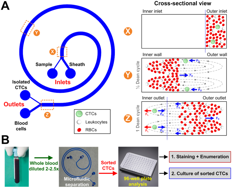
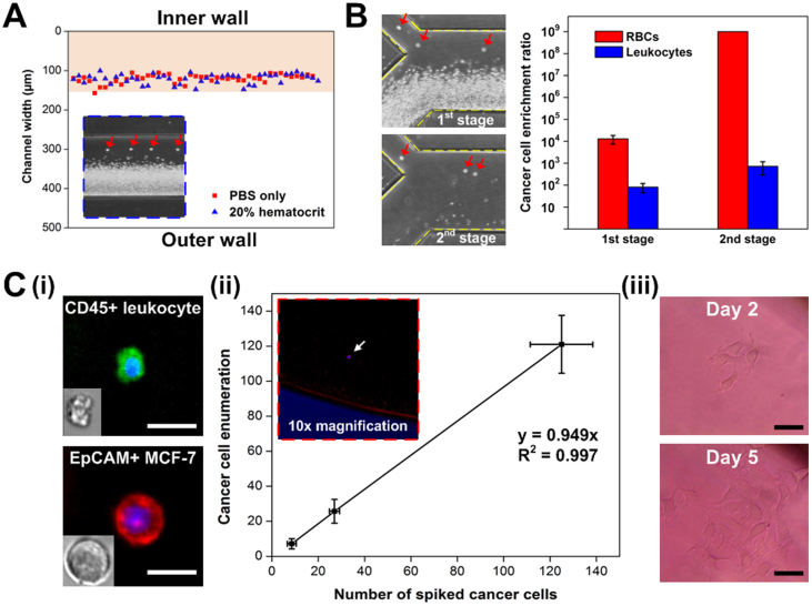
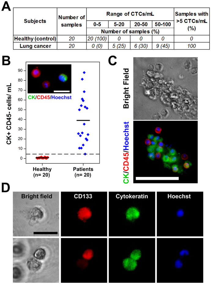
Presence and frequency of rare circulating tumor cells (CTCs) in bloodstreams of cancer patients are pivotal to early cancer detection and treatment monitoring. Here, we use a spiral microchannel with inherent centrifugal forces for continuous, size-based separation of CTCs from blood (Dean Flow Fractionation (DFF)) which facilitates easy coupling with conventional downstream biological assays. Device performance was optimized using cancer cell lines (> 85% recovery), followed by clinical validation with positive CTCs enumeration in all samples from patients with metastatic lung cancer (n = 20; 5-88 CTCs per mL). The presence of CD133⁺ cells, a phenotypic marker characteristic of stem-like behavior in lung cancer cells was also identified in the isolated subpopulation of CTCs. The spiral biochip identifies and addresses key challenges of the next generation CTCs isolation assay including antibody independent isolation, high sensitivity and throughput (3 mL/hr); and single-step retrieval of viable CTCs.


Conflict of interest statement

H.W.H., A.A.B., J.H. and C.T.L., along with others, have filed a patent application on the technology described here.

Figures

Figure 1
Figure 1
(A) Schematic illustration of the separation principle for high-throughput CTCs isolation using Dean Flow Fractionation (DFF). Blood sample and sheath fluid are pumped through the outer and inner inlets of the spiral device respectively. Under the influence of Dean drag forces (FD (blue arrows)), the smaller hematologic cells (RBCs and leukocytes) migrate along the Dean vortices towards the inner wall, then back to outer wall again (Dean cycle 1), while the larger CTCs experience additional strong inertial lift forces (FL (red arrows)) and focus along the microchannel inner wall, thus achieving separation. (B) Overall workflow of device operation and coupling with 96-well plate for various downstream applications such as CTCs enumeration and culture of sorted CTCs.
Figure 2
Figure 2. Device characterization using hematological cells and cancer cell lines at Dean Cycle 1 (DC 1).
(A) Averaged composite images and intensity plot illustrate broadening RBCs occupied regions (red dashed line) for increasing hematocrit prior outlet bifurcation. Yellow dotted lines indicate position of channel walls. (B) Linescan indicating lateral positions of leukocytes near the outlet at DC 1. The larger leukocytes behaved similar to RBCs and were transposed to outer half of the channel under the influence of Dean drag forces, resulting in minimal leukocytes entering the CTCs outlet (150 μm width). (C) Average composite images representing the focusing position of MCF-7 cells at corresponding locations of the spiral channel. Scale bar is 1 cm. (D) Histogram plot indicating a high separation efficiency of >85% for different cancer cell lines tested. (E) Images of sorted MCF-7 clusters stained with anti-EpCAM antibodies (red) and Hoechst dye (blue) to identify the cell membrane and nucleus respectively. Scale bar is 100 μm.
Figure 3
Figure 3. Isolation of cancer cells from blood in a 2-stage cascaded spiral system.
(A) Plot indicates similar focusing positions of MCF-7 cells suspended in PBS solution and 20% hematocrit blood samples within the pink shaded area (150 μm wide) which corresponds to the dimension of CTCs outlet. High speed image (6400 fps) captured at the channel outlet (blue dotted box) clearly illustrates focusing of larger MCF-7 cells at the inner wall (red arrows) while smaller RBCs occupied the outer channel region at DC 1. (B) Representative images highlighting separation of MCF-7 cells (red arrows) from 20% hematocrit samples at different stages in the cascaded system. Plot (log scale) indicates a high cancer cell enrichment ratio of 109 fold (over RBCs) and ~800 fold (over leukocytes) using the cascaded system. (C) (i) Bright field and fluorescence images of sorted CD45-positive (green) leukocyte and EpCAM-positive (red) MCF-7 cells on 96-well plate after immuno-staining. Both cell types were stained with Hoechst dye for nuclei identification. Scale bar is 20 μm. (ii) High cancer cell recovery (~95%) was achieved using 96-well plate analysis after sorting blood samples spiked with physiological relevant CTCs concentration (~10–100/mL). Inset image (red dotted box) shows a stained cancer cell (white arrow) in a well under 10× magnification. (iii) Images showing successful culture of sorted MCF-7 cells in 96-well plate. Scale bar is 50 μm.
Figure 4
Figure 4. Enumeration of CTCs from cancer patients.
(A) Summary of CTCs count per mL of blood for healthy samples and patients with metastatic lung cancer. (B) CTCs enumeration plot for healthy donors (red) and lung cancer patients (blue). Dashed line indicates threshold of 5 CTCs/mL. (Inset) Representative image (60× magnification) of isolated CTCs and leukocytes from lung cancer patients stained with antibodies against cytokeratin (green), CD45 (red) and Hoechst (blue). CTC morphology is characterized by high nuclear: cytoplasmic ratios. Scale bar is 20 μm. (c ) Optical images (60× magnification) of an isolated CTC microcluster using DFF. Scale bar is 50 μm. (D) Bright field and fluorescence images (60× magnification) of sorted CTCs stained for stem cell marker CD133 (red), cytokeratin (green) and Hoechst (blue). Scale bar is 20 μm.

Similar articles

Cited by

References

    1. Budd G. T. et al. Circulating tumor cells versus imaging - Predicting overall survival in metastatic breast cancer. Clin. Cancer Res. 12, 6403–6409 (2006). - PubMed
    1. Cristofanilli M. et al. Circulating Tumor Cells, Disease Progression, and Survival in Metastatic Breast Cancer. N Engl J Med 351, 781–791 (2004). - PubMed
    1. Hayes D. F. et al. Circulating tumor cells at each follow-up time point during therapy of metastatic breast cancer patients predict progression-free and overall survival. Clin. Cancer Res. 12, 4218–4224 (2006). - PubMed
    1. Punnoose E. A. et al. Molecular Biomarker Analyses Using Circulating Tumor Cells. PLoS ONE 5, e12517 (2010). - PMC - PubMed
    1. Maheswaran S. et al. Detection of Mutations in EGFR in Circulating Lung-Cancer Cells. New England Journal of Medicine 359, 366–377 (2008). - PMC - PubMed

Publication types