Review
. 2013 Mar;252(1):41-51.
doi: 10.1111/imr.12033.

NF-κB: roles and regulation in different CD4(+) T-cell subsets

Affiliations
Review

NF-κB: roles and regulation in different CD4(+) T-cell subsets

Hyunju Oh et al. Immunol Rev. 2013 Mar.

Abstract

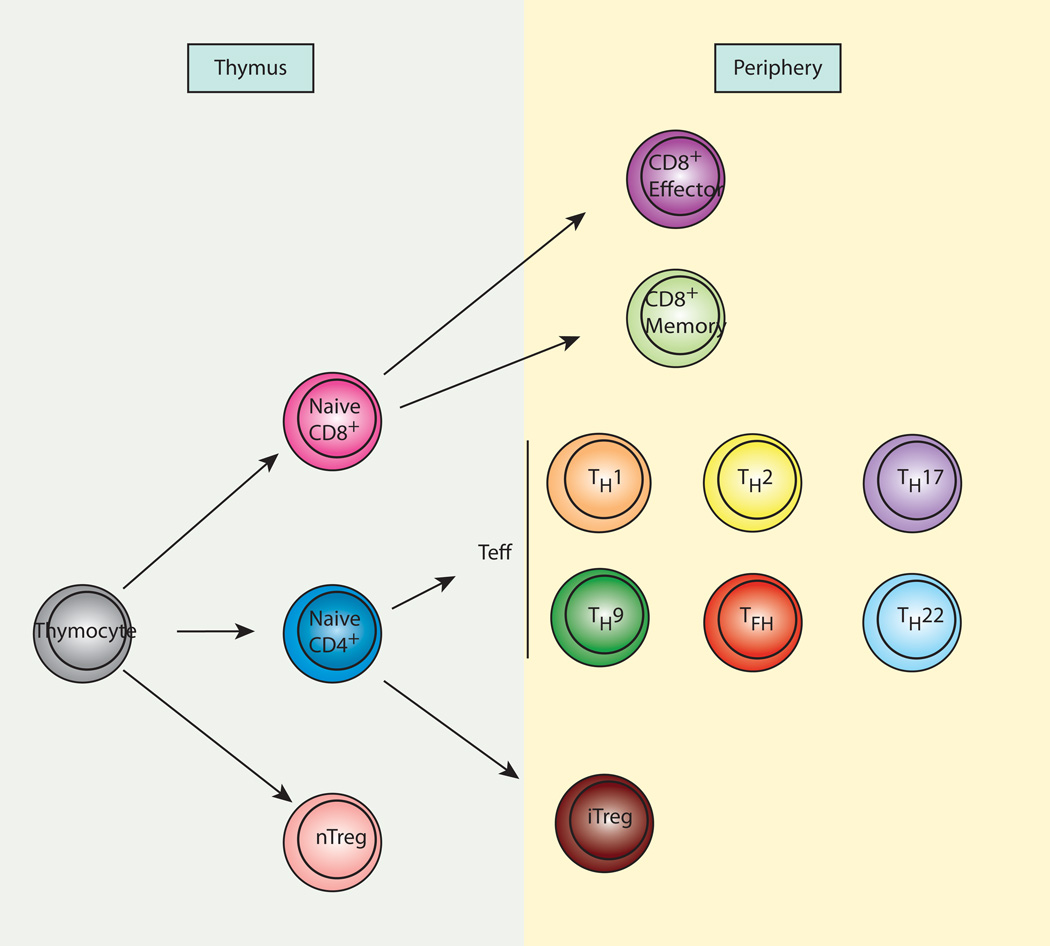
The nuclear factor-κB (NF-κB) family of transcription factors plays important roles in various biological processes including apoptosis, stress response, immunity, and inflammation. NF-κB signaling is involved in both immune cell development and function, and it is critical in modulation of the immune response through the transcriptional regulation of cytokine and chemokine expression. An area of great interest in T-cell-mediated adaptive immunity is the ability of naive CD4(+) T cells generated in the thymus to differentiate into various subsets including T-helper 1 (Th1), Th2, Th17, Th9, follicular helper T (Tfh), Th22, and regulatory T (Treg) cells, upon encountering different pathogens and microenvironments. In this review, we discuss the role of NF-κB pathway in the development and functional divergence of the different helper T-cell subsets as well as in regulatory T cells.


Conflict of interest statement

The authors have no conflicts of interest to declare.

Figures

Fig. 1
Fig. 1. CD4+ T-cell subsets
In the thymus, naive CD4+ and CD8+ T cells develop from thymocytes. Naive CD4+ T cells from the thymus migrate to the periphery, where they can differentiate to various subsets of effector cells upon encountering specific antigens. The subsets include Th1, Th2, Th17, Th9, Tfh, and Th22. Naive CD8+ T cells also migrate to the periphery and differentiate to effector cells or memory cells upon encountering antigens. A small population of thymocytes differentiates into regulatory T cells (nTreg) in the thymus. In addition, some naive CD4+ T cells differentiate into regulatory T cells (iTreg) in specific microenvironments.
Fig. 2
Fig. 2. Members of the NF-κB/Rel family
Schematic depiction of members of the NF-kB transcription factor family. RHD, Rel homolgy domain; TAD, transactivation domain; LZ, leucin zipper domain; GRR, glycine-rich region; DD, death domain; ANK, ankyrin repeats.
Fig. 3
Fig. 3. Canonical and noncanonical pathways of NF-κB activation
NF-κB activation occurs through two different pathways: canonical and noncanonical. In the canonical pathway, the IKK complex, composed of IKKα, IKKβ, and the regulatory subunit NEMO, phosphorylates IκB, leading to its ubiquitination and proteosomal degradation. A NF-κB dimer, which is comprised of p50, p65, or c-Rel, is released from IκB and translocates into the nucleus, where it binds to a κB-binding site for regulation of gene expression. In the noncanonical pathway, IKKα phosphorylates p100 resulting in its ubiquitination and partial processing into p52. A heterodimer of p52 and RelB translocates into the nucleus and binds to it binding sites. P, phosphorylation; Ub, ubiquitination.
Fig. 4
Fig. 4. TCR/CD28 signaling pathway
A TCR complex on the surface of T cells recognizes a specific antigen-MHC complex on an antigen-presenting cell (APC). At the same time, a costimulatory receptor CD28 interacts with CD80 or CD86 on the APC. Upon TCR/CD28 stimulation, PI3K activates PDK1 leading to subsequent activation of PKCθ. This results in the formation of the complex consisting of CARMA1, BCL10, and MALT1, that activates the IKK complex. Phosphorylation of IκB by the IKK complex leads to its degradation, resulting in the liberation of NF-κB dimers. NF-κB dimers translocate into the nucleus and regulate the expression of target genes. PDK1 activates the Akt/mTOR pathway as well as NF-κB. TCR stimulation also leads to activation of JNK, NFAT, and p38 MAPK in a PDK-independent manner.

References

    1. Hayden MS, Ghosh S. NF-kappaB in immunobiology. Cell Res. 2011;21:223–244. - PMC - PubMed
    1. Hsieh CS, Liang Y, Tyznik AJ, Self SG, Liggitt D, Rudensky AY. Recognition of the peripheral self by naturally arising CD25+ CD4+ T cell receptors. Immunity. 2004;21:267–277. - PubMed
    1. Dustin ML. The cellular context of T cell signaling. Immunity. 2009;30:482–492. - PMC - PubMed
    1. Gerondakis S, Siebenlist U. Roles of the NF-kappaB pathway in lymphocyte development and function. Cold Spring Harbor Persp Biol. 2010;2 a000182. - PMC - PubMed
    1. Oeckinghaus A, Hayden MS, Ghosh S. Crosstalk in NF-kappaB signaling pathways. Nat Immunol. 2011;12:695–708. - PubMed

Publication types

MeSH terms