. 2013 Jun;19(11-12):1386-97.
doi: 10.1089/ten.TEA.2012.0367. Epub 2013 Apr 4.

Human perivascular stem cells show enhanced osteogenesis and vasculogenesis with Nel-like molecule I protein

Affiliations

Human perivascular stem cells show enhanced osteogenesis and vasculogenesis with Nel-like molecule I protein

Asal Askarinam et al. Tissue Eng Part A. 2013 Jun.

Abstract

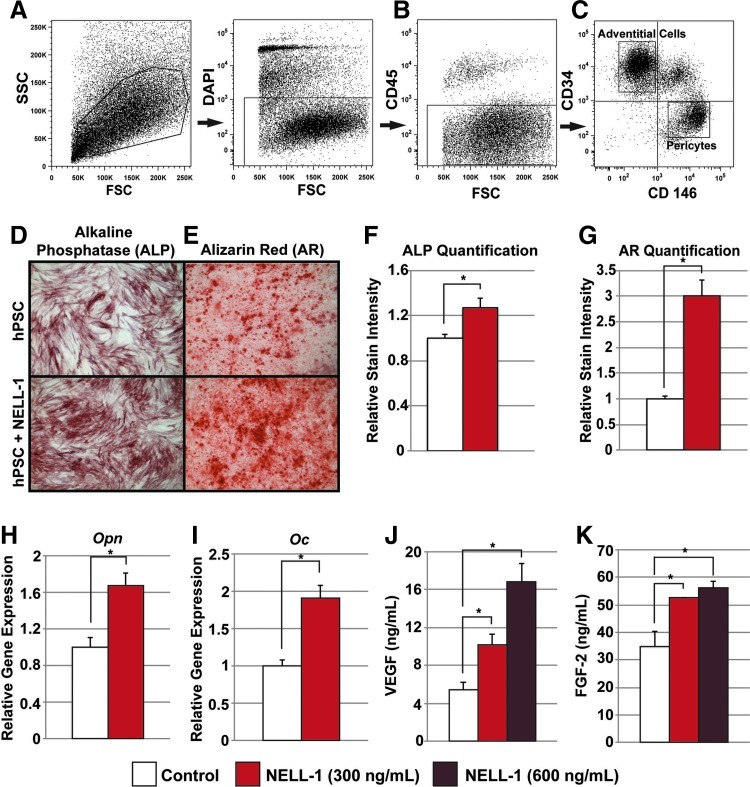
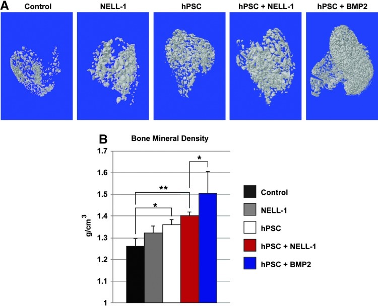
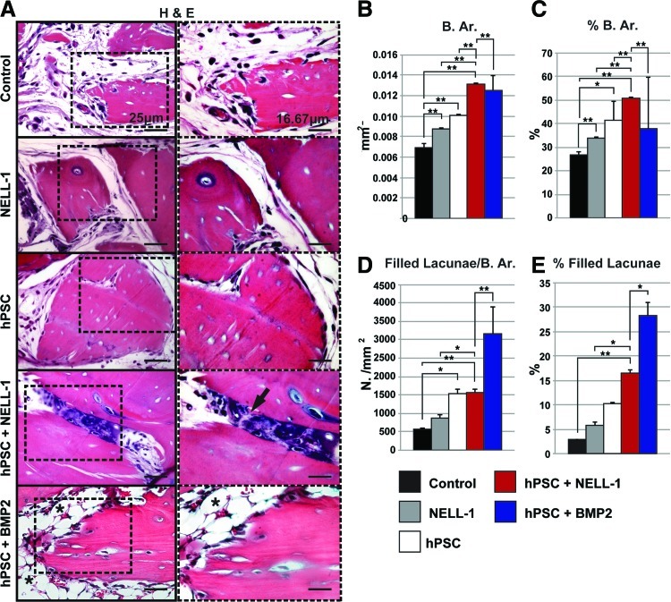
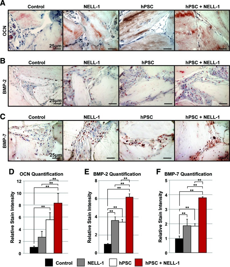
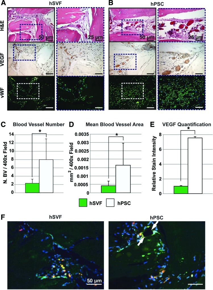
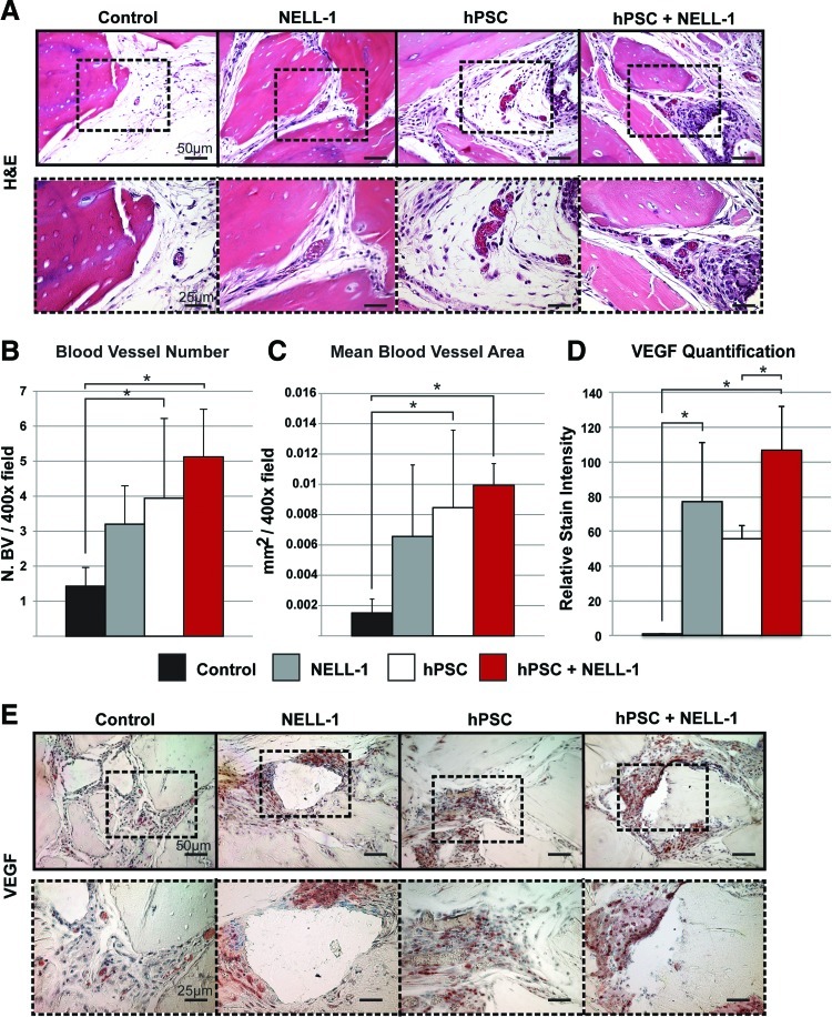
An ideal mesenchymal stem cell (MSC) source for bone tissue engineering has yet to be identified. Such an MSC population would be easily harvested in abundance, with minimal morbidity and with high purity. Our laboratories have identified perivascular stem cells (PSCs) as a candidate cell source. PSCs are readily isolatable through fluorescent-activated cell sorting from adipose tissue and have been previously shown to be indistinguishable from MSCs in the phenotype and differentiation potential. PSCs consist of two distinct cell populations: (1) pericytes (CD146+, CD34-, and CD45-), which surround capillaries and microvessels, and (2) adventitial cells (CD146-, CD34+, and CD45-), found within the tunica adventitia of large arteries and veins. We previously demonstrated the osteogenic potential of pericytes by examining pericytes derived from the human fetal pancreas, and illustrated their in vivo trophic and angiogenic effects. In the present study, we used an intramuscular ectopic bone model to develop the translational potential of our original findings using PSCs (as a combination of pericytes and adventitial cells) from human white adipose tissue. We evaluated human PSC (hPSC)-mediated bone formation and vascularization in vivo. We also examined the effects of hPSCs when combined with the novel craniosynostosis-associated protein, Nel-like molecule I (NELL-1). Implants consisting of the demineralized bone matrix putty combined with NELL-1 (3 μg/μL), hPSC (2.5×10(5) cells), or hPSC+NELL-1, were inserted in the bicep femoris of SCID mice. Bone growth was evaluated using microcomputed tomography, histology, and immunohistochemistry over 4 weeks. Results demonstrated the osteogenic potential of hPSCs and the additive effect of hPSC+NELL-1 on bone formation and vasculogenesis. Comparable osteogenesis was observed with NELL-1 as compared to the more commonly used bone morphogenetic protein-2. Next, hPSCs induced greater implant vascularization than the unsorted stromal vascular fraction from patient-matched samples. Finally, we observed an additive effect on implant vascularization with hPSC+NELL-1 by histomorphometry and immunohistochemistry, accompanied by in vitro elaboration of vasculogenic growth factors. These findings hold significant implications for the cell/protein combination therapy hPSC+NELL-1 in the development of strategies for vascularized bone regeneration.


Figures

FIG. 1.
FIG. 1.
Human perivascular stem cell (hPSC) isolation and in vitro osteogenic differentiation. (AC) Fluorescence-activated cell sorting (FACS) isolation method for hPSCs. (A) 4′,6-Diamidino-2-phenylindole (DAPI)+ dead cells and (B) CD45+ hematopoietic cells were excluded before hPSC isolation. (C) Purified hPSCs consist of distinct CD34− and CD146+ pericytes and CD34+ and CD146− adventitial cells. (DG) hPSCs were cultured under osteogenic conditions, treated with or without 300 ng/mL Nel-like Molecule I (NELL-1) protein. (D) Alkaline phosphatase (ALP) staining at 5 days of osteogenic differentiation. (E) Alizarin Red (AR) staining at 10 days of osteogenic differentiation. (F) Photographic semiquantitative analysis of ALP staining and (G) AR staining based on random n=3 (38.4×) images, using the Adobe Photoshop magic wand tool (tolerance of 32). (H) Osteopontin (Opn) expression by quantitative real time (RT)–polymerase chain reaction (qRT-PCR) at 7 days of differentiation. (I) Osteocalcin (Oc) expression by qRT-PCR at 7 days of differentiation. (J) Secreted vascular endothelial growth factor (VEGF) protein in the supernatant as assessed by enzyme-linked immunosorbent assay (ELISA) at 48-h treatment with NELL-1 (300–600 ng/mL). (K) Secreted FGF-2 protein in the supernatant as assessed by ELISA at 48-h treatment with NELL-1 (300–600 ng/mL). *p<0.05. Color images available online at www.liebertpub.com/tea
FIG. 2.
FIG. 2.
hPSCs form significantly more bone in vivo when combined with NELL-1. Implants consisting of the demineralized bone matrix (DBX) or DBX treated with hPSCs (2.5×10⁵ cells), NELL-1 (3 μg/μL), or both hPSC and NELL-1 were inserted intramuscularly in SCID mice. Bone morphogenetic protein-2 (BMP-2) protein was used as a comparison to NELL-1 (see Supplementary Table S1 for treatment group specifics). Mice were harvested 4 weeks post-implantation and evaluated for bone formation. (A) Representative three-dimensional microcomputed tomography images at Th120, and corresponding analysis of (B) mean bone mineral density (BMD, n=8) (Th50–120 used for analysis). *p<0.05; **p<0.01. Color images available online at www.liebertpub.com/tea
FIG. 3.
FIG. 3.
Histological evidence of enhanced osteogenic potential of combined hPSC-NELL-1-treatment. Hematoxylin and eosin (H&E) staining was performed on the five previously mentioned experimental groups (DBX, DBX+NELL-1, DBX+hPSC, DBX+hPSC+NELL-1, and DBX+hPSC+BMP2). (A) Representative H&E images. (BE) Histomorphometric semiquantitative analysis of random 200× H&E images: (B) Bone area (B. Ar), n=10 images per group, tolerance=50, (C) percent bone area (% B. Ar.), n=10 images per group, tolerance=50, (D) number osteocyte/bone area, (N. Oc/B. Ar.), n=15 images per group, tolerance=20, and (E) % filled lacunae, n=15 images per group, tolerance=20. *p<0.05; **p<0.01. Black arrow indicates endochondral ossification. Black asterisk indicated lipid accumulation. Color images available online at www.liebertpub.com/tea
FIG. 4.
FIG. 4.
Immunohistochemical analysis of ectopic bone formation. (AC) Immunohistochemistry was performed on the four previously mentioned experimental groups (DBX, DBX+NELL-1, DBX+hPSC, and DBX+hPSC+NELL-1). Semiquantitative analysis of the relative stain intensity was determined, using the magic wand tool of Adobe Photoshop on random 400× fields. (A) Representative images of Osteocalcin (OCN) immunohistochemistry and corresponding relative stain quantification (D), based on n=5 images per group and tolerance of 30. (B) Representative images of BMP-2 immunohistochemistry and corresponding relative stain quantification (E), based on n=8 images per group and tolerance of 30. (C) Representative images of bone morphogenetic protein-7 (BMP-7) immunohistochemistry and corresponding relative stain quantification (F), based on n=8 images per group and tolerance of 30. **p<0.01. Color images available online at www.liebertpub.com/tea
FIG. 5.
FIG. 5.
hPSC-treated implants show increased vascularization as compared to the human stromal vascular fraction (hSVF). Equal numbers (2.5×10⁵) of either hPSCs or hSVF cells loaded onto DBX putty were implanted intramuscularly. Samples were harvested 4 weeks postimplantation. Specimens were stained with H&E and for VEGF and von Willebrand Factor (vWF). Histomorphometric semiquantitative analysis of respective 400× images followed using the Adobe Photoshop magic wand tool. (A) H&E, VEGF, and vWF staining among hSVF-treated implants. (B) H&E, VEGF, and vWF staining among hPSC-treated implants. (C) Blood vessel number, based on n=10 H&E images per group. (D) Mean blood vessel area, based on n=10 H&E images per group at a tolerance of 30. (E) VEGF quantification, based on n=5 images per group and tolerance of 30. (F) CD31 immunofluorescent staining, appearing green with red fluorescent-labeled cells. DAPI is used as a nuclear counterstain, appearing blue. White arrow indicates close association of PSCs with a blood vessel. *p<0.05. Color images available online at www.liebertpub.com/tea
FIG. 6.
FIG. 6.
NELL-1 enhances hPSC-mediated vascularization in vivo. H&E staining and VEGF immunohistochemistry were performed on the four previously mentioned experimental groups (DBX, DBX+NELL-1, DBX+hPSC, and DBX+hPSC+NELL-1). Histomorphometric semiquantitative analysis of respective 200× images followed using the Adobe Photoshop magic wand tool. (A) Representative H&E images. (B) Blood vessel number, based on n=10 H&E images per group. (C) Mean blood vessel area, based on n=10 H&E images per group. (D) VEGF stain quantification, based on n=7 images per group at a tolerance of 30. (E) Representative image of VEGF immunohistochemistry. *p<0.05. Color images available online at www.liebertpub.com/tea

Similar articles

Cited by

References

    1. Giannoudis P.V. Dinopoulos H. Tsiridis E. Bone substitutes: an update. Injury. 2005;36(Suppl 3):S20. - PubMed
    1. Laurencin C.T. Ambrosio A.M. Borden M.D. Cooper J.A., Jr. Tissue engineering: orthopedic applications. Annu Rev Biomed Eng. 1999;1:19. - PubMed
    1. Laurie S.W. Kaban L.B. Mulliken J.B. Murray J.E. Donor-site morbidity after harvesting rib and iliac bone. Plast Reconstr Surg. 1984;73:933. - PubMed
    1. Frodel J.L., Jr. Marentette L.J. Quatela V.C. Weinstein G.S. Calvarial bone graft harvest. Techniques, considerations, and morbidity. Arch Otolaryngol Head Neck Surg. 1993;119:17. - PubMed
    1. Cancedda R. Mastrogiacomo M. Bianchi G. Derubeis A. Muraglia A. Quarto R. Bone marrow stromal cells and their use in regenerating bone. Novartis Found Symp. 2003;249:133. discussion 143, 170, 239. - PubMed

Publication types

MeSH terms

Substances