. 2013 Apr;19(4):262-8.
doi: 10.1111/cns.12066. Epub 2013 Feb 14.

Antimalarial drug artemisinin extenuates amyloidogenesis and neuroinflammation in APPswe/PS1dE9 transgenic mice via inhibition of nuclear factor-κB and NLRP3 inflammasome activation

Affiliations

Antimalarial drug artemisinin extenuates amyloidogenesis and neuroinflammation in APPswe/PS1dE9 transgenic mice via inhibition of nuclear factor-κB and NLRP3 inflammasome activation

Jian-Quan Shi et al. CNS Neurosci Ther. 2013 Apr.

Abstract

Background: The activation of nuclear factor-kappa B (NF-κB) and NLRP3 inflammasome is involved in neuroinflammation, which is closely linked to Alzheimer's disease (AD). In vivo and in vitro studies have suggested that artemisinin shows antiinflammatory effects in inflammation-related diseases. However, the impacts of artemisinin on AD have not been investigated.

Aims: In this study, 5-month-old APPswe/PS1dE9 transgenic mice were treated daily with 40 mg/kg artemisinin for 30 days by intraperitoneal injection to evaluate the effects of artemisinin on AD.

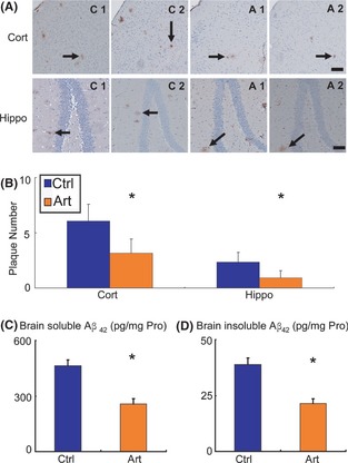
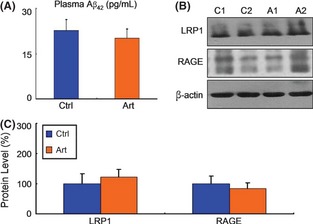
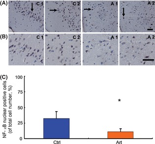
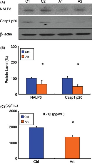
Results: We found that artemisinin treatment (1) decreased neuritic plaque burden; (2) did not alter Aβ transport across the blood-brain barrier; (3) regulated APP processing via inhibiting β-secretase activity; (4) inhibited NF-κB activity and NALP3 inflammasome activation in APPswe/PS1dE9 double transgenic mice.

Conclusions: The in vivo study clearly demonstrates that artemisinin has protective effects on AD pathology due to its effects on suppressing NF-κB activity and NALP3 inflammasome activation. Our study suggests that targeting NF-κB activity and NALP3 inflammasome activation offers a valuable intervention for AD.


Conflict of interest statement

The authors declare no conflict of interests.

Figures

Figure 1
Figure 1
Effect of artemisinin on neuritic plaque formation in APPswe/PS1dE9 double transgenic mice. APPswe/PS1dE9 transgenic mice aged 5 months were treated with 40 mg/kg artemisinin (Art) for 30 days, whereas age‐matched control APPswe/PS1dE9 mice received vehicle solution (Ctrl) as control. The mice were killed, and the brains were dissected, fixed, and sectioned. (A) Neuritic plaques were detected by immunohistochemistry using an Aβ1‐42 antibody. The plaques were visualized by microscopy with 200× magnification. Artemisinin significantly reduced the numbers of neuritic plaques in the cortex (Cort) and hippocampus (Hippo) of mice compared with controls. Black arrows point to plaques. Bars: 100 μm. (B) Quantification of neuritic plaques in APPswe/PS1dE9 mice in each group. An ELISA was conducted to measure brain‐soluble Aβ42 (C) and brain‐insoluble Aβ42 (D) levels in transgenic mice in each group. The numbers represent the mean ± SEM. n = 4 mice each (for immunohistochemistry). n = 3 mice each (for ELISA). *P < 0.05 by Student's t‐test.
Figure 2
Figure 2
Effect of Artemisinin on the transport of Aβ across the blood–brain barrier in APPswe/PS1dE9 double transgenic mice. An ELISA was conducted to measure peripheral blood Aβ42 (A) levels in transgenic mice in each group. Artemisinin did not alter the peripheral blood Aβ42 level. n = 3. *P < 0.05 by Student's t‐test. (B) Half brains from mice in each group were lysed in RIPA buffer. LRP1 and RAGE were detected by Western blotting using β‐actin as a loading control. (C) Quantification of LRP1 and RAGE in brains. Artemisinin had no influence on LRP1 and RAGE expression. n = 3. *, P < 0.05 by Student's t‐test.
Figure 3
Figure 3
Effect of artemisinin on APP processing pathways in APPswe/PS1dE9 double transgenic mice. (A) Brain tissues from APPswe/PS1dE9 mice were subjected to Western blotting to determine the levels of APP‐full length (FL), BACE1, PS1, Aph‐1a, and APPCTFs, with β‐actin as a loading control. (B) Quantification of APP‐FL, BACE1, PS1, Aph‐1a, and APPCTFs in brain tissues. Artemisinin significantly decreased BACE1 expression and had no influence on the levels of APP‐FL, PS1, Aph‐1a, and APPCTFs. n = 3. *P < 0.05 by Student's t‐test.
Figure 4
Figure 4
Effects of artemisinin on the nuclear translocation of nuclear factor‐kappa B (NF‐κB) p65 in APPswe/PS1dE9 double transgenic mice. Brain tissues from APPswe/PS1dE9 mice were subjected to immunohistochemistry to determine the nuclear translocation of NF‐κB p65. (A) Representative photographs of immunohistochemistry staining with anti‐NF‐κB p65 antibody (with 200× magnification). (B) Representative photographs of immunohistochemistry staining with anti‐NF‐κB p65 antibody (with 400× magnification). Artemisinin significantly reduced the ratio of NF‐κB p65 nuclear–positive cells. Black arrows point to NF‐κB p65 nuclear–positive cells. Bars: 50 μm. (C) Quantification of the ratio of NF‐κB p65 nuclear–positive cells in APPswe/PS1dE9 mice in each group; the numbers represent the mean ± SEM. n = 3 mice each. *P < 0.05 by Student's t‐test.
Figure 5
Figure 5
Effect of artemisinin on nuclear factor‐kappa B (NF‐κB) activity in APPswe/PS1dE9 double transgenic mice. (A) Brain tissues from APPswe/PS1dE9 mice were subjected to Western blotting to determine the levels of p‐NF‐κB p65, NF‐κB p65, I‐κBα, and p‐I‐κBα, with β‐actin as a loading control. (B) Quantification of p‐NF‐κB p65, NF‐κB p65, I‐κBα, and p‐I‐κBα. Artemisinin significantly decreased the levels of p‐NF‐κB p65, NF‐κB p65, and p‐I‐κBα and increased the level of I‐κBα. n = 3. *, P < 0.05 by Student's t‐test. (C) An ELISA was conducted to measure brain IL‐6 and TNF‐α level in the brains of mice in each group. Artemisinin significantly reduced IL‐6 and TNF‐α level in brains. n = 3. *, P < 0.05 by Student's t‐test.
Figure 6
Figure 6
Effects of artemisinin on NALP3 inflammasome activation in APPswe/PS1dE9 double transgenic mice. (A) Brain tissues from APPswe/ PS1dE9 mice were subjected to Western blotting to determine the levels of NALP3 and caspase‐1 p20 subunit, with b‐actin as a loading control. (B) Quantification of NALP3 and caspase‐1 p20 subunit. Artemisinin significantly decreased the levels of NALP3 and caspase‐1 p20 subunit. n = 3. *, P < 0.05 by Student's t‐test. (C) An ELISA was conducted to measure brain IL‐1b level in the brains of mice in each group. Artemisinin significantly reduced IL‐1b level in brains. n = 3. *, P < 0.05 by Student's t‐test.

References

    1. Cummings JL, Cole G. Alzheimer disease. JAMA 2002;287:2335–2338. - PubMed
    1. O'Brien RJ, Wong PC. Amyloid precursor protein processing and Alzheimer's disease. Annu Rev Neurosci 2011;34:185–204. - PMC - PubMed
    1. Wyss‐Coray T, Rogers J. Inflammation in Alzheimer disease‐a brief review of the basic science and clinical literature. Cold Spring Harb Perspect Med 2012;2:a006346. - PMC - PubMed
    1. He P, Zhong Z, Lindholm K, et al. Deletion of tumor necrosis factor death receptor inhibits amyloid beta generation and prevents learning and memory deficits in Alzheimer's mice. J Cell Biol 2007;178:829–841. - PMC - PubMed
    1. Lim JE, Kou J, Song M, et al. MyD88 deficiency ameliorates β‐amyloidosis in an animal model of Alzheimer's disease. Am J Pathol 2011;179:1095–1103. - PMC - PubMed

Publication types

MeSH terms