. 2013 Jul;19(13-14):1592-600.
doi: 10.1089/ten.TEA.2012.0394. Epub 2013 Apr 15.

Absence of immune responses with xenogeneic collagen and elastin

Affiliations

Absence of immune responses with xenogeneic collagen and elastin

Alexandra Bayrak et al. Tissue Eng Part A. 2013 Jul.

Abstract

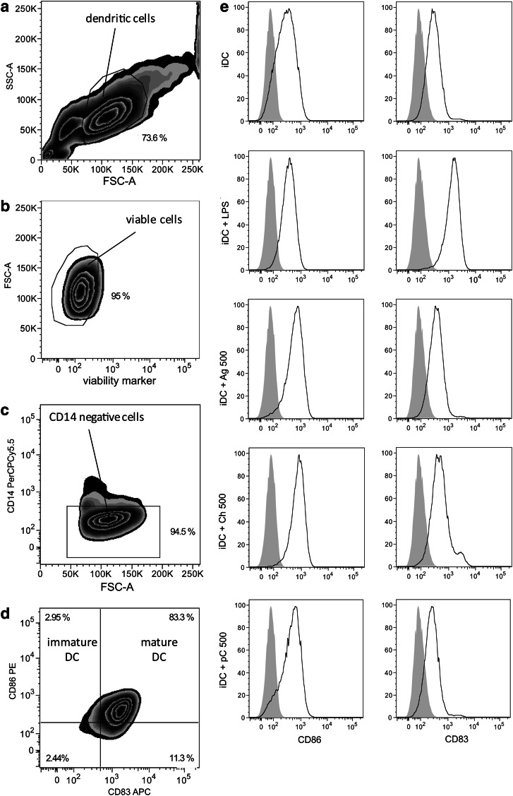
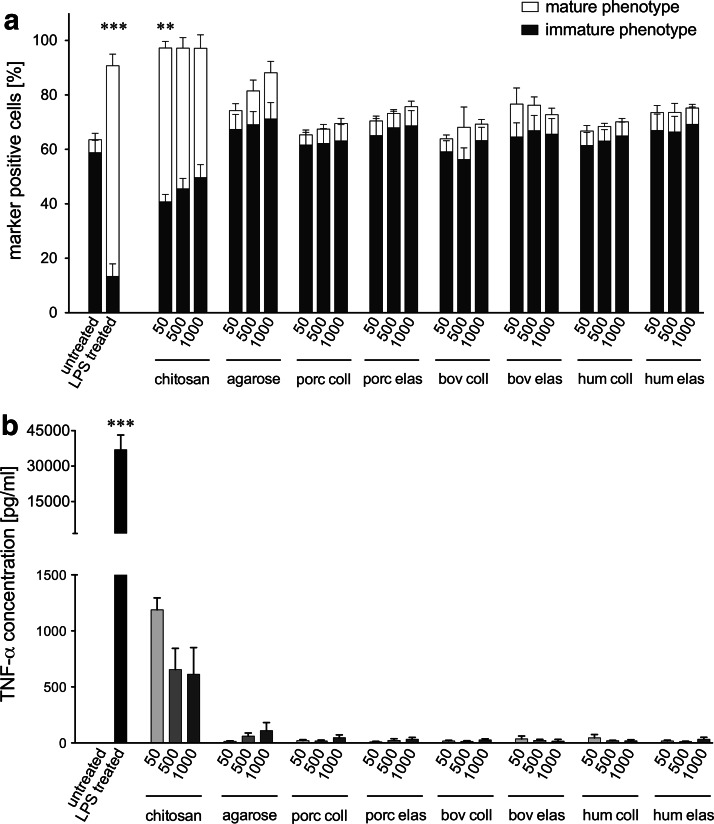
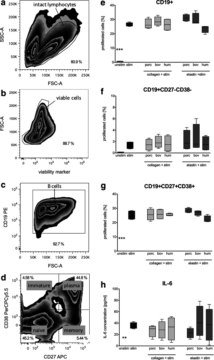
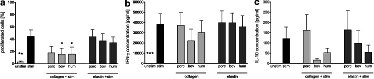
Novel tissue-engineering approaches for cardiovascular matrices based on xenogeneic extracellular matrix protein (ECMp) constituents require a detailed evaluation of their interaction with essential immune cell subsets playing a role in innate or adaptive immunity. Therefore, in this study, the effects of xenogeneic (porcine, bovine) collagen type I and elastin as the two main components of the heart valve ECM were analyzed in comparison to their human equivalents. First, their potential to induce maturation and cytokine secretion of human dendritic cells (DC) was tested by flow cytometry. Second, the influence on proliferation and cytokine release of purified human B and T cells was measured. We could demonstrate that xenogeneic collagen type I and elastin are not able to trigger the maturation of DC as verified by the lack of CD83 induction accompanied by a low tumor necrosis factor-α release. Moreover, both ECMp showed no effect on the proliferation and the interleukin-6 release of either unstimulated or prestimulated B cells. Additionally, anti-CD3-induced purified T cell proliferation and secretion of cytokines was not affected. All in vitro data verify the low immunogenicity of porcine and bovine collagen type I and elastin and favor their suitability for tissue-engineered scaffolds.


Figures

FIG. 1.
FIG. 1.
Flow cytometric gating strategy and phenotypical characterization of monocyte-derived human dendritic cells. Purified human CD14+ monocytes were cultured for 5 days in the presence of IL-4 and granulocyte-macrophage colony-stimulating factor to generate immature dendritic cells (iDC). Subsequent lipopolysaccharide (LPS) stimulation of iDC for 24 h generated mature DC (iDC+LPS). Gating was performed as follows: (a) DCs were pregated according to forward and sideward scatter, (b) verified viable, (c) and CD14-negative before (d) classification as immature (CD14− CD86+ CD83−) or mature DC (CD14− CD86+ CD83+). (e) Histograms show the expression of surface markers CD86 and CD83 (black lines) in comparison to unlabeled cells (solid gray curves) for untreated (iDC) or iDC treated with LPS (iDC+LPS), chitosan (500 μg/mL; iDC+Ch500), agarose (500 μg/mL; iDC+Ag500), and for porcine collagen (500 μg/mL; iDC+pC500). Representative data from 5 independent experiments are shown.
FIG. 2.
FIG. 2.
Influence of xenogeneic and human extracellular matrix protein (ECMp) on the maturation status and tumor necrosis factor-α (TNF-α) secretion of DC. (a) The percentage of DC with an immature CD14− CD86+ CD83− (black bars) or mature CD14− CD86+ CD83+ (white bars) phenotype is shown for untreated iDC, LPS treated iDC, as well as for iDC cocultured with agarose, chitosan, porcine collagen (porc coll), porcine elastin (porc elas), bovine collagen (bov coll), bovine elastin (bov elas), human collagen (hum coll), or human elastin (hum elas) at different concentrations (50, 500, 1000 μg/mL). (b) Supernatants of monocyte-derived iDC cocultured with xenogeneic and human ECMp or with the reference substances agarose and chitosan were collected after 24 h, and TNF-α secretion was quantified by enzyme-linked immunosorbent assay (ELISA). TNF-α release data are presented for all treatment groups described in (a). All data are given as mean±standard error of the mean (SEM) from independent experiments (n=5). Asterisks indicate a significant difference relative to untreated iDC (**p<0.01 or ***p<0.001).
FIG. 3.
FIG. 3.
Impact of xenogeneic and human ECMp on the proliferation of B cell subpopulations and IL-6 secretion. Purified B cells were 5,6-carboxyfluorescein diacetate N-succinimidyl ester (CFSE) stained and cocultured with porcine (porc), bovine (bov), or human (hum) collagens and elastins at a concentration of 500 μg/mL in the presence of CpG, IL-2, and IL-10 stimulation (stim) for 5 days. Harvested cells were costained with mouse anti-human B cell antibodies and the proliferation was determined by flow cytometry after gating on specific subsets. (a) Lymphocytes were gated based on their typical location in a scatter plot, (b) and verified viable (c) before B cells were selected based on the expression of the B cell marker CD19 and (d) classified into the following phenotypes; immature (CD19+ CD27− CD38+), naïve (CD19+ CD27− CD38−), memory B cells (CD19+ CD27+ CD38−), or plasma cells (CD19+ CD27+ CD38+). CFSE-based proliferation was measured for (e) all CD19+ cells, (f) CD19+ CD27-CD38- naïve B cells, and (g) CD19+ CD27+ CD38+ plasma cells. (h) Supernatants were analyzed after 5 days of coculture by a standard IL-6 ELISA. All data are represented as mean±SEM from independent experiments (n=4). Asterisks indicate a significant difference compared to the CpG/IL-2/IL-10 stimulated control (**p<0.01 or ***p<0.001).
FIG. 4.
FIG. 4.
Proliferation and cytokine secretion of purified T cells in coculture with ECMp. Purified CFSE-labeled T cells were cocultured without stimulation (unstim), or with low-dose anti-CD3 (stim), in uncoated plates or in plates coated with 50 μg/mL porcine (porc), bovine (bov), or human (hum) collagens or elastins for 5 days before supernatants were collected and cells were labeled with antibodies and analyzed by flow cytometry in a CFSE-based proliferation assay. The percentage of proliferated T cells (a) was determined by gating on all viable cells followed by gating on the CD3+ population and measuring divided cells based on dilution of the CFSE signal in a histogram. An ELISA was used to determine the amount of (b) IFN-γ and (c) IL-10 present in the supernatants. All data are given as mean±SEM from independent experiments (n=4). Asterisks indicate a significant difference compared to the a-CD3-stimulated control (*p<0.05 or **p<0.01 or ***p<0.001).

References

    1. Piterina A.V. Cloonan A.J. Meaney C.L. Davis L.M. Callanan A. Walsh M.T. McGloughlin T.M. ECM-based materials in cardiovascular applications: inherent healing potential and augmentation of native regenerative processes. Int J Mol Sci. 2009;10:4375. - PMC - PubMed
    1. Akhyari P. Minol P. Assmann A. Barth M. Kamiya H. Lichtenberg A. [Tissue engineering of heart valves] Chirurg. 2011;82:311. - PubMed
    1. Gilbert T.W. Sellaro T.L. Badylak S.F. Decellularization of tissues and organs. Biomaterials. 2006;27:3675. - PubMed
    1. Schoen F.J. Heart valve tissue engineering: quo vadis? Curr Opin Biotechnol. 2011;22:698. - PubMed
    1. Simon P. Kasimir M.T. Seebacher G. Weigel G. Ullrich R. Salzer-Muhar U. Rieder E. Wolner E. Early failure of the tissue engineered porcine heart valve SYNERGRAFT in pediatric patients. Eur J Cardiothorac Surg. 2003;23:1002. - PubMed

Publication types

LinkOut - more resources