. 2013 Feb 13:11:13.
doi: 10.1186/1741-7007-11-13.

Notch2 is required in somatic cells for breakdown of ovarian germ-cell nests and formation of primordial follicles

Affiliations

Notch2 is required in somatic cells for breakdown of ovarian germ-cell nests and formation of primordial follicles

Jingxia Xu et al. BMC Biol. .

Abstract

Background: In the mouse ovary, oocytes initially develop in clusters termed germ-cell nests. Shortly after birth, these germ-cell nests break apart, and the oocytes individually become surrounded by somatic granulosa cells to form primordial follicles. Notch signaling plays essential roles during oogenesis in Drosophila, and recent studies have suggested that Notch signaling also plays an essential role during oogenesis and ovary development in mammals. However, no in vivo loss-of-function studies have been performed to establish whether Notch family receptors have an essential physiological role during normal ovarian development in mutant mice.

Results: Female mice with conditional deletion of the Notch2 gene in somatic granulosa cells of the ovary exhibited reduced fertility, accompanied by the formation of multi-oocyte follicles, which became hemorrhagic by 7 weeks of age. Formation of multi-oocyte follicles resulted from defects in breakdown of the primordial germ-cell nests. The ovaries of the Notch2 conditional mutant mice had increased numbers of oocytes, but decreased numbers of primordial follicles. Oocyte numbers in the Notch2 conditional mutants were increased not by excess or extended cellular proliferation, but as a result of decreased oocyte apoptosis.

Conclusions: Our work demonstrates that Notch2-mediated signaling in the somatic-cell lineage of the mouse ovary regulates oocyte apoptosis non-cell autonomously, and is essential for regulating breakdown of germ-cell nests and formation of primordial follicles. This model provides a new resource for studying the developmental and physiological roles of Notch signaling during mammalian reproductive biology.


Figures

Figure 1
Figure 1
Generation of mice with ovary-specific Notch2 gene deletion. (A) Breeding scheme to generate mice with ovary-specific Notch2 gene deletion using the Amhr2-Cre driver line. (B, C) Efficiency and specificity of recombination of the Notch2flox allele by the Amhr2-Cre driver. (B) Southern-blot analysis of genomic DNA from ovaries obtained from: (lane 1) Notch2flox/+;+/+ control (mouse has no Cre driver); (lane 2) Notch2flox/Notch2null;Amhr2-Cre/+ mutant; (lanes 3 and 4), two individual Notch2flox/+;Amhr2-Cre/+ mice (heterozygous for the Notch2flox allele). All mice were four to five weeks old. (C) Southern blot of ovary and spleen DNA to test the specificity and efficiency of Amhr2-Cre-mediated recombination. DNA was isolated from two individual Notch2flox/+;Amhr2-Cre/+ control mice (heterozygous for the Notch2flox allele) at 9 weeks of age. There was almost complete deletion of the Notch2flox allele in ovary DNA, but not in spleen DNA. Note that the band resulting from Cre-mediated recombination (labeled 'Null') was present only in Amhr2-Cre/+ ovaries.
Figure 2
Figure 2
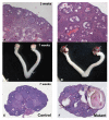
Ovaries of Amhr2-Cre/+;Notch2flox/- mice contain multi-oocyte follicles that become hemorrhagic. (A, C, E) Littermate controls; (B, D, F) Amhr2-Cre/+;Notch2flox/- mutants. (A, B) Multi-oocyte follicles were present in 3-week-old Amhr2-Cre/+;Notch2flox/- female mice (B. arrowheads). (C-F) Follicles in Amhr2-Cre/+;Notch2flox/- females became hemorrhagic by approximately 7 weeks of age (D, F, arrows).
Figure 3
Figure 3
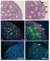
Germ-cell nests persisted in Amhr2-Cre/+;Notch2flox/- mice. (A, B) Germ-cell nests in (A) littermate control and (B) 8-day-old Amhr2-Cre/+;Notch2flox/- mouse. (B) In the mutant mouse, the germ-cell nests were still visible (black arrows), whereas in (A) the nests had broken down and the oocytes were assembled into primordial follicles. Slides were stained using the Periodic-Acid-Schiff (PAS) (C) By postnatal day (PND)3, most oocytes expressing the cytoplasmic germ-cell marker Vasa (orange) in the cortical region of the ovary had been recruited into primordial follicles in the control littermate mice. (D) However, Vasa-positive germ cells in the cortical region of the Amhr2-Cre/+;Notch2flox/- mice were still in germ-cell nests (white arrows) at PND3. Cuboidal granulosa cells of primary follicles in the medulla of both littermate and mutant ovaries expressed anti-Müllerian hormone (AMH; green). Cell nuclei were counterstained with 4',6-diamidino-2-phenylindole (DAPI) (blue). (E) Laminin immunostaining (green) showed that by PND3, primordial follicles in the cortex of the ovary of control littermate mice were surrounded by a layer of basement membrane. (F) However, in Amhr2-Cre/+;Notch2flox/- mice ovaries at the same stage, the oocytes remained in germ-cell cysts and were not individually surrounded by a basement membrane. Scale bar: (A, B): 20 μm; (C-F): 50 μm.
Figure 4
Figure 4
Assessment and quantification of follicle types in the ovaries of prepubertal Amhr2-Cre/+;Notch2flox/- and control littermate mice. (A-D) Follicle types were assessed and counted in serial sections of ovaries isolated from Amhr2-Cre/+;Notch2flox/- (white bars) and control littermate (black bars) mice at postnatal day 18. (A-C) Amhr2-Cre/+;Notch2flox/- ovaries exhibited a significant increase in the number of multi-oocyte follicles (containing four or more oocytes) of (A) the primordial, (B) primary and (C) secondary follicle types compared with littermate control ovaries. Amhr2-Cre/+;Notch2flox/- ovaries also exhibited a significantly reduced number of primordial follicles containing a single oocyte. * P < 0.05; ** P < 0.005.
Figure 5
Figure 5
Absence of extended oocyte proliferation in embryonic Amhr2-Cre/+;Notch2flox/- ovaries. Pregnant mice were injected with 5-bromo-2'-deoxyuridine (BrdU) twice daily between E16.5 through E18.5 (a total of six injections). Ovaries were isolated from Amhr2-Cre/+;Notch2flox/- and control littermate progeny on the day of birth (postnatal day 0; PND0), and sections were stained with anti-Vasa (red) antibodies to label oocytes, and with anti-BrdU (green) antibodies to identify proliferating cells. Sections were also counterstained with DAPI (blue) to stain cell nuclei. As shown in the merged figure on the right, at PND0, no oocyte in either (A) littermate control or (B) mutant ovaries had incorporated BrdU as a result of these injections. (C) Higher magnification view of the mutant ovary (boxed region in B).
Figure 6
Figure 6
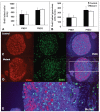
Absence of extended cell proliferation in neonatal Amhr2-Cre/+;Notch2flox/- ovaries. (A, B) Quantification of (A) total 5-bromo-2'-deoxyuridine (BrdU)-positive cells and (B) BrdU-positive pre-granulosa/granulosa cells (defined as a BrdU-positive somatic cell adjacent to a Vasa-positive oocyte). Ovaries were harvested from Amhr2-Cre/+;Notch2flox/- and control littermate mice after a 2-hour BrdU pulse administration on postnatal day (PND)1 or PND2. Data are presented as the mean ± SD of four ovaries from four individual mutant or control mice. There were no significant differences in the numbers of either (A) total proliferating cells or (B) proliferating pre-granulosa/granulosa cells between the control and the Amhr2-Cre/+;Notch2flox/- ovaries. (C, D) Representative micrographs showing BrdU incorporation from (C) control littermate and (D) Amhr2-Cre/+;Notch2flox/- ovaries at PND1. No oocyte in either (C) the control or (D) mutant ovary incorporated BrdU. BrdU-positive cells are in green; oocytes are in red (Vasa-positive), with nuclear DAPI staining in blue. (E) Higher magnification view of the mutant ovary (boxed region in D).
Figure 7
Figure 7
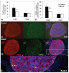
Decreased apoptotic cell death in Amhr2-Cre/+;Notch2flox/- ovaries. (A, B) Quantification of terminal deoxynucleotidyl transferase-mediated deoxyuridine triphosphate (TUNEL)-positive (A) oocytes and (B) pre-granulosa/granulosa cells (defined as a TUNEL-positive somatic cell adjacent to a Vasa-positive oocyte). Data are presented as the mean ± SD of four ovaries from four individual mutant or control mice. At postnatal day (PND)1, there were significant differences in the numbers of both apoptotic (that is, TUNEL-positive) oocytes and pre-granulosa/granulosa cells between the control and the Amhr2-Cre/+;Notch2flox/- ovaries. * P < 0.05; ** P < 0.01. (C, D) Representative micrographs showing TUNEL staining from (C) control littermate and/(D) Amhr2-Cre/+;Notch2flox ovaries at PND1. TUNEL-positive cells are in green; oocytes are in red (Vasa-positive), with nuclear DAPI staining in blue. (E) Higher magnification view of the mutant ovary (boxed region in D). Arrow: TUNEL-positive oocyte; arrowhead: two TUNEL-positive pre-granulosa/granulosa cells adjacent to a Vasa-positive oocyte.

Similar articles

Cited by

References

    1. Saga Y. Mouse germ cell development during embryogenesis. Curr Opin Genet Dev. 2008;18:337–341. doi: 10.1016/j.gde.2008.06.003. - DOI - PubMed
    1. Jagarlamudi K, Rajkovic A. Oogenesis: transcriptional regulators and mouse models. Mol Cell Endocrinol. 2012;356:31–39. doi: 10.1016/j.mce.2011.07.049. - DOI - PubMed
    1. Epifano O, Dean J. Genetic control of early folliculogenesis in mice. Trends Endocrinol Metab. 2002;13:169–173. doi: 10.1016/S1043-2760(02)00576-3. - DOI - PubMed
    1. Tingen C, Kim A, Woodruff TK. The primordial pool of follicles and nest breakdown in mammalian ovaries. Mol Hum Reprod. 2009;15:795–803. doi: 10.1093/molehr/gap073. - DOI - PMC - PubMed
    1. Pepling ME. Follicular assembly: mechanisms of action. Reproduction. 2012;143:139–149. doi: 10.1530/REP-11-0299. - DOI - PubMed

Publication types

LinkOut - more resources