Brain involvement in Alström syndrome
- PMID: 23406482
- PMCID: PMC3584911
- DOI: 10.1186/1750-1172-8-24
Brain involvement in Alström syndrome
Abstract
Background: Alström Syndrome (AS) is a rare ciliopathy characterized by cone-rod retinal dystrophy, sensorineural hearing loss, obesity, type 2 diabetes mellitus and cardiomyopathy. Most patients do not present with neurological issues and demonstrate normal intelligence, although delayed psychomotor development and psychiatric disorders have been reported. To date, brain Magnetic Resonance Imaging (MRI) abnormalities in AS have not been explored.
Methods: We investigated structural brain changes in 12 genetically proven AS patients (mean-age 22 years; range: 6-45, 6 females) and 19 matched healthy and positive controls (mean-age 23 years; range: 6-43; 12 females) using conventional MRI, Voxel-Based Morphometry (VBM) and Diffusion Tensor Imaging (DTI).
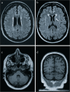
Results: 6/12 AS patients presented with brain abnormalities such as ventricular enlargement (4/12), periventricular white matter abnormalities (3/12) and lacune-like lesions (1/12); all patients older than 30 years had vascular-like lesions. VBM detected grey and white matter volume reduction in AS patients, especially in the posterior regions. DTI revealed significant fractional anisotropy decrease and radial diffusivity increase in the supratentorial white matter, also diffusely involving those regions that appeared normal on conventional imaging. On the contrary, axial and mean diffusivity did not differ from controls except in the fornix.
Conclusions: Brain involvement in Alström syndrome is not uncommon. Early vascular-like lesions, gray and white matter atrophy, mostly involving the posterior regions, and diffuse supratentorial white matter derangement suggest a role of cilia in endothelial cell and oligodendrocyte function.
Figures


References
-
- Marshall JD, Hinman EG, Collin GB, Beck S, Cerqueira R, Maffei P, Milan G, Zhang W, Wilson DI, Hearn T, Tavares P, Vettor R, Veronese C, Martin M, So WV, Nishina PM, Naggert JK. Spectrum of ALMS1 variants and evaluation of genotypephenotype correlations in Alström syndrome. Hum Mutat. 2007;28(11):1114–1123. doi: 10.1002/humu.20577. - DOI - PubMed
MeSH terms
LinkOut - more resources
Full Text Sources
Other Literature Sources
Research Materials

