. 2013 Mar;140(6):1262-71.
doi: 10.1242/dev.085316. Epub 2013 Feb 13.

Cofilin and Vangl2 cooperate in the initiation of planar cell polarity in the mouse embryo

Affiliations

Cofilin and Vangl2 cooperate in the initiation of planar cell polarity in the mouse embryo

James P Mahaffey et al. Development. 2013 Mar.

Abstract

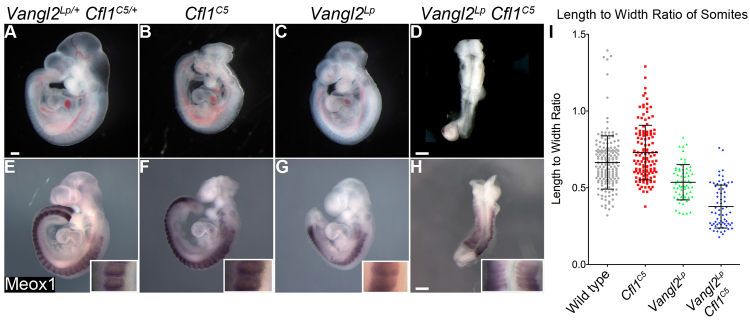
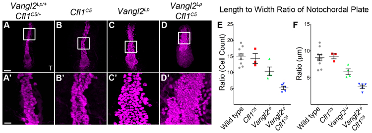
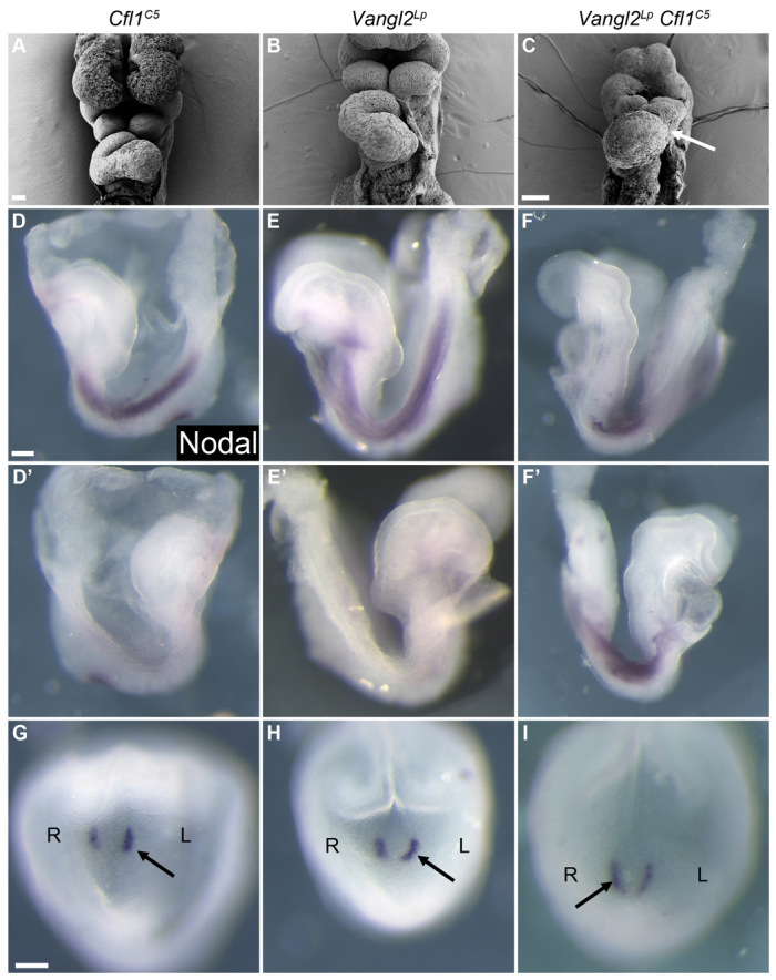
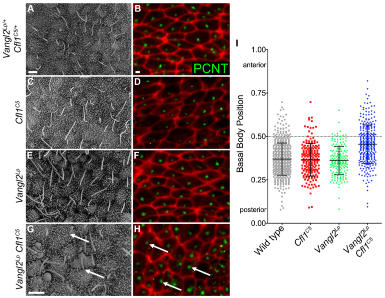
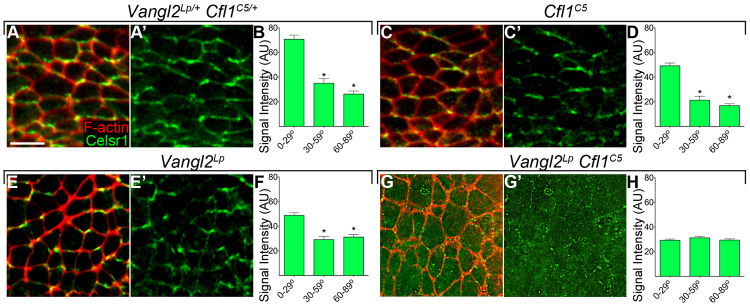
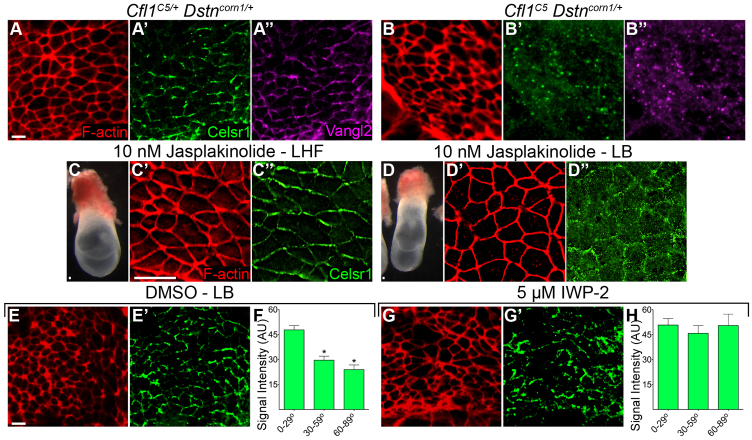
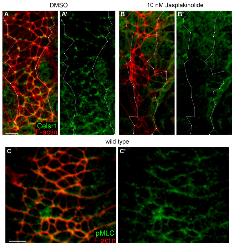
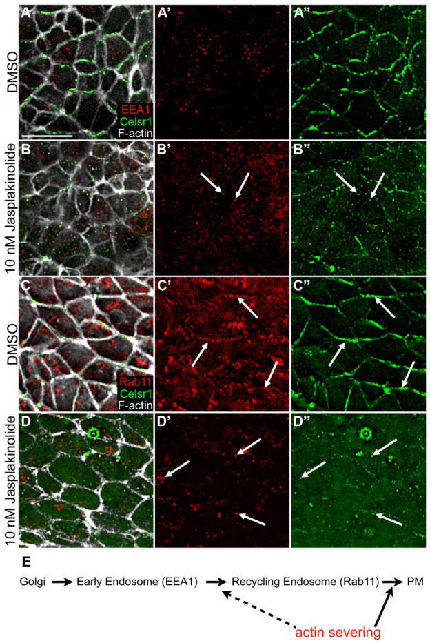
The planar cell polarity (PCP; non-canonical Wnt) pathway is required to orient the cells within the plane of an epithelium. Here, we show that cofilin 1 (Cfl1), an actin-severing protein, and Vangl2, a core PCP protein, cooperate to control PCP in the early mouse embryo. Two aspects of planar polarity can be analyzed quantitatively at cellular resolution in the mouse embryo: convergent extension of the axial midline; and posterior positioning of cilia on cells of the node. Analysis of the spatial distribution of brachyury(+) midline cells shows that the Cfl1 mutant midline is normal, whereas Vangl2 mutants have a slightly wider midline. By contrast, midline convergent extension fails completely in Vangl2 Cfl1 double mutants. Planar polarity is required for the posterior positioning of cilia on cells in the mouse node, which is essential for the initiation of left-right asymmetry. Node cilia are correctly positioned in Cfl1 and Vangl2 single mutants, but cilia remain in the center of the cell in Vangl2 Cfl1 double mutants, leading to randomization of left-right asymmetry. In both the midline and node, the defect in planar polarity in the double mutants arises because PCP protein complexes fail to traffic to the apical cell membrane, although other aspects of apical-basal polarity are unaffected. Genetic and pharmacological experiments demonstrate that F-actin remodeling is essential for the initiation, but not maintenance, of PCP. We propose that Vangl2 and cofilin cooperate to target Rab11(+) vesicles containing PCP proteins to the apical membrane during the initiation of planar cell polarity.


Figures

Fig. 1.
Fig. 1.
Genetic interaction between Vangl2Lp and Cfl1C5 at E9.5. (A) A Vangl2Lp/+ CflC5/+ embryo, which is indistinguishable from wild type. (B) Cfl1C5 homozygote. Homozygous mutants are exencephalic and die ∼E11.0. (C) Vangl2Lp homozygote. Homozygous mutant embryos have an open neural tube caudal to the midbrain-hindbrain junction. (D) Vangl2Lp CflC5 double mutant. These embryos have a completely open neural tube, a short body axis and abnormal somites. (E-H) Meox1 in situ hybridization (purple) highlights the somites; there are wide somites in the double mutant (H). Insets show a 4× higher magnification of somites. (I) The length to width ratio (LWR) of somites. Somites in Vangl2Lp mutants are significantly shorter and wider than wild-type or CflC5 embryos (P<0.001). Somites in Vangl2Lp CflC5 double mutants have significantly reduced LWR compared with wild type or either single mutant (P<0.001); Vangl2Lp CflC5 double mutants had 20-22 somite pairs and other genotypes had 22-28 at the stage analyzed. Scale bars: 250 μm. Data are mean±s.d.
Fig. 2.
Fig. 2.
Cfl1C5 enhances the convergent extension defect in the Vangl2Lp notochord. (A-D) Projections of flat-mounted E8.0 embryos stained for brachyury (T) (magenta). (A′-D′) Higher magnifications (4×) of midline cells from boxed regions of A-D highlight the arrangement of cells in each genotype. Wild type (A) and Cfl1C5 (B) have a similar arrangement of T+ cells in the midline, whereas the Vangl2Lp midline (C) is wider and shorter. The Vangl2Lp Cfl1C5 mutant midline (D) is wider and shorter than Vangl2Lp. (E,F) The LWR of the notochordal plate measured by the distribution of cells (E) or in μm (F). Scale bars: 100 μm in A-D; 25 μm in A′-D′. Data are mean±s.d.
Fig. 3.
Fig. 3.
Defects in left-right asymmetry in Vangl2Lp Cfl1C5 double mutant embryos. (A-C) A fraction of Vangl2Lp Cfl1C5 double mutants examined at E9.5 showed reversed heart looping (arrow), a phenotype not seen in either single mutant. (D-I) This phenotype is due to an early defect in left-right asymmetry, as some double mutants express Nodal (purple) bilaterally in the lateral plate mesoderm and show increased Nodal expression on the right side of the node. Arrows indicate the side of the node with stronger Nodal expression. Scale bars: 100 μm.
Fig. 4.
Fig. 4.
Nodal cilia are not posteriorly positioned in Vangl2Lp Cfl1C5 double mutants. (A,C,E) Scanning electron microscope images of the node show that cilia are polarized to the posterior of node cells in wild type, Cfl1C5 and Vangl2Lp single mutants. (B,D,F) Basal bodies, visualized by the centrosomal protein pericentrin (green), show a similar posterior position in the cells of wild type, Cfl1C5 and Vangl2Lp single mutants. F-actin at the cell borders is highlighted with phalloidin (red). (G) In Vangl2Lp Cfl1C5 double mutants, many cilia point anteriorly (arrows).(H) Many basal bodies are mislocalized to the anterior half of the cell in Vangl2Lp Cfl1C5 double mutants (arrows). Anterior is towards the top in all images. (I) The relative localization of basal bodies in individual central node cells was quantified from immunofluorescence images from at least three embryos per genotype. The graph shows that basal bodies in double mutants are not positioned on the posterior of cells; in addition, there is a greater variance in the placement of cilia compared with wild type or either single mutant. Data are mean±s.d. Scale bars: 2 μm.
Fig. 5.
Fig. 5.
Celsr1, a core component of the PCP pathway, is not planar polarized in Vangl2Lp Cfl1C5 double mutants. (A-B) Celsr1 (green) is enriched on the anterior/posterior faces of node cells in wild-type embryos. Cell boundaries are highlighted by phalloidin (red), anterior towards the top.(C-F) The anterior/posterior enrichment of Celsr1 is retained in Cfl1C5 mutants (C-D) and Vangl2Lp mutants (E-F). (G-H) By contrast, Celsr1 is not enriched at the apical membrane in Vangl2Lp Cfl1C5 double mutants (G-H). (B,D,F,H) The mean intensity of Celsr1 staining normalized to phalloidin intensity along different edges of the cell. Celsr1 intensity was measured along individual cell borders and binned based on the angle with respect to the mediolateral axis of the embryo, such that 0-29° (horizontal) lines correspond to anterior/posterior faces of the cell, and 60-90° (vertical) lines correspond to mediolateral faces of the cell. Scale bar: 10 μm. Data are mean±s.d. *P<0.001.
Fig. 6.
Fig. 6.
Decreased cofilin activity is sufficient to disrupt apical trafficking of the PCP proteins Celsr1 and Vangl2. (A-A″) The PCP proteins Celsr1 and Vangl2 colocalize to the anterior/posterior faces of node cells in wild-type embryos. (B-B″) Celsr1 and Vangl2 colocalize in cytoplasmic puncta in the compound Cfl1C5 Dstncorn1/+ mutant. (C-H) Effects of pharmacological inhibitors on the planar polarization of PCP proteins in cultured embryos. (C-C″) Late headfold (LHF) stage embryos, shown before culture (C), show normal Celsr1 localization after 14-18 hours of culture in 10 nM jasplakinolide (C″). (D-D″) When cultured in the presence of jasplakinolide under the same conditions, earlier embryos (late-bud stage, LB) phenocopy Vangl2Lp Cfl1C5 and Cfl1C5 Dstncorn1/+, as Celsr1 fails to become membrane localized (D″). (E,E′) DMSO control for the late-bud stage culture. (G,G′) Blocking Wnt ligand secretion does not prevent recruitment of Celsr1 to the membrane, as embryos cultured with 5 μM IWP2 (an inhibitor of Wnt secretion) retain membrane-localized Celsr1, although Celsr1 is present on all faces of node cells. (F,H) Quantification of the intensity of Celsr1 staining shows normal enrichment to the horizontal (anterior and posterior) edges of node cells in DMSO control (F), which is lost when embryos are cultured with IWP2 (H). Scale bars: 10 μm. Data are mean±s.d. *P<0.001.
Fig. 7.
Fig. 7.
Celsr1 and phospho-myosin light chain 2 are planar polarized in the axial midline. (A,A′) In wild-type embryos cultured for 14-18 hours in 0.1% DMSO, Celsr1 (green) is planar polarized to the anterior/posterior faces in the apical membrane of cells in the midline (outlined in white). (B,B′) Membrane enrichment of Celsr1 is lost when wild-type embryos are cultured in the presence of 10 nM jasplakinolide. (C,C′) In the midline of wild-type embryos, phospho-myosin light chain 2 (green) is also planar polarized at the apical membrane to the anterior/posterior faces of the cell. Scale bars: 10 μm.
Fig. 8.
Fig. 8.
Treatment with jasplakinolide inhibits plasma membrane association of Rab11 and Celsr1. (A-A″) In wild-type embryos cultured for 14 hours in DMSO, there is no colocalization between Celsr1 (green) and the early endosomal marker EEA1 (red). (B-B″) Treatment with 10 nM jasplakinolide inhibits plasma membrane association of Celsr1; some cytoplasmic Celsr1 puncta overlaps with EEA1 (arrows). (C-C″) In wild-type embryos cultured for 14 hours in DMSO, there is some overlap between Rab11 (red) and Celsr1 adjacent to the plasma membrane (arrows). (D-D″) Rab11 colocalizes with some Celsr1 puncta after treatment with jasplakinolide (arrows), although neither Rab11 nor Celsr1 localizes to the plasma membrane. Phalloidin staining (white) marks F-actin. (E) Model for trafficking of PCP proteins. Celsr1 and Vangl2 traffic to EEA1+ endosomes then enter Rab11+ vesicles and are delivered to planar polarized complexes at the plasma membrane (PM) at the initiation of PCP in the node. Actin dynamics are required for this last step and possibly for the movement/transition of EEA1+ endosomes to the Rab11 compartment. Scale bar: 10 μm.

Similar articles

Cited by

References

    1. Axelrod J. D. (2001). Unipolar membrane association of Dishevelled mediates Frizzled planar cell polarity signaling. Genes Dev. 15, 1182–1187 - PMC - PubMed
    1. Bastock R., Strutt H., Strutt D. (2003). Strabismus is asymmetrically localised and binds to Prickle and Dishevelled during Drosophila planar polarity patterning. Development 130, 3007–3014 - PubMed
    1. Bellomo D., Lander A., Harragan I., Brown N. A. (1996). Cell proliferation in mammalian gastrulation: the ventral node and notochord are relatively quiescent. Dev. Dyn. 205, 471–485 - PubMed
    1. Bernstein B. W., Bamburg J. R. (2010). ADF/cofilin: a functional node in cell biology. Trends Cell Biol. 20, 187–195 - PMC - PubMed
    1. Blair A., Tomlinson A., Pham H., Gunsalus K. C., Goldberg M. L., Laski F. A. (2006). Twinstar, the Drosophila homolog of cofilin/ADF, is required for planar cell polarity patterning. Development 133, 1789–1797 - PubMed

Publication types

MeSH terms

LinkOut - more resources