. 2013 Mar;140(6):1231-9.
doi: 10.1242/dev.084665. Epub 2013 Feb 13.

Myod and H19-Igf2 locus interactions are required for diaphragm formation in the mouse

Affiliations

Myod and H19-Igf2 locus interactions are required for diaphragm formation in the mouse

Maud Borensztein et al. Development. 2013 Mar.

Abstract

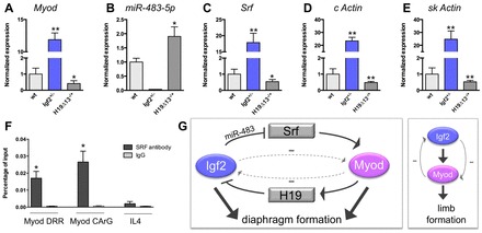
The myogenic regulatory factor Myod and insulin-like growth factor 2 (Igf2) have been shown to interact in vitro during myogenic differentiation. In order to understand how they interact in vivo, we produced double-mutant mice lacking both the Myod and Igf2 genes. Surprisingly, these mice display neonatal lethality due to severe diaphragm atrophy. Alteration of diaphragm muscle development occurs as early as 15.5 days post-coitum in the double-mutant embryos and leads to a defect in the terminal differentiation of muscle progenitor cells. A negative-feedback loop was detected between Myod and Igf2 in embryonic muscles. Igf2 belongs to the imprinted H19-Igf2 locus. Molecular analyses show binding of Myod on a mesodermal enhancer (CS9) of the H19 gene. Chromatin conformation capture experiments reveal direct interaction of CS9 with the H19 promoter, leading to increased H19 expression in the presence of Myod. In turn, the non-coding H19 RNA represses Igf2 expression in trans. In addition, Igf2 also negatively regulates Myod expression, possibly by reducing the expression of the Srf transcription factor, a known Myod activator. In conclusion, Igf2 and Myod are tightly co-regulated in skeletal muscles and act in parallel pathways in the diaphragm, where they affect the progression of myogenic differentiation. Igf2 is therefore an essential player in the formation of a functional diaphragm in the absence of Myod.


Figures

Fig. 1.
Fig. 1.
Diaphragm characteristics in E18.5 mutants. (A) Lungs were dissected out from the mouse embryos after caesarian delivery and dropped into water. Igf2+/- lungs float, whereas those from the Myod-/-;Igf2+/- DM sink to the bottom, which means that DM lungs were not inflated with air. (B) Sagittal sections of E18.5 diaphragms stained with H&E. (C) Immunostaining for laminin on E18.5 diaphragm sections was used to assess the distribution (%) of fiber cross-sectional area in the four genotypes. au, arbitrary units. The inset illustrates the curve graph of the histogram. (D) E18.5 diaphragm sections immunostained for laminin were used to assess muscle fiber number in the four genotypes. *P<0.05; error bars indicate s.e.m.
Fig. 2.
Fig. 2.
Electron microscopy of the mouse diaphragm. Representative micrographs showing ultrastructural details of the diaphragm of each genotype. (A-C) wt and single mutants show no anomalies of the sarcomeres. (D-F) Examples of abnormal sarcomeres and of zebra bodies (dashed outlines in E and F) in DM.
Fig. 3.
Fig. 3.
Diaphragm formation during mouse development. (A) Sagittal sections of E15.5 wt, Igf2+/-, Myod-/- and DM embryos were immunostained for Pax7 (α-Pax7, green) and laminin (α-Laminin, red). Myod-/- and DM diaphragms are thinner than those of wt and Igf2+/-. Stars indicate the position of the diaphragm. (B) Higher magnification of Pax7-stained wt and DM diaphragms. Scale bars: 25 μm. (C) The number of Pax7-positive cells is increased (3-fold) in Myod-/- and DM diaphragms compared with wt and Igf2+/-. (D) At E18.5, DM diaphragms continue to present a strong increase (4-fold) of Pax7-positive cells. (E) Sagittal sections of E15.5 wt, Igf2+/-, Myod-/- and DM embryos immunostained for Myog (α-Myogenin, red) and counterstained with DAPI (blue). Stars indicate the position of the diaphragm. (F) E15.5 DM embryos present a severe decrease (60%) in the number of Myog-positive cells in diaphragm compared with wt. *P<0.05, **P<0.01, ***P<0.001; error bars indicate s.e.m.
Fig. 4.
Fig. 4.
Myod interaction with the H19-Igf2 locus. (A) The expression level of Igf2 mRNA as assessed by RT-qPCR in diaphragm and limb muscle samples from wt, Myod-/-, Igf2+/- and H19Δ13-/+ mouse embryos. (B) Expression level of H19 RNA assessed by RT-qPCR in diaphragm and limb muscles from wt and Myod-/- embryos. (C) ChIP-Seq data showing the position of the peak of Myod binding in the H19 locus. The genes in this region are indicated by black boxes. Red boxes indicate the endodermal and mesodermal enhancers described in the literature. ICR, imprinting control region; HUC, H19 upstream conserved region; CS, conserved sequence. (D) 3C experiment showing the interaction between the mesodermal enhancer CS9 (located at +25 kb relative to the start of the H19 gene) and the H19 promoter. The location of the ICR, H19 gene and CS9 enhancer are shown by rectangles. *P<0.05, **P<0.01; error bars indicate s.e.m.
Fig. 5.
Fig. 5.
Control of Myod expression by Igf2. (A-E) Expression level of Myod, miR-483-5p, Srf, cardiac actin (c Actin) and skeletal actin (sk Actin) mRNA in limb muscle from wt, Igf2+/- and H19Δ13-/+ mouse embryos. (F) Srf ChIP assay in limb muscles extracted from wt newborns (P0) on the Myod distal regulatory region (DRR) including the CArG element. Values indicate the percentage of the input with the Srf antibody compared with normal IgG. Il4 is a negative control. *P<0.05, **P<0.01; error bars indicate s.e.m. (G) The negative-feedback loop (dashed arrows) postulated to occur between Igf2 and Myod via H19 and miR-483-Srf in diaphragm and limb muscles. The production of miR-483 by the Igf2 gene negatively controls the Srf transcript level. Srf is known to control Myod expression. In turn, Myod downregulates the Igf2 transcript level through activation of the non-coding H19 RNA. Myod and Igf2 act in two different pathways during diaphragm formation, whereas Igf2 acts upstream of Myod during limb development.

References

    1. Alzhanov D. T., McInerney S. F., Rotwein P. (2010). Long range interactions regulate Igf2 gene transcription during skeletal muscle differentiation. J. Biol. Chem. 285, 38969–38977 - PMC - PubMed
    1. Braem C., Recolin B., Rancourt R. C., Angiolini C., Barthès P., Branchu P., Court F., Cathala G., Ferguson-Smith A. C., Forné T. (2008). Genomic matrix attachment region and chromosome conformation capture quantitative real time PCR assays identify novel putative regulatory elements at the imprinted Dlk1/Gtl2 locus. J. Biol. Chem. 283, 18612–18620 - PubMed
    1. Bröhl D., Vasyutina E., Czajkowski M. T., Griger J., Rassek C., Rahn H. P., Purfürst B., Wende H., Birchmeier C. (2012). Colonization of the satellite cell niche by skeletal muscle progenitor cells depends on Notch signals. Dev. Cell 23, 469–481 - PubMed
    1. Buckingham M. (2007). Skeletal muscle progenitor cells and the role of Pax genes. C. R. Biol. 330, 530–533 - PubMed
    1. Cao Y., Yao Z., Sarkar D., Lawrence M., Sanchez G. J., Parker M. H., MacQuarrie K. L., Davison J., Morgan M. T., Ruzzo W. L., et al. (2010). Genome-wide MyoD binding in skeletal muscle cells: a potential for broad cellular reprogramming. Dev. Cell 18, 662–674 - PMC - PubMed

Publication types

MeSH terms