. 2013 Mar;140(6):1272-81.
doi: 10.1242/dev.087379. Epub 2013 Feb 13.

BRG1 promotes COUP-TFII expression and venous specification during embryonic vascular development

Affiliations

BRG1 promotes COUP-TFII expression and venous specification during embryonic vascular development

Reema B Davis et al. Development. 2013 Mar.

Abstract

Arteries and veins acquire distinct molecular identities prior to the onset of embryonic blood circulation, and their specification is crucial for vascular development. The transcription factor COUP-TFII currently functions at the top of a signaling pathway governing venous fate. It promotes venous identity by inhibiting Notch signaling and subsequent arterialization of endothelial cells, yet nothing is known about what regulates COUP-TFII expression in veins. We now report that the chromatin-remodeling enzyme BRG1 promotes COUP-TFII expression in venous endothelial cells during murine embryonic development. Conditional deletion of Brg1 from vascular endothelial cells resulted in downregulated COUP-TFII expression and aberrant expression of arterial markers on veins. BRG1 promotes COUP-TFII expression by binding conserved regulatory elements within the COUP-TFII promoter and remodeling chromatin to make the promoter accessible to transcriptional machinery. This study provides the first description of a factor promoting COUP-TFII expression in vascular endothelium and highlights a novel role for chromatin remodeling in venous specification.


Figures
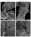
Fig. 1.
Fig. 1.
Brg1fl/fl:Tie2-Cre+ yolk sac veins are morphologically abnormal. (A-D) Anti-PECAM1 staining on flat-mounted E9.5 yolk sacs revealed Brg1fl/fl:Tie2-Cre+ veins were more abnormal than arteries. Whereas the arterial sides of control and Brg1fl/fl:Tie2-Cre+ yolk sacs contained branching networks of vessels, the venous side of Brg1fl/fl:Tie2-Cre+ yolk sacs failed to undergo normal branching and development. (C,D) Magnified views of the boxed regions in A,B, respectively, including vitelline veins (V.V.). Arrows in D indicate Brg1fl/fl:Tie2-Cre+ veins that failed to interconnect or underwent aberrant regression. Asterisks indicate round, non-vascular spaces that are characteristic of failed vascular plexus remodeling. Scale bars: 500 μm.
Fig. 2.
Fig. 2.
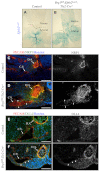
Brg1fl/fl:Tie2-Cre+ veins express arterial markers. (A,B) Control and Brg1fl/fl:Tie2-Cre+ embryos were crossed onto an Efnb2LacZ arterial reporter line and stained with X-gal solution to reveal sites of Efnb2 (LacZ) expression (blue). Flat-mounted E9.5 yolk sacs displayed aberrant Efnb2 expression in Brg1fl/fl;Efnb2LacZ:Tie2-Cre+ veins (B) compared with control veins (A). (C-F) E9.75 littermate control and Brg1fl/fl:Tie2-Cre+ embryos were cross-sectioned, and sections containing a dorsal aorta (D.A.) and cardinal vein (C.V.) were immunostained for arterial markers. (C,D) Sections were stained for the endothelial cell marker PECAM1 (red), the arterial marker NRP1 (green) and the nuclear marker Hoechst (blue). NRP1 was aberrantly upregulated on endothelial cells within Brg1fl/fl:Tie2-Cre+ cardinal veins (arrowheads in D). (E,F) Sections were stained for PECAM1 (red), DLL4 (green) and Hoechst (blue). Arterial DLL4 was likewise upregulated on Brg1fl/fl:Tie2-Cre+ cardinal veins (see arrowheads in F). Scale bars: 100 μm.
Fig. 3.
Fig. 3.
Brg1fl/fl:Tie2-Cre+ veins aberrantly recruit smooth muscle cells. Tissues from E10.5 littermate control and Brg1fl/fl:Tie2-Cre+ mutants were sectioned and immunostained for the endothelial cell marker PECAM1 (green), the smooth muscle cell marker α-smooth muscle actin (αSMA) (red) and the nuclear marker Hoechst (blue). (A,B) Extra-embryonic umbilical vessels, including umbilical veins (U.V.) and umbilical arteries (U.A.), were sectioned and stained. In control vessels, αSMA-positive cells predominantly accumulated around umbilical arteries (A). However, αSMA-positive cells accumulated around both umbilical arteries and veins in Brg1fl/fl:Tie2-Cre+ mutants (B). (C,D) Sections of embryos containing a dorsal aorta (D.A.) and cardinal vein (C.V.) were immunostained. αSMA-positive cells were detected around the Brg1fl/fl:Tie2-Cre+ C.V. (arrowheads in magnified inset of D) but not around the control C.V. (C). Scale bars: 100 μm.
Fig. 4.
Fig. 4.
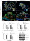
COUP-TFII expression is downregulated in Brg1-deficient endothelial cells. (A-D) Cryosections of littermate control and Brg1fl/fl:Tie2-Cre+ tissues were immunostained for the endothelial cell marker PECAM1 (red), COUP-TFII (green) and the nuclear marker Hoechst (blue). (A,B) Cross-sectioned E10.5 umbilical vessels were immunostained, and although COUP-TFII was expressed in umbilical vein (U.V.) endothelial cells in the control section (A), it was significantly diminished in Brg1fl/fl:Tie2-Cre+ venous endothelial cells (B). U.A., umbilical artery.(C,D) E9.75 embryos were cross-sectioned and immunostained. COUP-TFII was expressed in endothelial cells of the cardinal vein (C.V.) in the control section (C) but was downregulated in Brg1fl/fl:Tie2-Cre+ C.V. endothelial cells (D). D.A., dorsal aorta. For A-D, insets show magnified views of the boxed regions and arrowheads indicate individual endothelial cells. Scale bars: 100 μm. (E,F) Primary endothelial cells (ECs) were isolated from E10.5 control and Brg1fl/fl:Tie2-Cre+ tissues, RNA was purified and cDNA was synthesized. Samples from individual littermate control and Brg1fl/fl:Tie2-Cre+ yolk sacs (E) or embryos (F) were processed for qPCR analysis of Brg1 and COUP-TFII expression. Data from three independent experiments were combined and are presented as relative fold change over the expression levels in control cells±s.em. Significant differences were calculated using a two-tailed Student’s t-test (*P<0.05). (G,H) C166 endothelial cells were transfected with nonspecific (NS) or BRG1-specific siRNA for 24 hours. (G) RNA was isolated, cDNA was synthesized and qPCR for Brg1 or COUP-TFII was performed. Data from three independent experiments were combined and are presented as relative fold change over the expression levels in NS siRNA-treated cells±s.e.m. Significant differences were calculated using a two-tailed Student’s t-test (*P<0.05).(H) Protein samples were subjected to western blot analysis with antibodies that recognize BRG1, COUP-TFII or GAPDH.
Fig. 5.
Fig. 5.
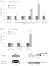
BRG1 binds to the COUP-TFII promoter in endothelial cells. (A) Alignment of the murine COUP-TFII promoter region with sequences from zebrafish, frog, opossum, dog, chimpanzee and human genomes from the NCBI DCODE website (http://www.dcode.org). Peak heights indicate degree of sequence homology; pink bars above peaks denote evolutionarily conserved regions; yellow represents the COUP-TFII 5′ untranslated region; blue indicates COUP-TFII exon 1. Boxed regions were selected for further analysis of BRG1 binding. Numbers above boxed regions denote approximate distances upstream of the COUP-TFII transcription start site (TSS). (B) Chromatin immunoprecipitation (ChIP) assays were performed on C166 endothelial cells using antibodies against BRG1 or isotype-matched non-specific IgG as a negative control. DNA was isolated and amplified by qPCR to determine whether BRG1 bound to various COUP-TFII promoter regions. Significant BRG1 binding was detected at the -0.3 kb, the -1.2 kb and the -4.7 kb promoter regions. A region upstream of the Fzd5 promoter (Fzd5 UP) was used as a negative control BRG1-binding region, and the Adamts1 promoter served as a positive control BRG1-binding region, as previously described (Griffin et al., 2011). Data from four independent experiments were combined and are presented as fold enrichment over the level of ChIP with negative control IgG antibodies±s.e.m. Significant differences were calculated using a two-tailed Student’s t-test (*P<0.05).
Fig. 6.
Fig. 6.
BRG1 remodels chromatin at the COUP-TFII promoter and influences accessibility of transcriptional machinery. (A,B) C166 endothelial cells were transfected with nonspecific (NS) or BRG1-specific siRNA for 24 hours prior to processing for ChIP assays. (A) ChIP with an antibody against total histone H3 was used to determine nucleosome density at various regions of the COUP-TFII promoter. Nucleosome density was significantly decreased at the -4.7 kb region of the COUP-TFII promoter but was significantly increased at the -1.2 kb and -0.3 kb promoter regions following BRG1 knockdown. (B) ChIP with an antibody against RNA polymerase II (RNAPolII) indicated its ability to bind the -0.3 kb region of the COUP-TFII promoter was significantly decreased following BRG1 knockdown. For A and B, a region upstream of the Fzd5 promoter (Fzd5 UP) was used as a negative control region, and the Adamts1 promoter served as a positive control region for BRG1-induced changes in nucleosome density or RNAPolII binding, respectively. Data from three independent experiments were combined and are presented as fold enrichment over the levels of ChIP with the H3 or RNAPolII antibodies in NS siRNA transfected cells±s.e.m. Significant differences were calculated using a two-tailed Student’s t test (*P<0.05). (C) Model of how BRG1 epigenetically promotes COUP-TFII expression. In wild-type endothelial cells, BRG1 binds the -4.7 kb region of the COUP-TFII promoter, where it mediates chromatin compaction. BRG1 also binds the -1.2 kb and -0.3 kb regions of the promoter, where it mediates chromatin decondensation, thereby allowing binding of RNAPolII close to the COUP-TFII transcription start site. These events promote the expression of COUP-TFII in wild-type cells. By contrast, in Brg1fl/fl:Tie2-Cre+ endothelial cells, which lack BRG1, the -4.7 kb COUP-TFII promoter region undergoes chromatin decondensation, potentially allowing for binding of a transcriptional repressor protein. Likewise, the -1.2 kb and -0.3 kb regions of the promoter undergo chromatin compaction, thereby inhibiting efficient RNAPolII binding and diminishing COUP-TFII expression.
Fig. 7.
Fig. 7.
BRG1 impacts expression of genes downstream of COUP-TFII signaling. (A) Primary endothelial cells (ECs) were isolated from E10.5 control and Brg1fl/fl:Tie2-Cre+ embryos, RNA was purified, and cDNA was synthesized. Expression levels of Brg1, the arterial markers (red) Nrp1, Hey1, Dll4, Hey2 and Foxc1, and the venous markers (blue) COUP-TFII, Ephb4 and Nrp2 were measured by qPCR. Data from three independent experiments were combined and are presented as relative fold change over the normalized expression level of each gene in control cells (dotted line) ±s.e.m. Significant differences were calculated using a two-tailed Student’s t-test (*P<0.05). (B) C166 cells were transfected with increasing amounts (0.02 ng, 0.2 ng, and 2 ng) of empty vector or comparable amounts of a BRG1 expression plasmid for 24 hours. RNA was isolated, cDNA was synthesized and qPCR for Brg1, COUP-TFII and Ephb4 was performed. Data from three independent experiments were combined and are presented as relative fold change over the normalized expression level of each gene in cells transfected with corresponding amounts of empty vector (dotted line). Bars represent ±s.e.m.; significant differences were calculated using a two-tailed Student’s t-test (*P<0.05). (C) C166 endothelial cells were transfected with nonspecific (NS) siRNA, BRG1-specific siRNA or BRG1 siRNA plus a COUP-TFII expression plasmid for 24 hours. RNA was isolated, cDNA was synthesized and qPCR for Hey2 was performed. Bars represent ±s.e.m. from three independent experiments; significant differences were calculated using a two-tailed Student’s t-test (*P<0.05). (D) Model of how BRG1 impacts venous specification. In arterial endothelial cells (ECs), Notch signaling promotes expression of arterial markers such as Ephrin B2. In venous ECs, BRG1 epigenetically promotes expression of COUP-TFII, presumably in cooperation with an unknown venous-specific co-regulatory protein or transcription factor. COUP-TFII directly inhibits Nrp1 and Foxc1, two upstream mediators of the Notch signaling pathway. COUP-TFII also directly inhibits the downstream Notch effector Hey2 (Chen et al., 2012). As a result of COUP-TFII-mediated Notch pathway inhibition, arterial marker expression is suppressed and the venous marker EPHB4 is expressed.

Similar articles

Cited by

References

    1. Adams R. H., Wilkinson G. A., Weiss C., Diella F., Gale N. W., Deutsch U., Risau W., Klein R. (1999). Roles of ephrinB ligands and EphB receptors in cardiovascular development: demarcation of arterial/venous domains, vascular morphogenesis, and sprouting angiogenesis. Genes Dev. 13, 295–306 - PMC - PubMed
    1. Brault V., Moore R., Kutsch S., Ishibashi M., Rowitch D. H., McMahon A. P., Sommer L., Boussadia O., Kemler R. (2001). Inactivation of the beta-catenin gene by Wnt1-Cre-mediated deletion results in dramatic brain malformation and failure of craniofacial development. Development 128, 1253–1264 - PubMed
    1. Carlson T. R., Yan Y., Wu X., Lam M. T., Tang G. L., Beverly L. J., Messina L. M., Capobianco A. J., Werb Z., Wang R. (2005). Endothelial expression of constitutively active Notch4 elicits reversible arteriovenous malformations in adult mice. Proc. Natl. Acad. Sci. USA 102, 9884–9889 - PMC - PubMed
    1. Chen X., Qin J., Cheng C. M., Tsai M. J., Tsai S. Y. (2012). COUP-TFII is a major regulator of cell cycle and Notch signaling pathways. Mol. Endocrinol. 26, 1268–1277 - PMC - PubMed
    1. Chi J. T., Chang H. Y., Haraldsen G., Jahnsen F. L., Troyanskaya O. G., Chang D. S., Wang Z., Rockson S. G., van de Rijn M., Botstein D., et al. (2003). Endothelial cell diversity revealed by global expression profiling. Proc. Natl. Acad. Sci. USA 100, 10623–10628 - PMC - PubMed

Publication types

MeSH terms

LinkOut - more resources