. 2013 Mar;140(6):1330-41.
doi: 10.1242/dev.086603. Epub 2013 Feb 13.

Conditional knockdown of DNA methyltransferase 1 reveals a key role of retinal pigment epithelium integrity in photoreceptor outer segment morphogenesis

Affiliations

Conditional knockdown of DNA methyltransferase 1 reveals a key role of retinal pigment epithelium integrity in photoreceptor outer segment morphogenesis

Igor O Nasonkin et al. Development. 2013 Mar.

Abstract

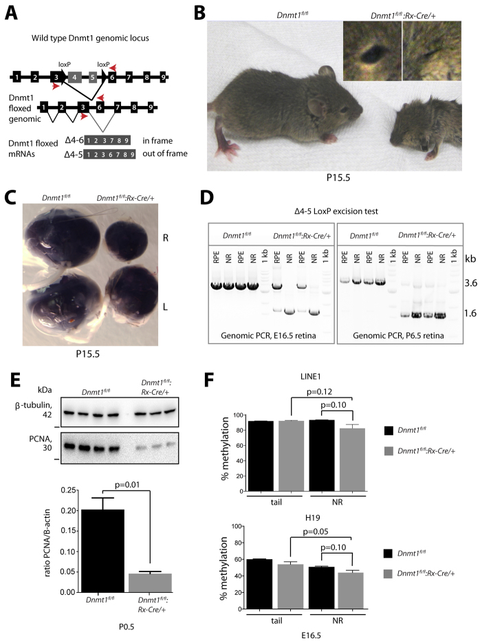
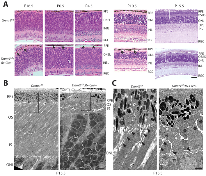
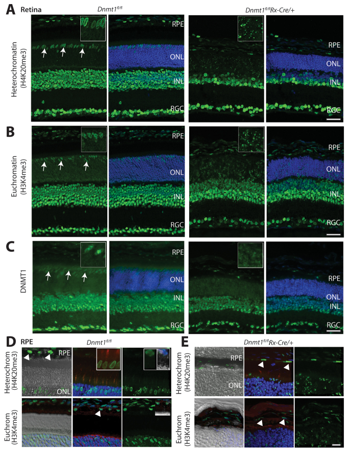
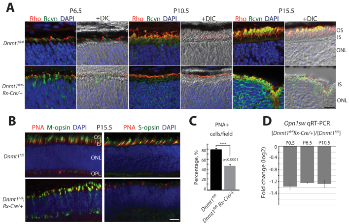
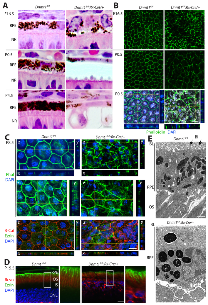
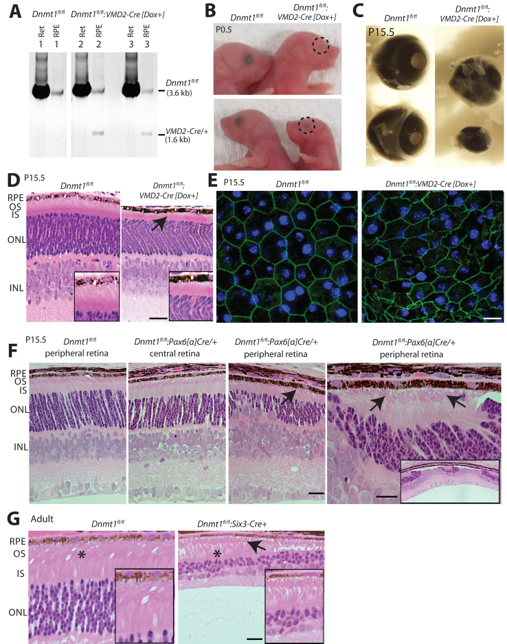
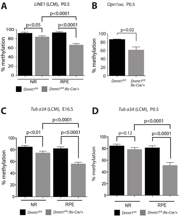
Dysfunction or death of photoreceptors is the primary cause of vision loss in retinal and macular degenerative diseases. As photoreceptors have an intimate relationship with the retinal pigment epithelium (RPE) for exchange of macromolecules, removal of shed membrane discs and retinoid recycling, an improved understanding of the development of the photoreceptor-RPE complex will allow better design of gene- and cell-based therapies. To explore the epigenetic contribution to retinal development we generated conditional knockout alleles of DNA methyltransferase 1 (Dnmt1) in mice. Conditional Dnmt1 knockdown in early eye development mediated by Rx-Cre did not produce lamination or cell fate defects, except in cones; however, the photoreceptors completely lacked outer segments despite near normal expression of phototransduction and cilia genes. We also identified disruption of RPE morphology and polarization as early as E15.5. Defects in outer segment biogenesis were evident with Dnmt1 exon excision only in RPE, but not when excision was directed exclusively to photoreceptors. We detected a reduction in DNA methylation of LINE1 elements (a measure of global DNA methylation) in developing mutant RPE as compared with neural retina, and of Tuba3a, which exhibited dramatically increased expression in mutant retina. These results demonstrate a unique function of DNMT1-mediated DNA methylation in controlling RPE apicobasal polarity and neural retina differentiation. We also establish a model to study the epigenetic mechanisms and signaling pathways that guide the modulation of photoreceptor outer segment morphogenesis by RPE during retinal development and disease.


Figures

Fig. 1.
Fig. 1.
Characterization of Dnmt1fl/fl:Rx-Cre/+ mutants. (A) Control and mutant Dnmt1 genomic region and mutant transcripts after Rx-Cre-mediated excision. Red arrows represent primers used to amplify genomic DNA (exons 3-6) and cDNAs for sequencing and PCR excision. Two Dnmt1 transcripts were detected in mutant mice: one translational out-of-frame (Δ4-5, as predicted, leading to premature truncation) and another creating an in-frame deletion (Δ4-6, due to exon 6 skipping). (B) P15.5 control and Dnmt1fl/fl:Rx-Cre/+ mice. The mutants are smaller and exhibit delayed eyelid opening. (C) The eye is smaller in mutants, with ∼15% having different left (L) and right (R) eye sizes. (D) Excision of exons 4 and 5 occurs early in both neural retina (NR) and retinal pigment epithelium (RPE) development. (E) Co-immunoprecipitation of proliferating cell nuclear antigen (PCNA) with DNMT1 from P0.5 NR. (Top) Tubulin antibody staining indicates equivalent protein loading. (Middle) Immunoblot of proteins precipitated with anti-DNMT1 antibody and probed with anti-PCNA. (Bottom) There is 3-4 times less PCNA binding in the Dnmt1 mutant. Dnmt1 Δ4-6 preserves the DNA methyltransferase catalytic domain but lacks most of the PCNA-binding domain. (F) Methylation of genomic DNA in LINE1 elements and of the imprinted gene H19 in P6.5 mutant and control NR, compared with Dnmt1fl/fl tail DNA. Average methylation of five (LINE1) or five (H19) CpG nucleotides is assayed in each sample; n=3-4 mice/assay. Error bars indicate s.e.m. P-values by two-tailed Student’s t-test.
Fig. 2.
Fig. 2.
Thin retinal layers and absence of photoreceptor outer segments in Dnmt1fl/fl:Rx-Cre/+ mice. (A) Retinal histology (H&E staining) in mutant and control retinas. Mutants display normal retinal lamination and reduced RPE/NR adhesion from E16.5 (arrows). At P8.5-P10.5, the ONL and INL become progressively thinner; the RGC layer shows fewer cells. By P15.5, mutant retinas show no photoreceptor OS and a disorganized, partially depolarized RPE cell layer with enlarged melanin granules. (B,C) P15.5 NR/RPE junction by EM at 3000× and 10,000× magnification, respectively, showing lack of photoreceptor OS and disorganized inner segments with aggregated mitochondria (asterisks) in mutant retinas; the presence of mitochondria between photoreceptor nuclei and RPE confirms the absence of OS. RPE microvilli (arrowheads) are disorganized in mutant retina. RPE, retinal pigment epithelium; ONBL, outer neuroblastic layer; INBL, inner neuroblastic layer; ONL, outer nuclear layer; INL, inner nuclear layer; RGC, retinal ganglion cell layer; OS, outer segment; IS, inner segment. Boxes in A and B indicate the photoreceptor/RPE junction area. Scale bars: 50 μm in A; 2 μm in B; 500 nm in C.
Fig. 3.
Fig. 3.
Altered distribution of heterochromatin, euchromatin and DNMT1 in Dnmt1 mutant retina. P15.5 cryosections immunostained with antibodies to heterochromatin marker H4K20me3 (A,D,E), euchromatin marker H3K4me3 (B,D,E) and DNMT1 (C). Nuclei are DAPI counterstained (blue). (A,B) Dramatic changes in heterochromatin and euchromatin distribution in the mutant ONL, especially in cones. Insets show an enlarged image of the ONL. Distinct cone chromatin patterns (arrows) are lacking in mutants, although cones are still present (red PNA staining, E; compare with D, top middle panel). (C) DNMT1 distribution in mutant retina lacks the typical cone nuclear staining (arrows). (D,E) Confocal images of heterochromatin and euchromatin (green) in control (D) and mutant (E) retina. Left panels, differential interference contrast (DIC) and chromatin staining (green); middle top panels, PNA staining of cone IS/OS (red) with DAPI (blue) and chromatin staining; right panels, chromatin staining only. (D) Inset, middle: H4K20me3 nuclear staining is cone specific, as red (PNA) and green (chromatin) originate from the same cells. (D) Inset, top right: strong nuclear H4K20me3 staining in control RPE. (D,E) Middle lower panels: the Müller glia marker glutamine synthetase (red) with DAPI and euchromatin staining. Euchromatin and heterochromatin patterns are preserved in mutant RPE regardless of changes in RPE polarity (flat and disorganized RPE cells, arrowheads). RPE, retinal pigment epithelium; ONL, outer nuclear layer; INL, inner nuclear layer; RGC, retinal ganglion cell layer. Scale bars: 50 μm in A-C; 20 μm in D,E.
Fig. 4.
Fig. 4.
Defects in photoreceptor maturation and lack of S-cones in Dnmt1 mutant retina. (A) DIC and immunofluorescence images with antibodies to photoreceptor OS (rhodopsin) and IS (recoverin) in control and mutant mice. OS development is halted in mutant retinas between P6.5 and P10.5, with rhodopsin and recoverin aggregated in IS. (B) Immunofluorescence staining of M- and S-cones in control and mutant P15.5 retinal sections. Left panels, PNA (red) and M-opsin antibody (green); right panels, PNA (red) and S-opsin antibody (green). There is reduced PNA and M-opsin in the mutant; S-cone staining was present in control littermates (9/9) but not in mutants (0/13). (C) Loss of cone cells in Dnmt1fl/fl:Rx-Cre/+ retinas. n=5 fields counted per graph. Error bars, s.e.m. P-values by two-tailed t-test. (D) Quantification of Opn1sw (short-wave cone opsin) expression in the NR of mutants, relative to control littermates (n=4 per genotype, each analyzed in triplicate). Rho, rhodopsin; Rcvn, recoverin; IS, inner segment; OS, outer segment; ONL, outer nuclear layer. Scale bars: 20 μm in A; 50 μm in B.
Fig. 5.
Fig. 5.
Early disruption of RPE polarity in Dnmt1 mutant retina. (A) H&E staining of sections of control and mutants retinas showing the RPE-NR junction. Abnormal RPE development was noted in mutants at E16.5, including poor NR/RPE adhesion, variable cell height and pigmentation, inconsistent nuclear size and variable heterochromatin compaction. (B) Disruption of the actin cytoskeleton in mutant RPE. Phalloidin staining (green) of filamentous actin in RPE flatmounts shows the lack of a cobblestone arrangement in the mutant RPE. Bottom panels are optical section z-stacks of P0.5 RPE flatmounts with DIC showing melanin distribution in RPE. z-stacks virtually resectioned in x and y planes reveal uniform pigmentation, cell height and position of junctional actin in control RPE, but not in mutant RPE. (C) Maximum projection z-stack images of flatmount RPE preparations at P8.5 prior to OS elongation showing staining with phalloidin (green) and for RPE polarity markers (red). z-stacks were virtually resectioned in the x and y planes (top panels) and compressed along the y-plane (middle panels). Mutant RPE shows variable cell height and position of junctional actin (top), reduced apical F-actin (middle) and disorganized apical microvilli and basal infoldings, as marked by ezrin (bottom). A similar pattern is observed for β-catenin, a lateral membrane marker. (D) Confocal images of vibratome sections demonstrate shorter microvilli (labeled with ezrin antibody, green) in mutant RPE. (E) EM of RPE (10,000×) revealed less well developed microvilli and basal infoldings (BI) in the mutant (arrows). The basal lamina (BL) appeared unaltered. Scale bars: 10 μm in A-C; 20 μm in D; 500 nm in E.
Fig. 6.
Fig. 6.
RPE-specific disruption of Dnmt1 prevents photoreceptor OS elongation. (A) Dnmt1 Δ4-5 excision demonstrates doxycycline-induced VMD2-Cre transgene activity in RPE but not NR (Ret). (B) Microphthalmia and severe loss of RPE pigmentation in P0 Dnmt1fl/fl:VMD2-Cre doxycycline-treated [Dox+] mice. (C) Smaller eyes in P9 Dnmt1fl/fl:VMD2-Cre [Dox+] mice, resembling those consistently observed in the Dnmt1fl/fl:Rx-Cre/+ model. (D) Methacrylate sections of Dnmt1fl/fl:VMD2-Cre [Dox+] mice and littermate controls (Dnmt1fl/fl), showing loss of OS in mutant mice and signs of poor adhesion between retina and RPE in mutants (arrow). (E) Compressed z-stacks of RPE flatmounts at P9 showing phalloidin (green) with nuclear DAPI counterstain (blue). Mutant RPE shows actin cytoskeleton abnormalities resembling those in Dnmt1fl/fl:Rx-Cre/+. (F) Methacrylate sections of control (Dnmt1fl/fl) and Dnmt1fl/fl:Pax6-[α]Cre littermates, in which Dnmt1 excision takes place in the peripheral but not central NR, resulting in degeneration of peripheral NR (third panel from the left). However, OSs remain (arrows) as long as some photoreceptors are preserved in the ONL. (G) Likewise, in Dnmt1fl/fl:Six3-Cre mice, OSs are preserved (arrow), despite Dnmt1 excision in NR and significant peripheral NR degeneration (shown). Asterisks indicate the areas enlarged in the insets. Scale bars: 50 μm in D; 10 μm in E; 20 μm in F,G.
Fig. 7.
Fig. 7.
Change in DNA methylation in Dnmt1fl/fl:Rx-Cre/+ NR and RPE. (A) DNA methylation in P0.5 control and mutant RPE and NR (n=3-4 for each cohort), isolated by laser capture microdissection (LCM). The five CpG sites in LINE1 that were analyzed in each sample showed more pronounced demethylation in mutant RPE than in mutant NR. A similar trend was found at E16.5 (not shown). (B) Quantification of Opn1sw promoter methylation at P0.5 (n=3), measured in NR using bisulfite pyrosequencing; methylation data from three CpG sites were averaged. (C,D) Demethylation of the proximal Tuba3a promoter in NR and RPE of mutant mice. Shown is the average methylation of five CpG sites in the Tuba3a promoter obtained from DNA of LCM samples of NR and RPE at E16.5 (C) and P0.5 (D) of control and mutant mice (n=3-4). Error bars, s.e.m. P-values by two-tailed Student’s t-test.

Similar articles

Cited by

References

    1. Arikawa K., Williams D. S. (1993). Acetylated alpha-tubulin in the connecting cilium of developing rat photoreceptors. Invest. Ophthalmol. Vis. Sci. 34, 2145–2149 - PubMed
    1. Bandyopadhyay M., Rohrer B. (2010). Photoreceptor structure and function is maintained in organotypic cultures of mouse retinas. Mol. Vis. 16, 1178–1185 - PMC - PubMed
    1. Bäumer N., Marquardt T., Stoykova A., Ashery-Padan R., Chowdhury K., Gruss P. (2002). Pax6 is required for establishing naso-temporal and dorsal characteristics of the optic vesicle. Development 129, 4535–4545 - PubMed
    1. Bernstein B. E., Meissner A., Lander E. S. (2007). The mammalian epigenome. Cell 128, 669–681 - PubMed
    1. Besharse J. C., Hollyfield J. G., Rayborn M. E. (1977). Turnover of rod photoreceptor outer segments. II. Membrane addition and loss in relationship to light. J. Cell Biol. 75, 507–527 - PMC - PubMed

Publication types

MeSH terms

Substances