Connectomics and epilepsy
- PMID: 23406911
- PMCID: PMC4064674
- DOI: 10.1097/WCO.0b013e32835ee5b8
Connectomics and epilepsy
Abstract
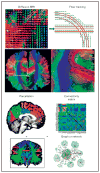
Purpose of review: Tremendous advances have occurred in recent years in elucidating basic mechanisms of epilepsy at the level of ion channels and neurotransmitters. Epilepsy, however, is ultimately a disease of functionally and/or structurally aberrant connections between neurons and groups of neurons at the systems level. Recent advances in neuroimaging and electrophysiology now make it possible to investigate structural and functional connectivity of the entire brain, and these techniques are currently being used to investigate diseases that manifest as global disturbances of brain function. Epilepsy is such a disease, and our understanding of the mechanisms underlying the development of epilepsy and the generation of epileptic seizures will undoubtedly benefit from research utilizing these connectomic approaches.
Recent findings: MRI using diffusion tensor imaging provides structural information, whereas functional MRI and electroencephalography provide functional information about connectivity at the whole brain level. Optogenetics, tracers, electrophysiological approaches, and calcium imaging provide connectivity information at the level of local circuits. These approaches are revealing important neuronal network disturbances underlying epileptic abnormalities.
Summary: An understanding of the fundamental mechanisms underlying the development of epilepsy and the generation of epileptic seizures will require delineation of the aberrant functional and structural connections of the whole brain. The field of connectomics now provides approaches to accomplish this.
Conflict of interest statement
There are no conflicts of interest.
Figures


References
-
- Sporns O. Discovering the human connectome. Boston: MIT Press; 2012. p. 240. Recent textbook on connectomics.
-
- Richardson MP. Large scale brain models of epilepsy: dynamics meets connectomics. J Neurol Neurosurg Psychiatry. 2012;83:1238–1248. A review of connectomics that considers epilepsy. - PubMed
-
- Basser PJ, Mattiello J, LeBihan D. Estimation of the effective self-diffusion tensor from the NMR spin echo. J Magn Reson B. 1994;103:247–254. - PubMed
-
- Stejskal EO, Tanner JE. Spin diffusion measurements: spin echoes in the presence of a time-dependent field gradient. J Chem Phys. 1965;42:288–292.
Publication types
MeSH terms
Grants and funding
- P41 EB015922/EB/NIBIB NIH HHS/United States
- R21 MH092647/MH/NIMH NIH HHS/United States
- K23 NS044936/NS/NINDS NIH HHS/United States
- P20 NS080181/NS/NINDS NIH HHS/United States
- R01 NS030549/NS/NINDS NIH HHS/United States
- R01 EB008432/EB/NIBIB NIH HHS/United States
- R37 NS033310/NS/NINDS NIH HHS/United States
- P01 NS002808/NS/NINDS NIH HHS/United States
- R01 NS033310/NS/NINDS NIH HHS/United States
- R01 NS65877/NS/NINDS NIH HHS/United States
- R01 NS075429/NS/NINDS NIH HHS/United States
- P01 NS02808/NS/NINDS NIH HHS/United States
- U01 NS042372/NS/NINDS NIH HHS/United States
- R01 A6040060/PHS HHS/United States
- R01 MH076994/MH/NIMH NIH HHS/United States
- R01 AG040060/AG/NIA NIH HHS/United States
- R01 NS065877/NS/NINDS NIH HHS/United States
- R01 080655/PHS HHS/United States
- R01 NS071048/NS/NINDS NIH HHS/United States
- R01 NS080655/NS/NINDS NIH HHS/United States
- R01 MH097268/MH/NIMH NIH HHS/United States
LinkOut - more resources
Full Text Sources
Other Literature Sources
Medical
Research Materials

