. 2013 Mar 5;110(10):3847-52.
doi: 10.1073/pnas.1300596110. Epub 2013 Feb 13.

Elasticity, structure, and relaxation of extended proteins under force

Affiliations

Elasticity, structure, and relaxation of extended proteins under force

Guillaume Stirnemann et al. Proc Natl Acad Sci U S A. .

Abstract

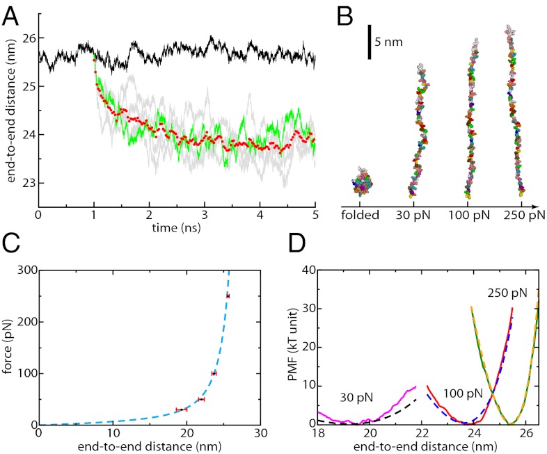
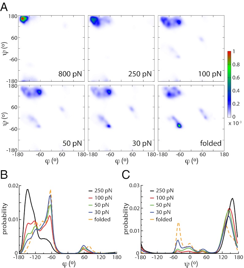
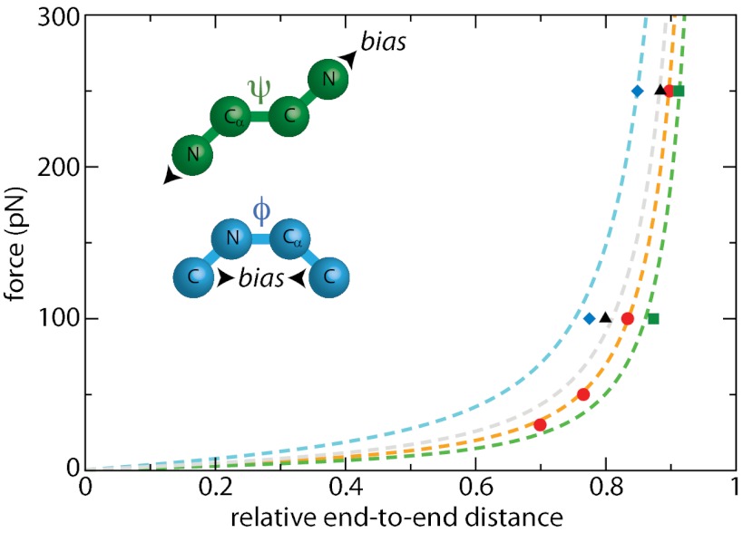
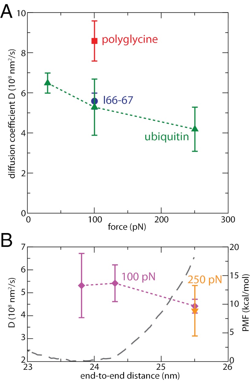
Force spectroscopies have emerged as a powerful and unprecedented tool to study and manipulate biomolecules directly at a molecular level. Usually, protein and DNA behavior under force is described within the framework of the worm-like chain (WLC) model for polymer elasticity. Although it has been surprisingly successful for the interpretation of experimental data, especially at high forces, the WLC model lacks structural and dynamical molecular details associated with protein relaxation under force that are key to the understanding of how force affects protein flexibility and reactivity. We use molecular dynamics simulations of ubiquitin to provide a deeper understanding of protein relaxation under force. We find that the WLC model successfully describes the simulations of ubiquitin, especially at higher forces, and we show how protein flexibility and persistence length, probed in the force regime of the experiments, are related to how specific classes of backbone dihedral angles respond to applied force. Although the WLC model is an average, backbone model, we show how the protein side chains affect the persistence length. Finally, we find that the diffusion coefficient of the protein's end-to-end distance is on the order of 10(8) nm(2)/s, is position and side-chain dependent, but is independent of the length and independent of the applied force, in contrast with other descriptions.


Conflict of interest statement

The authors declare no conflict of interest.

Figures

Fig. 1.
Fig. 1.
(A) End-to-end distance as a function of time for ubiquitin under an applied force of 250 pN (black curve). Relaxation trajectories are obtained by quenching force from 250 to 100 pN (gray curves, one highlighted in green) and averaged over five such trajectories (red circles). Equilibrium data are then accumulated once the average end-to-end distance has reached a plateau. A similar procedure is applied at other forces. (B) Snapshots of ubiquitin in a folded configuration (Left) and at different forces (30, 100, and 250 pN). Solvent molecules are not represented. (C) Force-extension profile at each force (black squares, average; red bars, standard deviation) and the corresponding WLC fit (dashed blue line). (D) PMF as a function of end-to-end distance at different forces (30 pN, magenta curve; 100 pN, red curve; 250 pN, green curve) and comparison with the WLC predictions (30 pN, dashed black curve; 100 pN, dashed blue curve; 250 pN, dashed yellow curve).
Fig. 2.
Fig. 2.
(A) Ramachandran plots of ubiquitin (Upper, from Left to Right: 800 pN, 250 pN, 100 N; Lower, from Left to Right: 50 pN, 30 pN, and folded at no force). (B) Distribution of the ϕ dihedral angle for unfolded ubiquitin at different forces and compared with the distribution for the folded, equilibrium protein at no force. (C) Same plot for the ψ dihedral angle.
Fig. 3.
Fig. 3.
Effect of dihedral angles and side chains on the WLC behavior (red, regular potentials; blue, biasing the ϕ potential toward 0°; green, biasing the ψ dihedral potential toward 180°; black, polyglycine analog of ubiquitin); points represent simulation data, and dashed lines show the best WLC fits using Lc = 28.4 nm.
Fig. 4.
Fig. 4.
Diffusion coefficients D along the end-to-end distance. (A) D as a function of force, for regular ubiquitin (green triangles), its polyglycine analog at 100 pN (red square) and the longer I66-67 (blue circle) at 100 pN. Here, D is calculated for each force at the corresponding average end-to-end distance L. Error bars are estimated after block averaging. (B) D as a function of length at 100 pN (violet diamonds); D at F = 250 pN and L = 25.5 nm, the equilibrium position at this force, is given for comparison. The PMF at 100 pN is also shown in gray dashed line.

References

    1. Bustamante C, Marko JF, Siggia ED, Smith S. Entropic elasticity of lambda-phage DNA. Science. 1994;265(5178):1599–1600. - PubMed
    1. Rief M, Gautel M, Oesterhelt F, Fernandez JM, Gaub HE. Reversible unfolding of individual titin immunoglobulin domains by AFM. Science. 1997;276(5315):1109–1112. - PubMed
    1. Neuman KC, Nagy A. Single-molecule force spectroscopy: Optical tweezers, magnetic tweezers and atomic force microscopy. Nat Methods. 2008;5(6):491–505. - PMC - PubMed
    1. Perez-Jimenez R, et al. Diversity of chemical mechanisms in thioredoxin catalysis revealed by single-molecule force spectroscopy. Nat Struct Mol Biol. 2009;16(8):890–896. - PMC - PubMed
    1. Merkel R, Nassoy P, Leung A, Ritchie K, Evans E. Energy landscapes of receptor-ligand bonds explored with dynamic force spectroscopy. Nature. 1999;397(6714):50–53. - PubMed

Publication types