. 2013 Sep;133(9):2191-201.
doi: 10.1038/jid.2013.66. Epub 2013 Feb 13.

Remodeling of three-dimensional organization of the nucleus during terminal keratinocyte differentiation in the epidermis

Affiliations

Remodeling of three-dimensional organization of the nucleus during terminal keratinocyte differentiation in the epidermis

Michal R Gdula et al. J Invest Dermatol. 2013 Sep.

Abstract

The nucleus of epidermal keratinocytes (KCs) is a complex and highly compartmentalized organelle, whose structure is markedly changed during terminal differentiation and transition of the genome from a transcriptionally active state seen in the basal and spinous epidermal cells to a fully inactive state in the keratinized cells of the cornified layer. Here, using multicolor confocal microscopy, followed by computational image analysis and mathematical modeling, we demonstrate that in normal mouse footpad epidermis, transition of KCs from basal epidermal layer to the granular layer is accompanied by marked differences in nuclear architecture and microenvironment including the following: (i) decrease in the nuclear volume; (ii) decrease in expression of the markers of transcriptionally active chromatin; (iii) internalization and decrease in the number of nucleoli; (iv) increase in the number of pericentromeric heterochromatic clusters; and (v) increase in the frequency of associations between the pericentromeric clusters, chromosomal territory 3, and nucleoli. These data suggest a role for nucleoli and pericentromeric heterochromatin clusters as organizers of nuclear microenvironment required for proper execution of gene expression programs in differentiating KCs, and provide important background information for further analyses of alterations in the topological genome organization seen in pathological skin conditions, including disorders of epidermal differentiation and epidermal tumors.


Figures

Figure 1
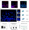
Figure 1. Remodelling of the nuclei during terminal keratinocyte differentiation
(a) 3D reconstruction of nuclei in the mouse footpad based on confocal microscopy of the tissue samples stained with marker of proliferating cells (antibody against Ki67) and DAPI. (b) Changes in nuclear shape measured as the ratio of long axis to the average of two shorter axes during keratinocyte differentiation. (c) Keratinocyte nuclear volume in the different epidermal layers. Data in b and c represent means with 95% confidence interval; 30 nuclei in every group were measured; p-values: * <0.05, **<0.01, ***<0.001.
Figure 2
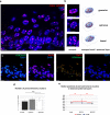
Figure 2. Remodelling of transcriptionally active chromatin domains and nucleoli during terminal keratinocyte differentiation
Enrichment of the markers of actively transcribed chromatin in the basal and spinous layers and decrease of their level in the granular layer: phospho-serine-2 RNA Pol II (elongating form) (a), H3K4me3 (b), H3K56ac (c). (d) Representative 200 nm single optical section of the epidermis stained with antibody against nucleolar marker nucleophosmin/B23. Epidermal/dermal border is indicated by dotted line. (e) Representative nuclei from the distinct epidermal layers (left) and geometrical models of the average keratinocyte nuclei form distinct epidermal layers generated on the basis of 3D-reconstruction of the 70-80 confocal stacks of the whole nuclei (right). Decrease in number of nucleoli (f), change in individual and total nucleolar volume (g) and in radial positions of the nucleoli (h) during terminal keratinocyte differentiation. Data in c, d and e represent means with 95% confidence interval; 30 nuclei in every group were measured, p-values: * <0.05, **<0.01, ***<0.001.Note that in geometrical models of the average keratinocyte nuclei (e), orientations of the nuclei from distinct epidermal layers were purportedly changed to provide a better visual appreciation of the topological differences between them. Scale bars = 10 um.
Figure 3
Figure 3. Remodelling of the pericentromeric heterochromatin clusters during terminal differentiation of keratinocytes in the murine epidermis
(a) Representative 200 nm single optical section images of the mouse epidermis after 3D FISH with the major satellite repeat probe. Epidermal/dermal border is indicated by dotted line. (b) Representative nuclei from the distinct epidermal layers: FISH images (left) and geometrical models of the average keratinocyte nucleus generated on the basis of 3D-reconstruction of the 70-80 confocal stacks of the whole nuclei (right). Note that in geometrical models of the average keratinocyte nuclei, orientation of the nuclei from distinct epidermal layers was purportedly changed to provide a better visual appreciation of the topological differences between them. (c) Centromeric clusters have heterochromatic character and express makers of silent chromatin like H3K9me3 which recruits protein HP1 taking part in chromatin compaction. (d) Changes of number of centromeric clusters during epidermal keratinocyte differentiation. (e) Change in radial positions of the pericentromeric clusters during terminal keratinocyte differentiation. Data in c and d represent means with 95% confidence interval; 30 nuclei in every group were measured p-values: * <0.05, **<0.01, ***<0.001. Scale bars = 10 um.
Figure 4
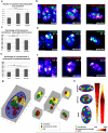
Figure 4. Changes in the spatial association between pericentromeric heterochromatin clusters, nucleoli and the chromosomal territory 3 during epidermal keratinocyte differentiation
(a, b). Spatial associations between the pericentromeric heterochromatin clusters and nucleoli (white arrows). (c, d). Spatial associations between the chromosome territory 3 and nucleolus (white arrows) (e, f). Spatial associations between the chromosome 3 and pericentromeric clusters located either at the peripheral (p) or internal (i) parts of the nucleus. (g) 3D reconstruction of a representative nucleus from the epidermal basal layer. (h) Summary: remodelling of keratinocyte nucleus during epidermal differentiation is likely to contribute to global changes in the transcriptional landscape in differentiating keratinocytes. Data in a, c, and e represent means with 95% confidence interval; 30 nuclei in every group were measured, p-values: * <0.05, **<0.01, ***<0.001. Scale bars = 5 um.

Comment in

References

    1. Alcobia I, Dilao R, Parreira L. Spatial associations of centromeres in the nuclei of hematopoietic cells: evidence for cell-type-specific organizational patterns. Blood. 2000;95:1608–1615. - PubMed
    1. Aleixandre C, Miller DA, Mitchell AR, Warburton DA, Gersen SL, Disteche C, et al. p82H identifies sequences at every human centromere. Hum Genet. 1987;77:46–50. - PubMed
    1. Bartova E, Horakova AH, Uhlirova R, Raska I, Galiova G, Orlova D, et al. Structure and epigenetics of nucleoli in comparison with non-nucleolar compartments. J Histochem Cytochem. 2010;58:391–403. - PMC - PubMed
    1. Bazzi H, Fantauzzo KA, Richardson GD, Jahoda CA, Christiano AM. Transcriptional profiling of developing mouse epidermis reveals novel patterns of coordinated gene expression. Dev Dyn. 2007;236:961–970. - PubMed
    1. Blanpain C, Fuchs E. Epidermal homeostasis: a balancing act of stem cells in the skin. Nat Rev Mol Cell Biol. 2009;10:207–217. Epub 2009 Feb 2011. - PMC - PubMed

Publication types

LinkOut - more resources