Review
. 2013 Mar;86(1023):20120163.
doi: 10.1259/bjr.20120163.

Quantitative parametric MRI of articular cartilage: a review of progress and open challenges

Affiliations
Review

Quantitative parametric MRI of articular cartilage: a review of progress and open challenges

D A Binks et al. Br J Radiol. 2013 Mar.

Abstract

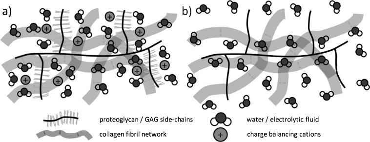
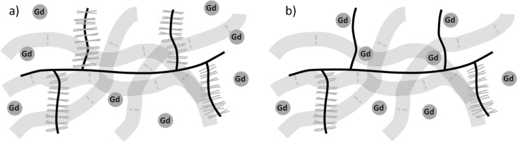
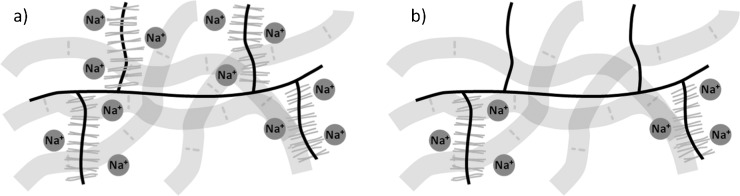
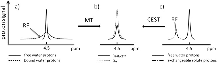
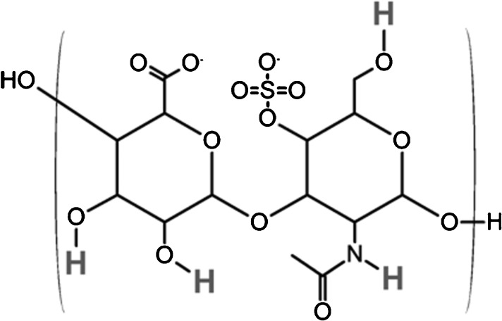
With increasing life expectancies and the desire to maintain active lifestyles well into old age, the impact of the debilitating disease osteoarthritis (OA) and its burden on healthcare services is mounting. Emerging regenerative therapies could deliver significant advances in the effective treatment of OA but rely upon the ability to identify the initial signs of tissue damage and will also benefit from quantitative assessment of tissue repair in vivo. Continued development in the field of quantitative MRI in recent years has seen the emergence of techniques able to probe the earliest biochemical changes linked with the onset of OA. Quantitative MRI measurements including T(1), T(2) and T(1ρ) relaxometry, diffusion weighted imaging and magnetisation transfer have been studied and linked to the macromolecular structure of cartilage. Delayed gadolinium-enhanced MRI of cartilage, sodium MRI and glycosaminoglycan chemical exchange saturation transfer techniques are sensitive to depletion of cartilage glycosaminoglycans and may allow detection of the earliest stages of OA. We review these current and emerging techniques for the diagnosis of early OA, evaluate the progress that has been made towards their implementation in the clinic and identify future challenges in the field.


Figures

Figure 1.
Figure 1.
(a) The macromolecular composition of cartilage. The collagen fibril network provides the structural framework for cartilage and confers resistance to shear and tensile forces. Proteoglycans are embedded within the collagen network and consist of a central protein core and covalently attached negatively charged glycosaminoglycan (GAG) side chains. The negatively charged GAGs increase the local concentration of cationic species such as Na+ and help to maintain fluid within the tissue, bestowing stiffness and resistance to compressive forces. (b) In proteoglycan-depleted cartilage, the loss of negatively charged GAGs and the corresponding reduction in mobile cation concentration diminish the ability of the cartilage macromolecular matrix to constrain fluid, reducing its capacity to withstand compression.
Figure 2.
Figure 2.
Conventional parameter-weighted MR images of a cadaveric knee joint. (a) Two-dimensional coronal intermediate-weighted spin echo image used to assess gross joint alignment, collateral ligaments and medial and lateral menisci, as well as cartilage morphology and the presence or absence of subchondral cysts. (b) Three-dimensional (3D) T2* weighted gradient echo image with selective water excitation; a 3D acquisition which allows the cartilage thickness and volume to be measured as well as providing information about bone attrition and osteophyte formation.
Figure 3.
Figure 3.
Quantitative MR parameter mapping. A pixel-by-pixel map of a single MR property is displayed on top of an anatomical image, showing the variation of that particular parameter in a region of interest. This particular image shows the variation in T2 relaxation time in the femoral articular cartilage and patellar cartilage of the knee joint.
Figure 4.
Figure 4.
Distribution of gadolinium diethylenetriamine penta-acetic acid [Gd(DTPA)2−] in (a) healthy and (b) glycosaminoglycan (GAG)-depleted cartilage extracellular matrices. The local concentration of the administered gadolinium contrast agent is inversely proportional to cartilage GAG content owing to the electrostatic repulsion between negatively charged GAGs and the negatively charged contrast agent. Water proton T1 relaxation times are reduced in the vicinity of the paramagnetic contrast agent and can therefore be used to measure GAG concentration.
Figure 5.
Figure 5.
Distribution of sodium (Na+) ions in (a) healthy and (b) glycosaminoglycan (GAG)-depleted cartilage extracellular matrices. The negative fixed charge density of GAG is balanced by cationic Na+ ions. GAG-depleted regions have lower negative fixed charge densities and therefore fewer Na+ ions. MRI techniques can measure the Na+ concentration, allowing the fixed charge density and GAG concentration to be calculated.
Figure 6.
Figure 6.
Saturation transfer effects between protons in the free and bound water pools and exchangeable protons of solute molecules. (a) In magnetisation transfer (MT), an off-resonance radiofrequency (RF) pulse saturates the broad proton resonance of low-mobility bound water molecules. Proton exchange between bound water molecules and the free water pool results in saturation transfer to the free water pool and a detectable reduction in the signal intensity of the free water resonance. (b) The magnetisation transfer ratio (MTR) is defined as MTR=1−SMT/S0, where S0 is the signal intensity recorded without a preparatory saturation pulse and SMT is the signal intensity observed with the inclusion of a preparatory saturation pulse. (c) In the chemical exchange-dependent saturation transfer (CEST) technique, solute protons are selectively saturated by using an RF pulse. Chemical exchange of the solute protons with water protons again results in saturation transfer to the free water pool and a measurable reduction in water proton signal intensity.
Figure 7.
Figure 7.
Molecular structure of one disaccharide unit of chondroitin-4-sulphate, one of the constituent glycosaminoglycans (GAGs) of proteoglycan. Exchangeable protons that contribute to the chemical exchange-dependent saturation transfer (CEST) effects seen using the GAG-CEST technique are highlighted. H, hydrogen; N, nitrogen; O, oxygen; S, sulphur.

References

    1. Burstein D, Gray ML. Is MRI fulfilling its promise for molecular imaging of cartilage in arthritis? Osteoarthritis Cartilage 2006;14:1087–90 - PubMed
    1. Williams F, Spector T. Biomarkers in osteoarthritis. Arthritis Res Ther 2008;10:101. - PMC - PubMed
    1. Buckwalter JA, Mankin HJ. Articular cartilage: tissue design and chondrocyte-matrix interactions. Instr Course Lect 1998;47:477–86 - PubMed
    1. Mow VC, Holmes MH, Lai WM. Fluid transport and mechanical properties of articular cartilage: a review. J Biomech 1984;17:377–94 - PubMed
    1. Buckwalter JA, Mankin HJ. Articular cartilage: degeneration and osteoarthritis, repair, regeneration, and transplantation. Instr Course Lect 1998;47:487–504 - PubMed

Publication types