. 2013 Feb 13;3(2):120164.
doi: 10.1098/rsob.120164.

The structure of F₁-ATPase from Saccharomyces cerevisiae inhibited by its regulatory protein IF₁

Affiliations

The structure of F₁-ATPase from Saccharomyces cerevisiae inhibited by its regulatory protein IF₁

Graham C Robinson et al. Open Biol. .

Abstract

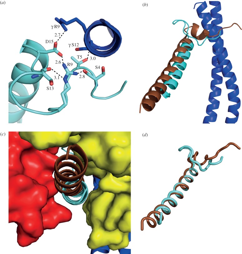
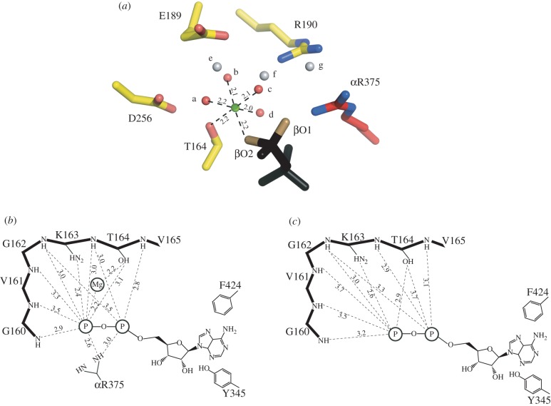
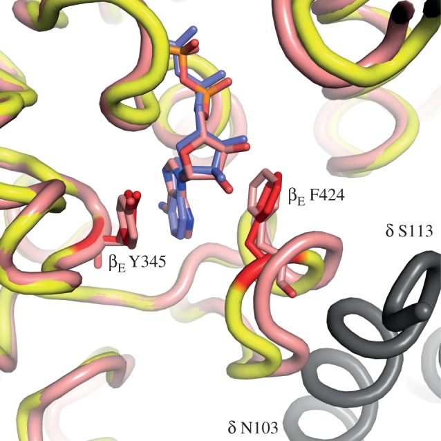
The structure of F₁-ATPase from Saccharomyces cerevisiae inhibited by the yeast IF₁ has been determined at 2.5 Å resolution. The inhibitory region of IF₁ from residues 1 to 36 is entrapped between the C-terminal domains of the α(DP)- and β(DP)-subunits in one of the three catalytic interfaces of the enzyme. Although the structure of the inhibited complex is similar to that of the bovine-inhibited complex, there are significant differences between the structures of the inhibitors and their detailed interactions with F₁-ATPase. However, the most significant difference is in the nucleotide occupancy of the catalytic β(E)-subunits. The nucleotide binding site in β(E)-subunit in the yeast complex contains an ADP molecule without an accompanying magnesium ion, whereas it is unoccupied in the bovine complex. Thus, the structure provides further evidence of sequential product release, with the phosphate and the magnesium ion released before the ADP molecule.


Figures

Figure 1.
Figure 1.
Alignment of the sequences of residues 1–60 of bovine IF1, and the equivalent region of yeast IF1, with the same regions from other species. The purple, green and yellow stripes denote identical, highly conserved and poorly conserved residues, respectively. The alignment was performed with ClustalW. The bars above the sequences denote α-helical regions in the bovine protein. The yIF1 used in crystallization experiments contained the mutation E21A.
Figure 2.
Figure 2.
Gel filtration chromatography of yeast and bovine F1-ATPase-IF1 complexes. The yeast and bovine enzymes were inhibited with the inhibitor protein from S. cerevisiae (yF1) and with bovine IF1 (bIF1), respectively. (a) Column profiles of yF1–yIF1, active yF1 and bF1–bIF1 complexes, respectively. V0 is the void volume of the column. (b) SDS–PAGE analysis of peaks 1, 2 and 3 from (a).
Figure 3.
Figure 3.
The structure of the F1-I1–53 complex from S. cerevisiae. The α-, β-, γ-, δ- and ε-subunits are depicted in ribbon form in red, yellow, dark blue, green and magenta, respectively, and residues 1–36 of I1–53 are light blue. (a) Overall view of the complex viewed from the side with IF1 shown in solid representation. (b) View (upwards from the foot of the central stalk) along the axis of the γ-subunit showing the position of the α-helix of yeast IF1 in ribbon representation relative to the C-terminal domains of α- and β-subunits.
Figure 4.
Figure 4.
The binding site for yeast I1–53 in the structure of yeast F1-ATPase. (a) The N-terminal loop region of the inhibitor protein (light blue) in juxtaposition with the C-terminal helix of the γ-subunit (dark blue). Dotted lines represent possible interactions with distances in angstrom. (b) View from the side of the central stalk showing the orientations of the yeast and bovine inhibitor proteins (light blue and brown, respectively) relative to the central stalk. (c) View from outside the F1-domain towards the γ-subunit of the enzyme (dark blue) of the deep cleft between the C-terminal domains of the αDP- and βDP-subunits (red and yellow, respectively) where the α-helical region of I1–53 (light blue) is bound. The position of the equivalent region of the bovine inhibitor protein in the structure of bovine F1-I1–60 is shown in brown. (d) View of the bovine inhibitor (brown) superposed onto the yeast inhibitor protein (light blue) via residues 22–25 and 17–20 in the bovine and yeast inhibitors, respectively.
Figure 5.
Figure 5.
Influence of point mutation of selected residues in yI1–53 on its inhibition of yeast F1-ATPase. The quantitative data from which the figure is derived are given in table 2. Kiwt and Kimut are the dissociation constants for the wild-type and mutant proteins, respectively.
Figure 6.
Figure 6.
Change in the coordination of a magnesium ion in the active site of yeast F1-ATPase. (a) Hexacoordination of the magnesium ion (green) in the catalytic site of the βDP-subunit. The ligands are provided by water molecule a–d (red), by the oxygen atom βO2 of ADP (or ATP), and by the β-hydroxy-group of βDP-T164. The water molecules a–d are themselves hydrogen-bonded to other water molecules e–g (grey), by the oxygen atom βO1 of the nucleotide, and by the side chain functionalities of residues βDP-E189, βDP-R190 and βDP-D256. (b) Schematic of the disposition of the magnesium ion and bound ADP in the catalytic site of the βDP-subunit. The P-loop sequence (upper left) helps to bind the magnesium ion and the nucleotide. The adenosine moiety of the nucleotide is bound in the hydrophobic pocket between the side chains of residues βDP-Y345 and βDP-F424. Distances are given in angstrom, and those to the phosphate group are to the closest oxygen atom. (c) Rotation of γ-subunit driven by hydrolysis of ATP has opened the nucleotide binding domain of the subunit, converting the βDP-site to the βE-site. The catalytic ‘arginine finger’ residue, αR375 becomes disordered, and the coordination sphere of the magnesium ion is disrupted, releasing the metal ion. In the structure of yF1-I1–53, the inhibitor protein has arrested the conversion of the site just before the formation of a fully ‘empty’ or ‘open’ site. The adenosine binding pocket is still intact, and ADP remains bound to the subunit. In the final step of the conversion of the site to a fully formed βE-site (as observed in ‘ground state’ structures of F1-ATPase), the side chain of βE-F424 rotates through 90°, releasing the ADP molecule.
Figure 7.
Figure 7.
Comparison of the nucleotide binding pockets in the βE-subunits in the structures of yF1-I1–53 and bovine F1-PH. The yeast and bovine protein backbones are coloured yellow and pink, respectively. The side chains of residues βE-F424 and βE-Y345 are red in the yeast enzyme and pink in the bovine enzyme. They provide the pocket for binding the adenosine moieties of the ADP molecules (blue and pink, respectively in the yeast and bovine enzymes). In grey is shown α-helix b (residues 102–110) in the δ-subunit of an adjacent yeast F1-complex in the crystal lattice of yeast F1-I1–53. It approaches to within 4 Å of α-helix C3 carrying βE-F424 in the yeast structure. Thus, it makes a crystal contact that may influence the position of α-helix C3 in the βE-subunit of the yeast enzyme.

References

    1. Pullman ME, Penefsky H, Datta A, Racker E. 1960. Partial resolution of the enzymes catalysing oxidative phosphorylation. Purification and properties of soluble, dinitrophenol-stimulated adenosine triphosphatase. J. Biol. Chem. 235, 3322–3329 - PubMed
    1. Runswick MJ, Bason JV, Montgomery MG, Robinson GC, Fearnley IM, Walker JE. 2013. The affinity purification of ATP synthase complexes from mitochondria. Open Biol. - PMC - PubMed
    1. Frangione B, Rosenwasser E, Penefsky HS, Pullman ME. 1981. Amino acid sequence of the protein inhibitor of mitochondrial adenosine triphosphatase. Proc. Natl Acad. Sci. USA 78, 7403–7407 10.1073/pnas.78.12.7403 (doi:10.1073/pnas.78.12.7403) - DOI - PMC - PubMed
    1. Walker JE, Gay NJ, Powell SJ, Kostina M, Dyer MR. 1987. ATP synthase from bovine mitochondria: sequences of imported precursors of oligomycin sensitivity conferral protein, factor 6, and adenosinetriphosphatase inhibitor protein. Biochemistry 26, 8613–8619 10.1021/bi00400a018 (doi:10.1021/bi00400a018) - DOI - PubMed
    1. Cabezon E, Butler PJ, Runswick MJ, Walker JE. 2000. Modulation of the oligomerization state of the bovine F1-ATPase inhibitor protein, IF1, by pH. J. Biol. Chem. 275, 25 460–25 464 10.1074/jbc.M003859200 (doi:10.1074/jbc.M003859200) - DOI - PubMed

Publication types