. 2013 Feb 13;33(7):2873-88.
doi: 10.1523/JNEUROSCI.3357-12.2013.

Replication protein a links cell cycle progression and the onset of neurogenesis in Drosophila optic lobe development

Affiliations

Replication protein a links cell cycle progression and the onset of neurogenesis in Drosophila optic lobe development

Liya Zhou et al. J Neurosci. .

Abstract

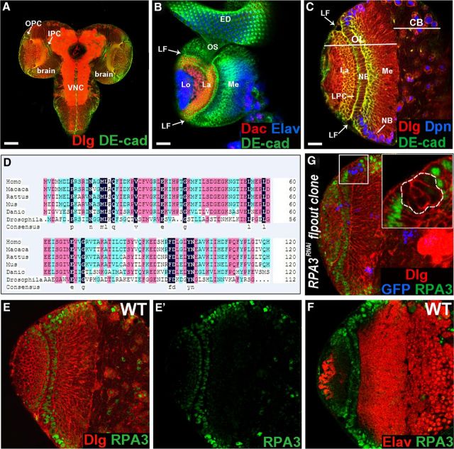
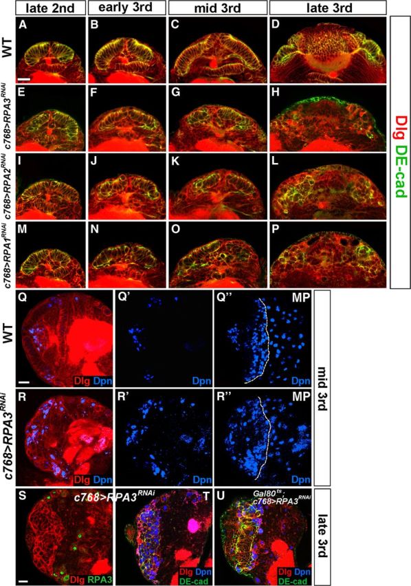
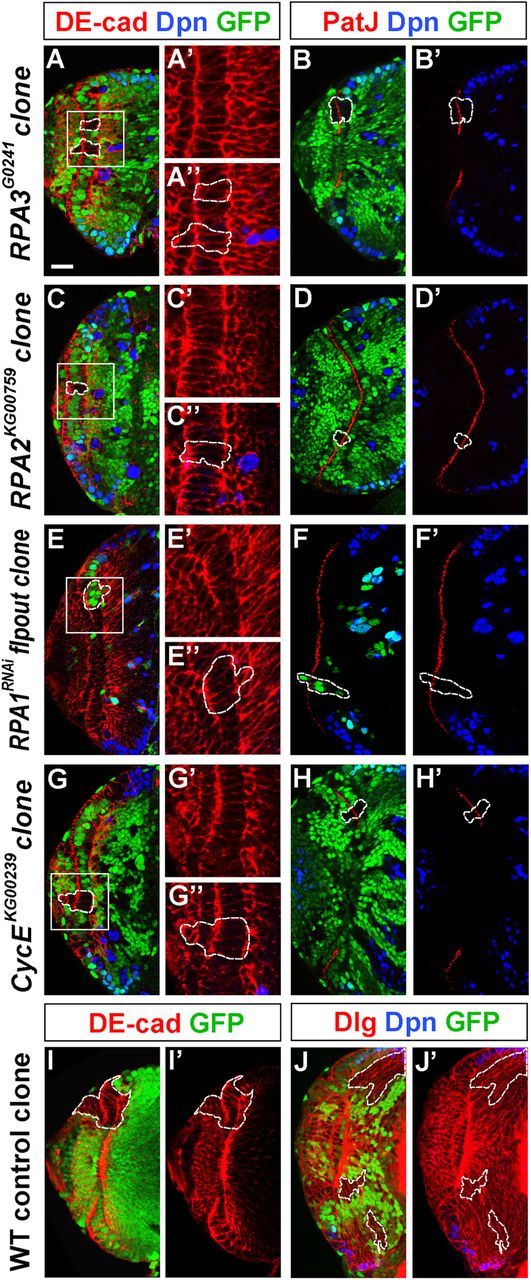
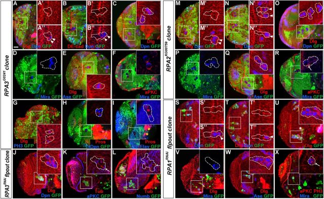
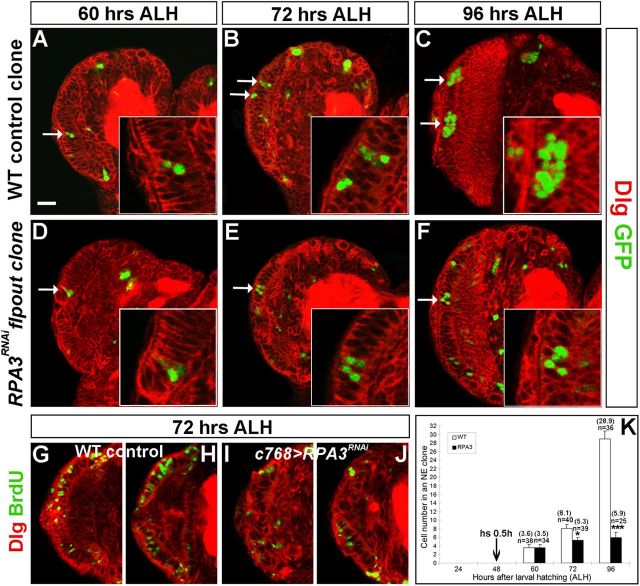
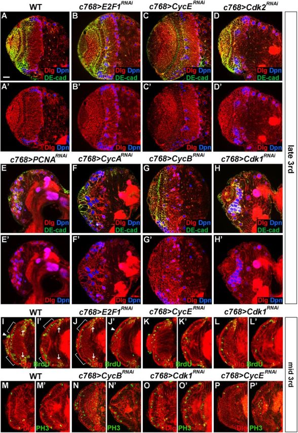
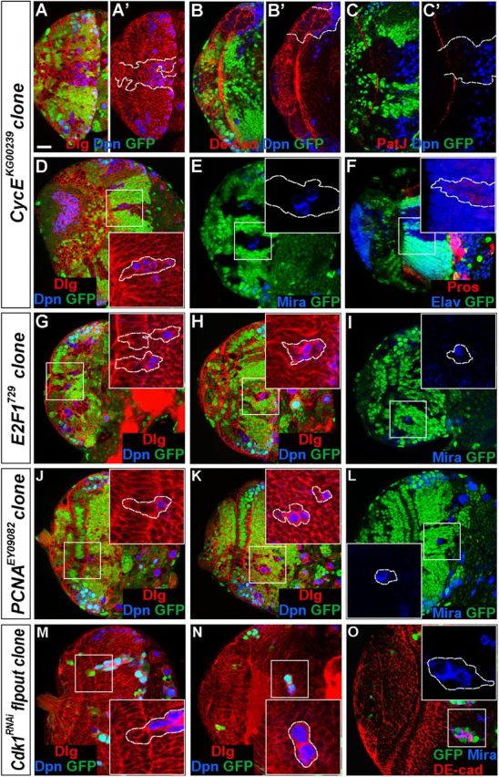
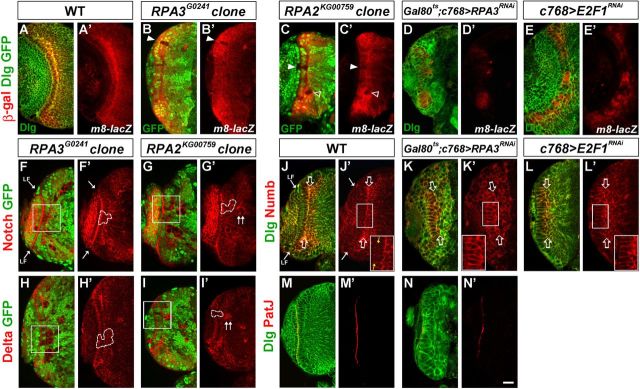
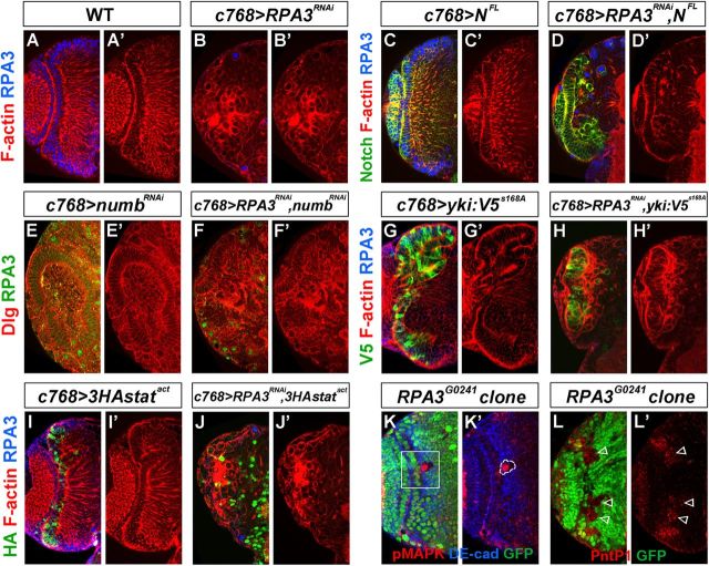
Stem cell self-renewal and differentiation must be carefully controlled during development and tissue homeostasis. In the Drosophila optic lobe, neuroepithelial cells first divide symmetrically to expand the stem cell population and then transform into asymmetrically dividing neuroblasts, which generate medulla neurons. The mechanisms underlying this cell fate transition are not well understood. Here, we show a crucial role of some cell cycle regulators in this transition. We find that loss of function in replication protein A (RPA), which consists of three highly conserved protein subunits and functions in DNA replication, leads to disintegration of the optic lobe neuroepithelium and premature differentiation of neuroepithelial cells into medulla neuroblasts. Clonal analyses of RPA loss-of-function alleles indicate that RPA is required to prevent neuroepithelial cells from differentiating into medulla neuroblasts. Inactivation of the core cell cycle regulators, including the G1/S regulators E2F1, Cyclin E, Cdk2, and PCNA, and the G2/M regulators Cyclin A, Cyclin B, and Cdk1, mimic RPA loss-of-function phenotypes, suggesting that cell cycle progression is required for both maintaining neuroepithelial cell identity and suppressing neuroblast formation. We further find that RPA or E2F1 inactivation in the neuroepithelial cells correlates with downregulation of Notch signaling activity, which appears to result from Numb mislocalization. Thus, we have shown that the transition from neuroepithelial cells to neuroblasts is directly regulated by cell cycle regulators and propose a model in which the inhibition of neuroepithelial cell cycle progression downregulates Notch signaling activity through Numb, which leads to the onset of neurogenesis.


Figures

Figure 1.
Figure 1.
RPA3 is expressed in the optic lobe neuroepithelia. A, The larval CNS. B, Lateral view of the optic lobe, which harbors three optic ganglia: the medulla (me), lamina (la), and lobula complex (lo). LF, lamina furrow; ED, eye imaginal disc; os, optic stalk. Anterior is to the right. C, Frontal view of the brain lobe with the laterally located optic lobe (OL) and the medially located central brain (CB). Along the lateral–medial axis, symmetrically dividing NE cells give rise to asymmetrically dividing NBs at the medial edge. LPC, lamina precursor cells. D, A multiple sequence alignment between Drosophila melanogaster CG15220, Homo sapiens RPA3, Macaca mulatta RPA3, Rattus norvegicus RPA3, Mus musculus RPA3, and Danio rerio RPA3. Conserved and similar amino acid residues are shaded in black, red, and blue. E, E', RPA3 protein is expressed in the NE cells, medulla and central brain NBs, GMCs, lamina precursor cells, and immature lamina neurons. RPA3 staining is shown in green. F, RPA3 is not expressed in medulla neurons. G, RPA3 expression was greatly reduced or eliminated in RPA3RNAi flip-out clones. Scale bars: A, 70 μm; B, 40 μm; C, 20 μm (for C, E, E', F, G).
Figure 2.
Figure 2.
RPA is essential for lamina and medulla development in the optic lobe. Late-third instar larval brains were stained with the antigens indicated. A, A', B, Wild-type brain lobes showing the crescent-shaped lamina (A, A') and the dome-shaped medulla (B). The border between the medulla and the central brain is revealed by strong Prospero (Pros) staining of central brain neurons. C, C', D, c768-Gal4/UAS-RPA3RNAi brain. The lamina is absent (C, C', arrow indicates lobular complex cells) and the medulla is smaller compared with wild type (D, compare with B). E, E', F, c768-Gal4/UAS-RPA2RNAi brains. The lamina is absent (E, E') and the medulla (F) is severely reduced in size. G, G', H, c768-Gal4/UAS-RPA1RNAi brains. The mutant brain also did not develop the lamina (G, G') and had a smaller medulla (H, compare with B). Anterior is to the left and dorsal up. Scale bar, 20 μm.
Figure 3.
Figure 3.
RPA maintains neuroepithelial integrity in the optic lobe. Markers are as indicated. A–P, Time course studies of optic lobe neuroepithelial growth and expansion. A–D, Wild-type brain lobes at late-second (A), early-third (B), mid-third (C), and late-third instar (D). E–P, Brain lobes in which RPA3 (E–H), RPA2 (I–L), or RPA1 (M–P) expression was knocked down by RNAi using the c768-Gal4 driver. Loss of RPA3, RPA2, or RPA1 activity led to the gradual loss of NE cells; the NE cells were partially lost by mid-third instar and completely lost by late-third instar. Lateral is up and medial down. Scale bar: A, 20 μm (for A–P). Q–R”, Loss of RPA3 activity leads to premature differentiation of NE cells into medulla NBs. Mid-third instar brains were analyzed. Q, Q', Q”, Wild-type brain lobe stained with Dpn and Dlg. MP, maximal projection. R, R', R”, c768-Gal4/UAS-RPA3RNAi brain lobe. Dpn+ NBs formed prematurely in RPA3 mutant brains compared with wild-type brains. Lateral is to the left and dorsal up. Scale bar: Q, 20 μm (for Q–R”). S–U, RPA3RNAi brain lobes from late-third instar larvae showing disintegration of the optic lobe neuroepithelium and their differentiation into NBs. S, RPA3 expression was specifically knocked down in the optic lobe NE cells using the c768-Gal4 driver. T, This led to the loss of NE cells and premature differentiation of NE cells into medulla NBs. U, Gal80ts; c768-Gal4/UAS-RPA3RNAi brains retained more NE cells by late-third instar stages compared with c768-Gal4/UAS-RPA3RNAi brain lobes. Lateral is to the left and dorsal up. Scale bar: S, 20 μm (for S–U).
Figure 4.
Figure 4.
RPA and CycE mutant NE cells in small clones do not disrupt epithelial cell integrity. Brain lobes from late-third instar larvae were stained for Dpn and DE-cad or PatJ. A–F', RPA3 mosaic clones (A–B'), RPA2 mosaic clones (C–D'), and RPA1RNAi flip-out clones (E–F'). RPA mutant cells in epithelial clones retained columnar NE cell morphology, expressed the apical marker PatJ, and did not express the NB marker Dpn. G–H', CycE mosaic clones. CycE mutant cells in small NE clones also retained epithelial morphology and identity, and did not express Dpn. I–J', Wild-type control clones. The epithelial cells in the wild-type clone always stayed in the OPC neuroepithelium (I, I') and those clones entirely localized in the medulla do not ectopically express Dpn (J, J'). Lateral is to the left and medial to the right. Scale bar, 20 μm.
Figure 5.
Figure 5.
Loss of RPA activity in epithelial cell clones causes extrusion of mutant epithelial cells and premature NB formation. Brain lobes from late-third instar larvae were stained with the antigens indicated; selected clones are identified by dashed lines. A–I, RPA3 mosaic clones. A–B, RPA3 mutant cells in epithelial clones were segregating from surrounding wild-type neuroepithelium and entering the medulla cortex. Some mutant cells located in the more medial region of the clone became NBs (Dpn+, arrowheads). C–I, Mutant clones entirely localized in the medulla cortex. The ectopic NBs expressed Dpn (C), Mira (D), and Ase (E); had asymmetric aPKC and Mira localization during mitosis (F); and had the ability to divide (G) to generate GMC progeny (Pros+) and neurons (Elav+) (H, I). J–L, RPA3RNAi flip-out clones. The mutant cells in RPA3RNAi flip-out clones located in the medulla expressed NB markers (J). Mitotic cells in the mutant clone underwent asymmetric division as Numb or aPKC was localized in one pole of the mitotic cell perpendicular to the spindle orientation (K, L, arrows). M–R, RPA2 mosaic clones. S–X, RPA1RNAi flip-out clones. Like RPA3 mutant clones, mutant NE cells in RPA2 (M, N) and RPA1 clones (S, T) were extruded from the OPC neuroepithelium and entered the medulla cortex, causing some of the mutant cells located in the more medial region of the clones to express Dpn. In medulla clones (O–R; U–X), ectopic NBs were generated that underwent asymmetric divisions and generated GMCs and neurons. Lateral is to the left and dorsal up. Scale bar, 20 μm.
Figure 6.
Figure 6.
RPA3 mutant NE cells have a proliferative defect. Dlg outlines all cell cortices in larval brains and GFP marks flip-out clones. Clones were induced at late-second instar and assayed at early-third (A, D), mid-third (B, E), or late-third instar stage (C, F). A–C, Wild-type control clones. The NE cell in a clone proliferated to expand the epithelial cell pool. D–F, RPA3RNAi flip-out clones. Mutant epithelial cells in RPA3RNAi clones proliferated poorly. Arrow indicates NE clone. G–J, BrdU labeling of wild-type (G, H) and RPA3RNAi (I, J) mutant brains at mid-third instar stage. Two sections were shown for each brain lobe. Fewer RPA3 mutant epithelial cells incorporated BrdU compared with wild type. K, Quantification of cell numbers per epithelial cell clone for wild-type (WT; white bars) and RPA3RNAi flip-out brains (RPA3; black bars). n, total number of clones analyzed; the average number of cells per clone is shown between brackets. *p < 0.05, ***p < 0.001; Student's t test. Error bars represent SDs. Lateral is to the left and dorsal up. Scale bar, 20 μm.
Figure 7.
Figure 7.
Cell cycle regulators are required for the maintenance and expansion of NE cells in the optic lobe. Brain lobes from late-third instar (A–H') or mid-third (I–P') instar larvae were stained with the antigens indicated. A, A', Wild type. B, B', c768-Gal4/UAS-E2F1RNAi brains. C, C', c768-Gal4/UAS-CycERNAi brains. D, D', c768-Gal4/UAS-Cdk2RNAi brains. E, E', c768-Gal4/UAS-PCNARNAi brains. F, F', c768-Gal4/UAS-CycARNAi brains. G, G', c768-Gal4/UAS-CycBRNAi brains. H, H', c768-Gal4/UAS-Cdk1RNAi brains. In brains mutant for each of these cell cycle genes, some Dpn+ NBs formed prematurely. For PCNA (E, E'), CycA (F, F'), and Cdk1(H, H') mutant brains, the NE cells were largely lost by late-third instar and most mutant cells in the optic lobe differentiated into NBs (Dpn+). I–L', BrdU labeling of NE cells at the mid-third instar stage. I, I', Wild type. J, J', c768-Gal4/UAS-E2F1RNAi brains. K, K', c768-Gal4/UAS-CycERNAi brains. L, L', c768-Gal4/UAS-Cdk1RNAi brains. In the mutant NE cells, compared with wild-type NE cells, BrdU labeling was sharply reduced. Two sections are shown for each brain lobe. The region of NE cells is indicated by brackets. Arrow indicates medulla NBs and GMCs incorporating BrdU. Filled arrowheads indicate BrdU labeling of lamina precursor cells and/or NBs from the IPC. M–P', Anti-phospho-histone H3 labeling of NE cells at the mid-third instar stage. M–M', Wild type. N, N′, c768-Gal4/UAS-CycBRNAi brains. O, O', c768-Gal4/UAS-Cdk1RNAi brains. P, P', c768-Gal4/UAS-CycERNAi brains. Compared with wild-type NE cells (M, M'), PH3 labeling in NE cells was significantly increased in c768-Gal4/UAS-CycBRNAi (N, N′) or c768-Gal4/UAS-Cdk1RNAi (O, O') brains, while the number of PH3-labeled NE cells in c768-Gal4/UAS-CycERNAi (P, P') brains was close to that in wild type. Two sections are shown for each brain lobe. Lateral is to the left and dorsal up. Scale bar, 20 μm.
Figure 8.
Figure 8.
Loss of cell cycle regulator function in mutant clones causes premature differentiation of NE cells into medulla NBs. Brain lobes from late-third instar larvae were stained with the antigens indicated; selected clones are identified by dashed lines. A–F, CycEKG00239 mosaic clones. A–C', Mutant clones that had both NE cells and cells that entered the medulla cortex. The mutant NE cells in the clone expressed PatJ (C'), but not Dpn (A', B'), while a number of the cells that entered the medulla expressed Dpn (A–C). In large mutant clones, the NE cells had reduced or lost PatJ expression (C'). D–F, CycE mutant clones entirely localized in the medulla. Ectopic NBs formed (D, E) that generated medulla neurons (F). G–O, E2F1729 mosaic clones (G–I), PCNAEY09082 mosaic clones (J–L), and Cdk1RNAi flip-out clones (M–O). The mutant epithelial cells in the clones were being extruded from the neuroepithelium (G, J, M) and entered the medulla cortex where ectopic NBs formed prematurely. Lateral is to the left and dorsal up. Scale bar, 20 μm.
Figure 9.
Figure 9.
Notch signaling activity is downregulated in RPA and E2F1 mutant NE cells. Brain lobes from late-third instar larvae were stained with the antigens indicated; the clones are marked by the lack of GFP expression and by dashed lines. Arrow indicates the lamina furrow (LF). A, A', E(spl)m8-lacZ expression in wild type. B–C', E(spl)m8-lacZ expression was dramatically downregulated in RPA3 (B, B') or RPA2 (C, C') mosaic NE clones (filled arrowheads). There is also a medulla clone immediately underneath the NE that had lost E(spl)m8-lacZ expression (C', open arrowhead). D–E', E(spl)m8-lacZ expression in RPA3RNAi brains (D, D') and E2F1RNAi brains (E, E'). E(spl)m8-lacZ expression was downregulated in RPA3 or E2F1 mutant brains. F–G', Notch expression was not affected in RPA3 (F, F') or RPA2 (G, G') mutant epithelial clones. H–I', Delta expression was not influenced in RPA3 (H, H') or RPA2 (I, I') mutant epithelial clones. Note that in mutant clones located entirely in the medulla, there was an increase of both Notch and Delta expression (G', I', double arrowheads). J–L', Numb localization in wild type (J, J'; yellow arrows indicate the apical membrane domain of NE cells), RPA3RNAi brains (K, K'), and E2F1RNAi brains (L, L'). Open arrows indicate the basal domain of NE cells. Numb is mislocalized and strongly concentrated along the subapical region and the lateral region of the NE cells in RPA3 and E2F1 mutant brains. M–N′, PatJ expression in wild-type (M, M') and RPA3RNAi brains (N, N′). The apical marker PatJ was reduced or almost eliminated in RPA3RNAi brains (N, N′). Lateral is to the left and dorsal up. Scale bar, 20 μm.
Figure 10.
Figure 10.
RPA3 interacts with several signaling pathways in the optic lobe. Brain lobes from late-third instar larvae were stained with the antigens indicated, as well as phalloidin, which stains F-actin in the cell cortex; the clones are marked by the lack of GFP expression and by dashed lines. A–B', Compared with wild type (A, A'), there are very few NE cells left in the c768-Gal4/UAS-RPA3RNAi brain lobes (B, B'). C–D', c768-Gal4/UAS-NFL (C, C') and c768-Gal4/UAS-RPA3RNAi,UAS-NFL brains (D, D'). The loss of NE cells in c768-Gal4/UAS-RPA3RNAi brains was significantly rescued by ectopically expressing full-length Notch (NFL) (D, D'). E–F', c768-Gal4/UAS-numbRNAi (E, E') and c768-Gal4/UAS-RPA3RNAi,UAS-numbRNAi brains (F, F'). Loss of numb activity led to neuroepithelial overgrowth (E, E'); when RPA3 and numb RNAs were simultaneously knocked down, the loss of NE cells in RPA3 RNAi brains was significantly rescued (F, F'). G–H', c768-Gal4/UAS-yki:V5s168A (G, G') and c768-Gal4/UAS-RPA3RNAi,UAS-yki:V5s168A brains (H, H'). Ectopic Yorkie activation led to neuroepithelial overgrowth (G, G'), while the loss of NE cells in RPA3 RNAi brains was significantly rescued by coexpression of activated Yorkie (H, H'). I–J', c768-Gal4/UAS-3HA-statACT (I, I') and c768-Gal4/UAS-RPA3RNAi,UAS-3HA-statACT brains (J, J'). Overexpression of activated stat92E (UAS-3HA-statACT [= UAS-3HA-stat92EΔNΔC]) led to neuroepithelial overgrowth (I, I'); however, the loss of NE cells in RPA3 RNAi brains was not rescued by coexpression of activated stat92E (J, J'). K–L', RPA3 mosaic clones. Loss of RPA3 in mosaic clones led to MAP kinase activation, revealed by anti-dpMAP kinase staining (K, K'). Correspondingly, Pointed P1 (Pnt1) expression in RPA3 mosaic clones was upregulated (L, L'). Open arrows indicate ectopic Pnt1 expression in RPA3 mutant cells in the medulla adjacent to the NE cells. Lateral is to the left and dorsal up. Scale bar, 20 μm.

Similar articles

Cited by

References

    1. Artegiani B, Lindemann D, Calegari F. Overexpression of cdk4 and cyclinD1 triggers greater expansion of neural stem cells in the adult mouse brain. J Exp Med. 2011;208:937–948. - PMC - PubMed
    1. Betschinger J, Mechtler K, Knoblich JA. Asymmetric segregation of the tumor suppressor brat regulates self-renewal in Drosophila neural stem cells. Cell. 2006;124:1241–1253. - PubMed
    1. Binz SK, Sheehan AM, Wold MS. Replication protein A phosphorylation and the cellular response to DNA damage. DNA Repair. 2004;3:1015–1024. - PubMed
    1. Bochkarev A, Bochkareva E. From RPA to BRCA2: lessons from single-stranded DNA binding by the OB-fold. Curr Opin Struct Biol. 2004;14:36–42. - PubMed
    1. Brand AH, Livesey FJ. Neural stem cell biology in vertebrates and invertebrates: more alike than different? Neuron. 2011;70:719–729. - PubMed

Publication types

MeSH terms

LinkOut - more resources