. 2013 Feb 13;33(7):3059-66.
doi: 10.1523/JNEUROSCI.2445-12.2013.

Preventing formation of reticulon 3 immunoreactive dystrophic neurites improves cognitive function in mice

Affiliations

Preventing formation of reticulon 3 immunoreactive dystrophic neurites improves cognitive function in mice

Qi Shi et al. J Neurosci. .

Abstract

Neuritic dystrophy is one of the important pathological features associated with amyloid plaques in Alzheimer's disease (AD) and age-dependent neuronal dysfunctions. We reported previously that reticulon-3 (RTN3) immunoreactive dystrophic neurites (RIDNs) are abundantly present in the hippocampus of AD patients, in AD mouse models, and in aged wild-type mice. Transgenic mice overexpressing the human RTN3 transgene spontaneously develop RIDNs in their hippocampi, and the formation of RIDNs correlates with the appearance of RTN3 aggregation. To further elucidate whether the formation of RIDNs is reversible, we generated transgenic mice expressing wild-type human RTN3 under the control of a tetracycline-responsive promoter. Treatment with doxycycline for 2 months effectively turned off expression of the human RTN3 transgene, confirming the inducible nature of the system. However, the formation of hippocampal RIDNs was dependent on whether the transgene was turned off before or after the formation of RTN3 aggregates. When transgenic human RTN3 expression was turned off at young age, formation of RIDNs was essentially eliminated compared with the vehicle-treated transgenic mice. More importantly, a fear conditioning study demonstrated that contextual associative learning and memory in inducible transgenic mice was improved if the density of RIDNs was lowered. Additional mechanistic study suggested that a reduction in BDNF levels in transgenic mice might contribute to the reduced learning and memory in transgenic mice overexpressing RTN3. Hence, we conclude that age-dependent RIDNs cannot be effectively cleared once they have formed, and we postulate that successful prevention of RIDN formation should be initiated before RTN3 aggregation.


Figures

Figure 1.
Figure 1.
Generation and characterization of TetOff-inducible RTN3 Tg mice. A, DNA construct of TRE–CMV–hRTN3 (tcRTN3) transgene contains sequences of Tet-responsive PhCMV*-1 promoter (TRE–CMV), human RTN3 cDNA, and β-globin poly(A) region. B, Genotyping PCR product of 565 bp is specific to the human RTN3 cDNA. A representative image shows positive PCR products present in four founder lines (lanes 3, 6–8). C, RTN3TetOff mouse contained both hRTN3 and tTA transgenes. A representative Western blot shows production of human RTN3 protein detected with R454, which is specific to human RTN3 epitope. RTN3 from human HEK293 cells serves as a positive control. Treatment of RTN3TetOff mouse with Dox turns off expression of the human RTN3 transgene but did not affect the endogenous human RTN3 expression in the HEK293 cells. The tcRTN3 mice did not express human RTN3 protein because they lacked the TetOff trans-activator.
Figure 2.
Figure 2.
RTN3 transgene was effectively turned off by Dox treatment. A, Equal amounts of hippocampal proteins from indicated genotypes of 12-month-old (12m) mice were analyzed by Western blotting. RTN3TetOff mice were treated with either Dox or 2% sucrose (vehicle) for 2 months (2m). Antibody R454 exclusively recognizes the N terminus of Tg RTN3 with human origin. Antibody R458 recognizes both C termini of Tg human RTN3 and endogenous mouse RTN3. In the representative Western blot image, wild-type (WT) and Tg RTN3 served as controls (lanes 1, 2). The proteins from RTN3TetOff mice treated with vehicle are shown in lanes 3 and 4, whereas the proteins from RTN3TetOff mice treated with Dox are shown in lanes 5 and 6. B, The relative protein levels of RTN3 over the loading control calnexin in each genotype group were shown in bar graphs. Tg RTN3 mice expressed Tg human RTN3 constitutively at a level fourfold higher than that of wild-type mice, whereas the total hippocampal RTN3 protein of human and mouse in RTN3TetOff mice was increased by twofold compared with wild-type mice (n = 4, 189.3 ± 14.3 vs 94.2 ± 15.4, *** p < 0.001, Tukey's multiple comparison test). Two-month Dox treatment completely turned off expression of RTN3 transgene in RTN3TetOff mice as shown in Western blotting with R454.
Figure 3.
Figure 3.
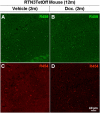
RIDNs remained in the hippocampus of RTN3TetOff mice after Dox treatment. Sagittal mouse brain sections (14 μm) were reacted with antibody R458 and anti-rabbit Alexa Fluor 488 secondary antibody (A, B) or with antibody R454 and anti-rabbit Alexa Fluor 568 secondary antibody (C, D). RIDNs in hippocampal CA1 region were visible by using confocal immunofluorescence microscopy. Scale bar, 40 μm. As shown in the confocal images of hippocampal CA1 region, the number of RIDNs was not significantly reduced in Dox-treated RTN3TetOff mice (B, D) compared with the vehicle-treated littermates (A, C). 2m, Two months; 12m, 12 months.
Figure 4.
Figure 4.
Turning off expression of RTN3 transgene in young Tg RTN3TetOff mice. A, RTN3TetOff Tg mice treated with either vehicle or Dox for 4 months (4m), and prepared hippocampal protein extracts were analyzed by Western blotting analysis. In vehicle-treated RTN3TetOff mice, RTN3 transgene was highly expressed as revealed by antibody R454 (lanes 3–7), whereas no RTN3 transgene was detected using R454 after 4 months Dox treatment (lanes 8–12). 9m, Nine months. B, The relative expression of RTN3 was normalized by β-actin expression. After normalization with β-actin, the total level of hippocampal RTN3 protein was reduced by 46% in the Dox-treated mice compared with that of vehicle-treated mice (n = 5, p < 0.001, Tukey's multiple comparison test), whereas its level was not significantly different from that of the tcRTN3 Tg mice (n = 5, p > 0.05, Tukey's multiple comparison test). One-way ANOVA statistical analysis indicated that there was significant variance among the three groups of mice (p < 0.001, n = 5).
Figure 5.
Figure 5.
Elimination of RIDNs in RTN3TetOff mice with early Dox treatment. Sagittal mouse brain sections were labeled by antibody R454 (A–C) or antibody R458 (D–F). Anti-rabbit Alexa Fluor 488 secondary is the secondary antibody. Antibody R458 recognizes both Tg RTN3 and endogenous mouse RTN3. To detect the presence of only RTN3 transgene in RIDNs, antibody R454 was used for detection. When antibody R458 was applied, hippocampal RIDNs were easily detected in the vehicle-treated RTN3TetOff mice, but only sparse hippocampal RIDNs were detected in both the tcRTN3 mice (D) and the Dox-treated RTN3TetOff mice (F). Scale bar, 40 μm. G, The hippocampal RIDN counts in the vehicle-treated RTN3TetOff mice were significantly higher than in the tcRTN3 mice (118 ± 21.5 vs 2.6 ± 0.7 in 6 sections per mouse, n = 5, p < 0.01, Tukey's multiple comparison test). After 4 months of Dox treatment, hippocampal RIDN counts were significantly reduced (4.6 ± 1.5 in sections per mouse, n = 5, p < 0.01, Tukey's multiple comparison test).
Figure 6.
Figure 6.
Reduction of APP processing by overexpressed RTN3. A, Hippocampal protein lysates were analyzed by Western blotting with antibody specific to APP C terminus (antibody A8717). APP C99 is the BACE1-cleaved product from APP, whereas APP C83 is the product from α-secretase cleavage of APP. β-Actin antibody was used to verify the loading. B, Relative BACE1 activity was assessed by the ratio of C99 over C83 or C99 over APP. Statistic significance was calculated by one-way ANOVA statistical analysis, which showed significance (p < 0.05 or p < 0.01, n = 5). 4m, Four months; 9m, 9 months.
Figure 7.
Figure 7.
Reducing formation of RIDNs improves contextual associative learning and memory in RTN3TetOff mice. A, Fear conditioning assay of learning and memory included a testing procedure of 3 d in a fear conditioning chamber. There was no difference in the percentage of freezing time before conditioning on day 1 (Pre-con). On day 2, contextual associative learning and memory of the mice were analyzed. Percentage of total freeze time of mice on day 2 was recorded as an ability of contextual associative learning and memory. The percentage of total freeze time was significantly improved from 24.5 ± 5.5 to 63.2 ± 5.0% after treatment of RTN3TetOff mice with Dox (n = 12, p < 0.01). B, The relationship between the RIDN counts and the contextual associated learning and memory was examined by statistical correlation analysis. Percentage of total freeze was plotted against the counts of RIDNs in hippocampal CA1 region. Their correlation coefficient r = −0.816, R2 = 0.666 (n = 12, p < 0.01), indicating a negative correlation between the hippocampal RIDN formation and the contextual associated learning and memory in the mice.
Figure 8.
Figure 8.
Reduction of BDNF in the hippocampus of the RTN3 Tg mice. ELISA was performed to determine the levels of BDNF in protein extracts that were prepared from either RTN3TetOff mice (A) or Tg RTN3 mice (B) and their littermate controls. The levels of hippocampal BDNF were significantly reduced in hippocampi of both mouse models (n = 6 in RTN3TetOff mice group and n = 6 in Tg RTN3 mice group; ***p < 0.01). However, the levels of cortical BDNF between Tg RTN3 mice and their control littermates showed no statistical difference (n = 6; p > 0.05). All results are presented as a mean ± SE. C, Immuno-confocal microscopy revealed that the positive BDNF immunity in the CA1 pyramidal neurons of the control mice (WT), whereas the BDNF immunity was mostly reduced in the Tg RTN3 mice. The mouse anti-BDNF antibody and the goat anti-mouse Alexa Fluor 488 antibody were used to label sagittal mouse brain sections (14 μm) in the immunofluorescent study.

Similar articles

Cited by

References

    1. Adachi N, Kohara K, Tsumoto T. Difference in trafficking of brain-derived neurotrophic factor between axons and dendrites of cortical neurons, revealed by live-cell imaging. BMC Neurosci. 2005;6:42. - PMC - PubMed
    1. Aicardi G, Argilli E, Cappello S, Santi S, Riccio M, Thoenen H, Canossa M. Induction of long-term potentiation and depression is reflected by corresponding changes in secretion of endogenous brain-derived neurotrophic factor. Proc Natl Acad Sci U S A. 2004;101:15788–15792. - PMC - PubMed
    1. Araki W, Oda A, Motoki K, Hattori K, Itoh M, Yuasa S, Konishi Y, Shin RW, Tamaoka A, Ogino K. Reduction of β-amyloid accumulation by reticulon 3 in transgenic mice. Curr Alzheimer Res. 2013 in press. - PubMed
    1. Brigadski T, Hartmann M, Lessmann V. Differential vesicular targeting and time course of synaptic secretion of the mammalian neurotrophins. J Neurosci. 2005;25:7601–7614. - PMC - PubMed
    1. Chow VW, Savonenko AV, Melnikova T, Kim H, Price DL, Li T, Wong PC. Modeling an anti-amyloid combination therapy for Alzheimer's disease. Sci Transl Med. 2010;2:13ra1. - PMC - PubMed

Publication types

MeSH terms

Substances