. 2013 Feb 13;33(7):3202-11.
doi: 10.1523/JNEUROSCI.2532-12.2013.

Segregated encoding of reward-identity and stimulus-reward associations in human orbitofrontal cortex

Affiliations

Segregated encoding of reward-identity and stimulus-reward associations in human orbitofrontal cortex

Miriam Cornelia Klein-Flügge et al. J Neurosci. .

Erratum in

Abstract

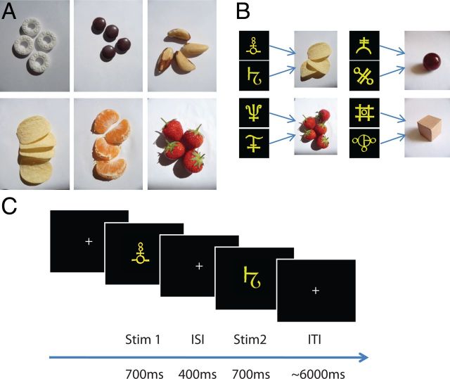
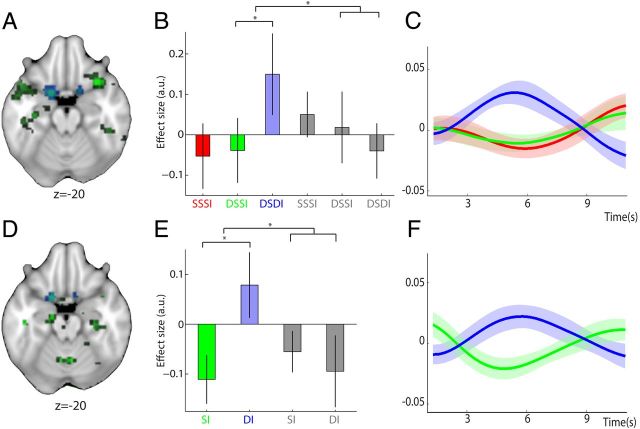
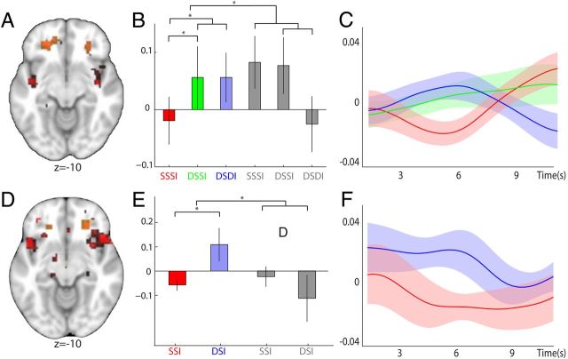
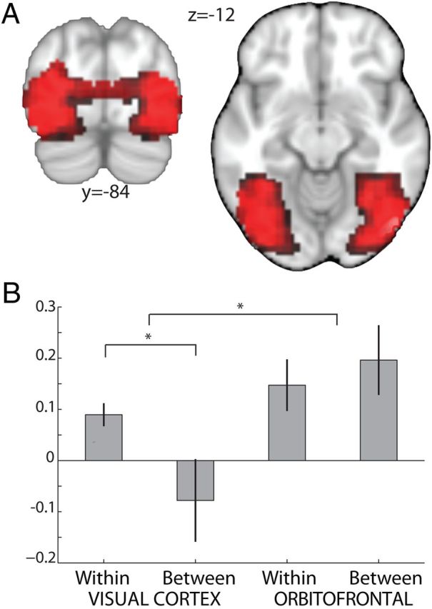
A dominant focus in studies of learning and decision-making is the neural coding of scalar reward value. This emphasis ignores the fact that choices are strongly shaped by a rich representation of potential rewards. Here, using fMRI adaptation, we demonstrate that responses in the human orbitofrontal cortex (OFC) encode a representation of the specific type of food reward predicted by a visual cue. By controlling for value across rewards and by linking each reward with two distinct stimuli, we could test for representations of reward-identity that were independent of associative information. Our results show reward-identity representations in a medial-caudal region of OFC, independent of the associated predictive stimulus. This contrasts with a more rostro-lateral OFC region encoding reward-identity representations tied to the predicate stimulus. This demonstration of adaptation in OFC to reward specific representations opens an avenue for investigation of more complex decision mechanisms that are not immediately accessible in standard analyses, which focus on correlates of average activity.


Figures

Figure 1.
Figure 1.
Experimental design. A, For each participant, two highly rated food rewards were chosen out of six possible food items. Food quantities (1–6 pieces) were adjusted individually so that the subjective values for the two food items were matched. B, Two abstract visual stimuli were assigned to each of the two food rewards and to two additional nonrewarding neutral objects (cardboard box and marble). Before scanning, participants were required to learn to associate each of the eight predictive stimuli with the corresponding food or neutral item. C, During scanning, participants were shown two consecutive stimuli on each trial and had to immediately elicit a representation of the associated item. In two trial types, participants were required to imagine the same item twice, in response to either the same stimulus presented repeatedly (SSSI), or two different stimuli predicting the same item (DSSI). A third trial type required participants to imagine two different items based on two different stimuli (DSDI).
Figure 2.
Figure 2.
Reward–identity representations independent of predictive stimulus. A, D, Regions representing the identity of rewarding food items were identified using repetition suppression, revealing a bilateral cluster in caudal OFC. Trials in which participants repeatedly imagined the same food reward in response to two different predictive stimuli were contrasted with those in which participants imagined two different food rewards (DSSIf vs DSDIf), and the resulting effect compared with that of neutral items ((DSDIf − DSSIf) − (DSDIn − DSSIn)). Whereas this analysis enabled identification of within-trial adaptation to the identity of food rewards (A), a second contrast was used to analyze adaptation effects of repeated representation of food rewards across adjacent trials to give a measure of between-trial adaptation (D) ((DIf − SIf) − (DIn − SIn)). In both contrasts, adaptation to reward–identity is found in caudal OFC (both p < 0.05) and shown in green; the extracted ROIs in caudal OFC are overlaid for visualization and shown in blue. B, Parameter estimates (± SEM) and (C) peristimulus BOLD time courses (shaded area shows SEM) demonstrate within-trial adaptation to reward–identity for food rewards (red, SSSIf; green, DSSIf; blue, DSDIf), with relative suppression of the BOLD signal to the representation of two identical food rewards (SSSIf or DSSIf) versus two different food rewards (DSDIf). As demonstrated by the parameter estimates, no such effect is present in the neutral conditions (gray, SSSIn, DSSIn, and DSDIn). Crucially, parameter estimates and time courses were extracted from an orthogonal ROI defined based on the between-trial adaptation contrast. E, F, For those trials where the same food reward was repeatedly elicited across adjacent trials, a similar suppression in the BOLD response can be observed, providing evidence for adaptation to reward–identity between trials. Parameter estimates and time courses are shown as in B and C, except that the orthogonal ROI from which they were extracted was defined from the within-trial contrast.
Figure 3.
Figure 3.
Representations of stimulus–reward associations. A, D, A bilateral region in lOFC was found to represent stimulus–reward information for food rewards. Trials with repeated elicitation of the same stimulus–reward mapping (SSSI) were compared against trials with two different predictive stimuli (DSSI and DSDI). To control for effects of mere visual stimulus adaptation, trials with food items were further contrasted against trials with neutral items (A) (((DSDIf + DSSIf) − (2 × SSSIf)) − ((DSDIn + DSSIn) − (2 × SSSIn))). In a similar way, we also analyzed between-trial adaptation effects by contrasting all trials that were preceded by a common stimulus–reward mapping, to those in which the stimulus–reward pairing of the preceding trial differed (D) ((DSIf − SSIf) − (DSIn − SSIn)). Regions demonstrating adaptation to stimulus–reward encoding in both the within- and between-trial contrast are shown (both p < 0.05 for visualization). This reveals that for both contrasts a region in rostro-lateral OFC adapted to the pairing of stimulus–reward information (shown in red); the extracted ROIs are overlaid for visualization and are shown in orange. B, C, E, F, Parameter estimates and time courses are shown as in Figure 2 (red, SSSIf; green, DSSIf; blue, DSDIf; gray, SSSIa, DSSIa, and DSDIa). B, C, Adaptation in lOFC to stimulus–reward information within trials, extracted from an orthogonal ROI defined by the equivalent between-trial contrast. E, F, Similar adaptation effects between trials in lOFC, extracted from the orthogonal within-trial ROI.
Figure 4.
Figure 4.
Sustained reward representations in OFC. To investigate the time window of repetition during which adaptation effects can be elicited in different brain regions, the average adaptation to reward–identity and stimulus–reward information in OFC was compared with adaptation to stimulus features in visual regions. A region of interest in visual cortex was extracted from the main effect of any visual event (p < 0.05, uncorrected). A, We observed adaptation to stimulus features for the within-trial contrast (shown is (DSDI + DSSI) − 2 × SSSI for food and neutral trials at p < 0.01 masked by the ROI): the BOLD signal was suppressed on trials with repeated presentation of the same stimulus compared with trials showing two different stimuli. B, Extraction of parameter estimates from this unbiased ROI demonstrates that this effect was restricted to the shorter within-trial repetition time. The adaptation effect was not observed between trials where repetition of a stimulus occurred on the consecutive trial. This finding is in contrast with adaptation effects in OFC, which occurred both within and between trials and thus suggests that information particular to the OFC can be retained across longer time intervals than information specific to visual processing regions.

References

    1. Balleine BW, Dickinson A. Goal-directed instrumental action: contingency and incentive learning and their cortical substrates. Neuropharmacology. 1998;37:407–419. - PubMed
    1. Bar M, Tootell RB, Schacter DL, Greve DN, Fischl B, Mendola JD, Rosen BR, Dale AM. Cortical mechanisms specific to explicit visual object recognition. Neuron. 2001;29:529–535. - PubMed
    1. Bar M, Kassam KS, Ghuman AS, Boshyan J, Schmid AM, Schmidt AM, Dale AM, Hämäläinen MS, Marinkovic K, Schacter DL, Rosen BR, Halgren E. Top-down facilitation of visual recognition. Proc Natl Acad Sci U S A. 2006;103:449–454. - PMC - PubMed
    1. Behrens TE, Woolrich MW, Walton ME, Rushworth MF. Learning the value of information in an uncertain world. Nat Neurosci. 2007;10:1214–1221. - PubMed
    1. Birn RM, Diamond JB, Smith MA, Bandettini PA. Separating respiratory-variation-related fluctuations from neuronal-activity-related fluctuations in fMRI. Neuroimage. 2006;31:1536–1548. - PubMed

Publication types

MeSH terms

LinkOut - more resources