. 2013 Apr 1;304(7):R553-65.
doi: 10.1152/ajpregu.00249.2012. Epub 2013 Feb 13.

Reversal of mitochondrial proteomic loss in Type 1 diabetic heart with overexpression of phospholipid hydroperoxide glutathione peroxidase

Affiliations

Reversal of mitochondrial proteomic loss in Type 1 diabetic heart with overexpression of phospholipid hydroperoxide glutathione peroxidase

Walter A Baseler et al. Am J Physiol Regul Integr Comp Physiol. .

Abstract

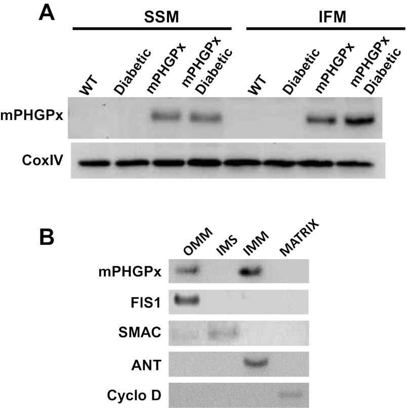
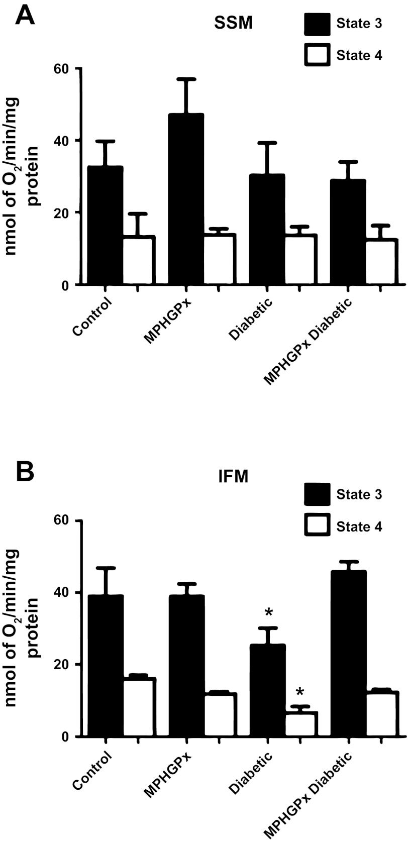
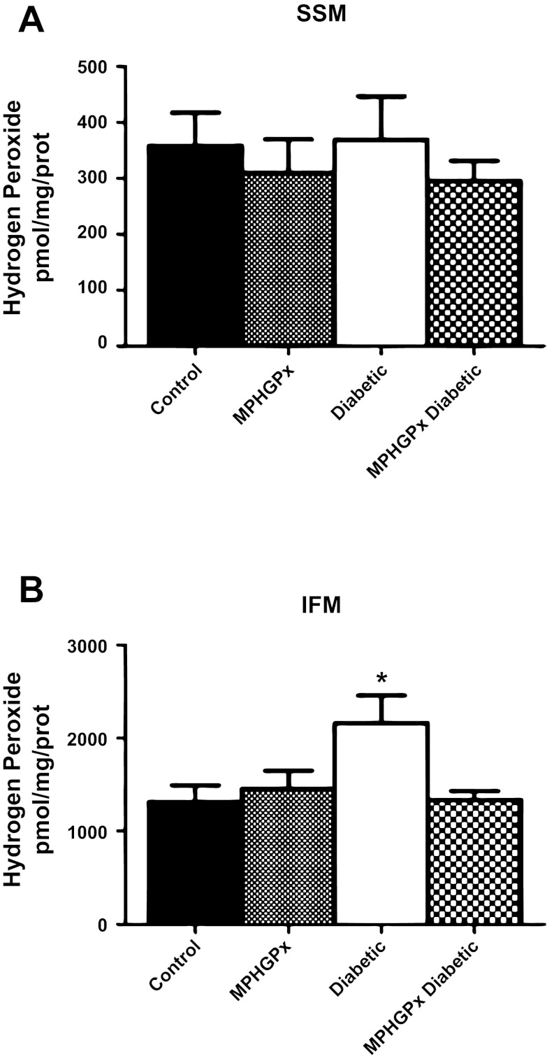
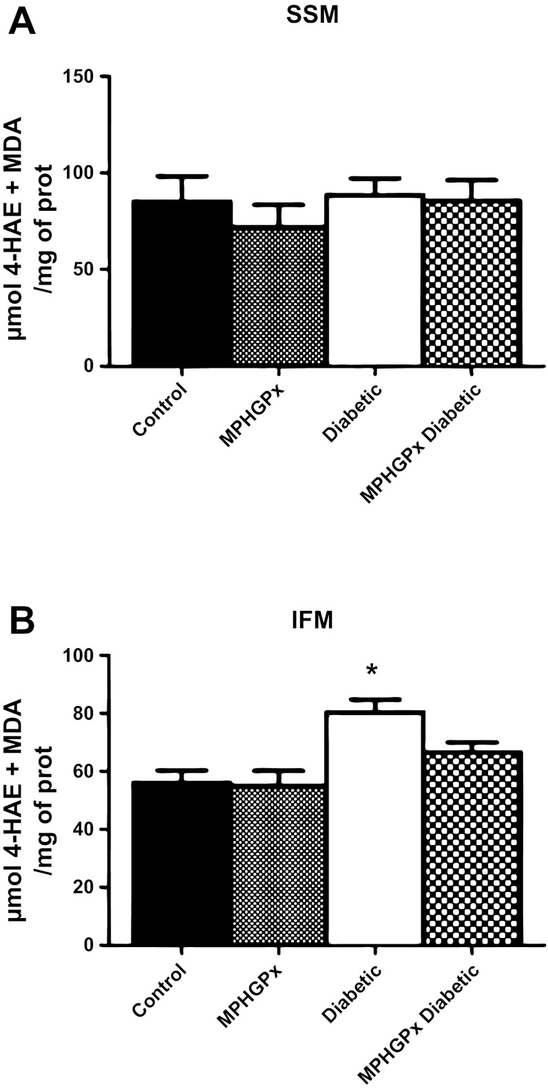
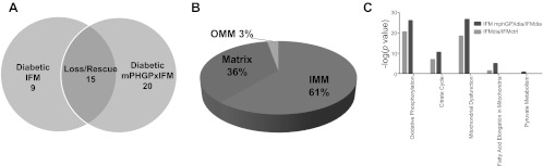
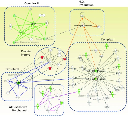
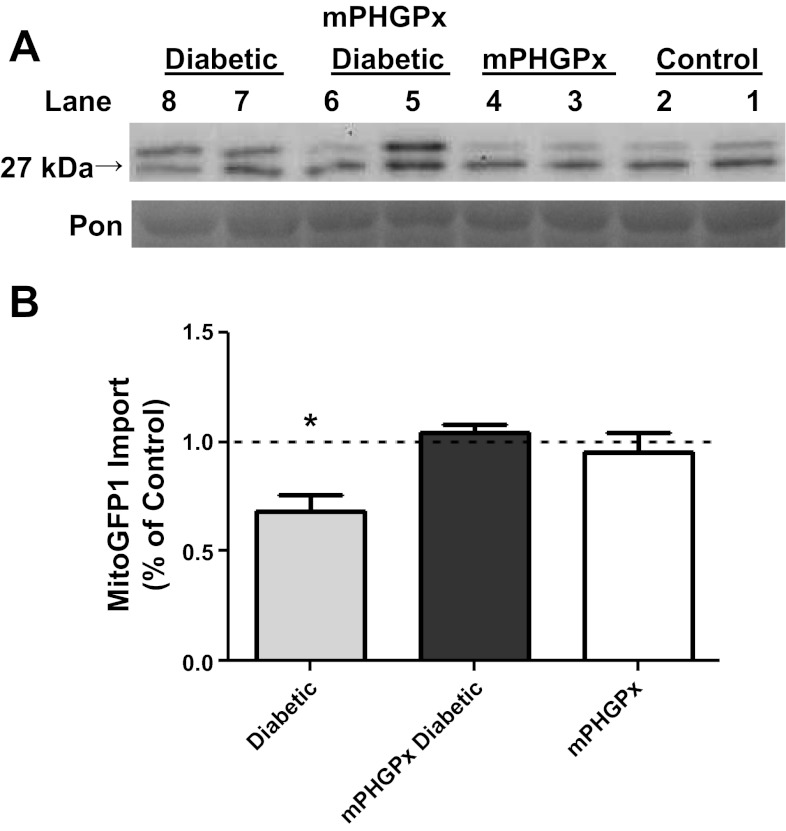
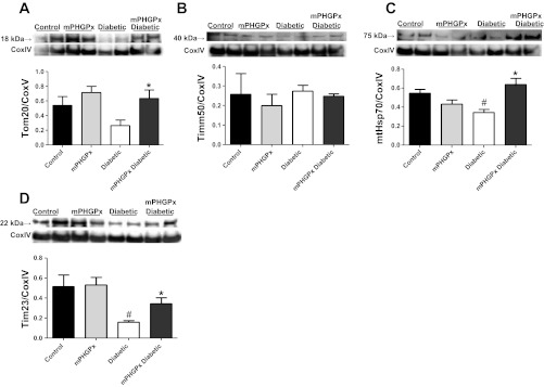
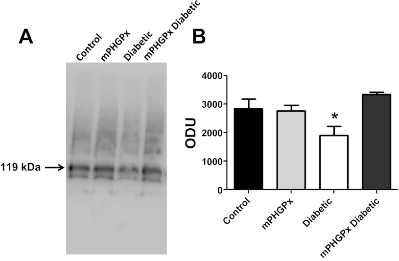
Mitochondrial dysfunction is a contributor to diabetic cardiomyopathy. Previously, we observed proteomic decrements within the inner mitochondrial membrane (IMM) and matrix of diabetic cardiac interfibrillar mitochondria (IFM) correlating with dysfunctional mitochondrial protein import. The goal of this study was to determine whether overexpression of mitochondria phospholipid hydroperoxide glutathione peroxidase 4 (mPHGPx), an antioxidant enzyme capable of scavenging membrane-associated lipid peroxides in the IMM, could reverse proteomic alterations, dysfunctional protein import, and ultimately, mitochondrial dysfunction associated with the diabetic heart. MPHGPx transgenic mice and controls were made diabetic by multiple low-dose streptozotocin injections and examined after 5 wk of hyperglycemia. Five weeks after hyperglycemia onset, in vivo analysis of cardiac contractile function revealed decreased ejection fraction and fractional shortening in diabetic hearts that was reversed with mPHGPx overexpression. MPHGPx overexpression increased electron transport chain function while attenuating hydrogen peroxide production and lipid peroxidation in diabetic mPHGPx IFM. MPHGPx overexpression lessened proteomic loss observed in diabetic IFM. Posttranslational modifications, including oxidations and deamidations, were attenuated in diabetic IFM with mPHGPx overexpression. Mitochondrial protein import dysfunction in diabetic IFM was reversed with mPHGPx overexpression correlating with protein import constituent preservation. Ingenuity Pathway Analyses indicated that oxidative phosphorylation, tricarboxylic acid cycle, and fatty acid oxidation processes most influenced in diabetic IFM were preserved by mPHGPx overexpression. Specific mitochondrial networks preserved included complex I and II, mitochondrial ultrastructure, and mitochondrial protein import. These results indicate that mPHGPx overexpression can preserve the mitochondrial proteome and provide cardioprotective benefits to the diabetic heart.


Figures

Fig. 1.
Fig. 1.
Mitochondria phospholipid hydroperoxide glutathione peroxidase 4 (mPHGPx) transgenic mouse screening. A: representative Western blot analysis of mPHGPx protein expression in individual mitochondrial subpopulations of mPHGPx transgenic (mPHGPx) and wild-type control (WT) mouse hearts. Pon, Ponceau staining loading control. B: representative Western blot analysis of mPHGPx protein expression in mitochondrial subcompartments. Mitochondrial subcompartments were fractionated and probed for mPHGPx presence. Individual fractions were probed with subcompartment-specific proteins to determine purity of the subfractionation process, outer mitochondrial membrane (OMM) (mitochondrial fission 1 protein, FIS1), inner membrane space (IMS) (second mitochondria-derived activator of caspases, SMAC), inner mitochondrial membrane (IMM) (adenine nucleotide translocase, ANT), and matrix (cyclophilin D).
Fig. 2.
Fig. 2.
Mitochondrial respiration. State 3 and state 4 respiration rates with glutamate-malate as substrates in subsarcolemmal membrane (SSM) (A) and interfibrillar mitochondria (IFM) (B). State 3 and state 4 respiration rates were determined in the presence of the substrates glutamate-malate, and state 3 respiration was examined on addition of ADP. Values are ± SE. *P < 0.05 for diabetic vs. all groups.
Fig. 3.
Fig. 3.
H2O2 production. H2O2 production in SSM (A) and IFM (B) was determined using the oxidation of the fluorogenic indicator amplex red in the presence of horseradish peroxidase. H2O2 production was initiated in mitochondria using glutamate-malate as substrates. Standard curves were obtained by adding known amounts of H2O2 to the assay medium in the presence of the substrates amplex red and horseradish peroxidase. Final values were calculated as pmol/mg of protein. Values are expressed as means ± SE. *P < 0.05 for diabetic vs. all groups.
Fig. 4.
Fig. 4.
Lipid peroxidation by-products. Oxidative damage to lipids was assessed in SSM (A) and IFM (B) by measuring lipid peroxidation by-products malondialdehyde (MDA) and 4-hydroxyalkenals (4-HAE) using a colorimetric assay and compared against a standard curve of known 4-HAE and MDA concentrations. Values for lipid peroxidation by-products are expressed as means ± SE. *P < 0.05 for diabetic vs. all groups.
Fig. 5.
Fig. 5.
Proteomic alterations in diabetic and mPHPGx diabetic IFM. A: total number of proteins decreased solely in the diabetic IFM (left), proteins decreased in diabetic IFM and preserved within mPHGPx diabetic IFM (middle), and proteins increased solely within the mPHGPx diabetic IFM (right). B: breakdown in approximate percentages of protein contents lost in diabetic IFM and subsequently preserved in the mPHGPx diabetic IFM. C: mitochondrial canonical pathways identifyed using Ingenuity Pathway Analysis (IPA) software most negatively impacted by diabetic insult and subsequently restored by mPHGPx overexpression. Oxidative phosphorylation, tricarboxylic acid cycle (citric acid cycle), fatty acid metabolism, and fatty acid elongation were negatively affected (light gray shade) by diabetes mellitus. Conversely, these pathways as well as pyruvate metabolism were preserved (dark gray shade) in diabetic IFM with the overexpression of mPHGPx. Diab., diabetic; Ctrl, control.
Fig. 6.
Fig. 6.
Mitochondrial protein networks. Identification using IPA software, of mitochondrial proteomic networks negatively influenced in diabetic IFM and preserved within mPHGPx diabetic IFM. Six networks, including electron transport chain (ETC) complexes I and II, H2O2 production, structure, ATP-sensitive K+ channel, and mitochondrial protein import were identified. Mitochondrial protein import network is the central node linking all other networks. Green indicates proteins preserved within mPHGPx diabetic IFM. Red indicates proteins positively enhanced in mPHGPx diabetic IFM. White indicates proteins unchanged by diabetes mellitus but are part of their respective networks.
Fig. 7.
Fig. 7.
Mitochondrial protein import. Effect of Type 1 diabetes mellitus and mPHGPx overexpression on MitoGFP1 import in IFM. A: representative Western blots from IFM protein import assay. Control for protein loading was confirmed by Ponceau (Pon) staining. B: graphical representation of mitochondrial protein import performed in control diabetic, mPHGPx, and mPHGPx diabetic. The relative amount of imported MitoGFP1 was determined by densitometry. Dashed line denotes control levels. Import efficiency was based off of percent control at the 1- and 2-min time points. Both time point percentages were then averaged to show total import efficiency. Values are presented as means ± SE; *P < 0.05 for Diabetic vs. all groups. N = 5 per group.
Fig. 8.
Fig. 8.
Mitochondrial protein import constituents. Western blot analysis of protein import constituents in IFM. Key proteins involved in mitochondrial protein import were assessed using Western blot analysis. Representative Western blot and densitometry analysis for Tom20 (A), Tim50 (B), mtHsp70 (C), and Tim23 (D) from control, mPHGPx, diabetic, and mPHGPx diabetic. Control for protein loading was confirmed by CoxIV staining. Values are presented as means ± SE; *P < 0.05 for mPHGPx Diabetic vs. Diabetic; #P < 0.05 for Control vs. Diabetic. N = 4 per group.
Fig. 9.
Fig. 9.
mtHsp70 protein complexing. Effect of Type 1 diabetes mellitus and mPHGPx overexpression on mtHsp70-Tim44 complexing in isolated IFM from control, diabetic, mPHGPx, and mPHGPx diabetic. IFM were subjected to BN-PAGE, and protein complexes were probed for mtHSP70. A: complex of mtHsp70 and Tim44 exists at 119 kDa. B: mtHsp70 content was analyzed through densitometry. *P < 0.05 for Diabetic vs. all groups. N = 4 per group.

References

    1. Anderson S, Bankier AT, Barrell BG, de Bruijn MH, Coulson AR, Drouin J, Eperon IC, Nierlich DP, Roe BA, Sanger F, Schreier PH, Smith AJ, Staden R, Young IG. Sequence and organization of the human mitochondrial genome. Nature 290: 457–465, 1981 - PubMed
    1. Baseler WA, Dabkowski ER, Williamson CL, Croston TL, Thapa D, Powell MJ, Razunguzwa TT, Hollander JM. Proteomic alterations of distinct mitochondrial subpopulations in the type 1 diabetic heart: contribution of protein import dysfunction. Am J Physiol Regul Integr Comp Physiol 300: R186–R200, 2011 - PMC - PubMed
    1. Becker D, Krayl M, Strub A, Li Y, Mayer MP, Voos W. Impaired interdomain communication in mitochondrial Hsp70 results in the loss of inward-directed translocation force. J Biol Chem 284: 2934–2946, 2009 - PubMed
    1. Bohnert M, Rehling P, Guiard B, Herrmann JM, Pfanner N, van der Laan M. Cooperation of stop-transfer and conservative sorting mechanisms in mitochondrial protein transport. Curr Biol 20: 1227–1232, 2010 - PubMed
    1. Bradford MM. A rapid and sensitive method for the quantitation of microgram quantities of protein utilizing the principle of protein-dye binding. Anal Biochem 72: 248–254, 1976 - PubMed

Publication types

MeSH terms

Substances

LinkOut - more resources