

Renal sensory and sympathetic nerves reinnervate the kidney in a similar time-dependent fashion after renal denervation in rats
- PMID: 23408032
- PMCID: PMC3627950
- DOI: 10.1152/ajpregu.00599.2012
Renal sensory and sympathetic nerves reinnervate the kidney in a similar time-dependent fashion after renal denervation in rats
Abstract
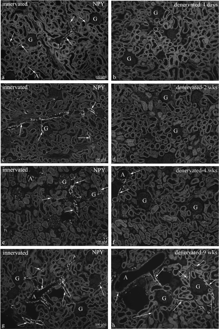
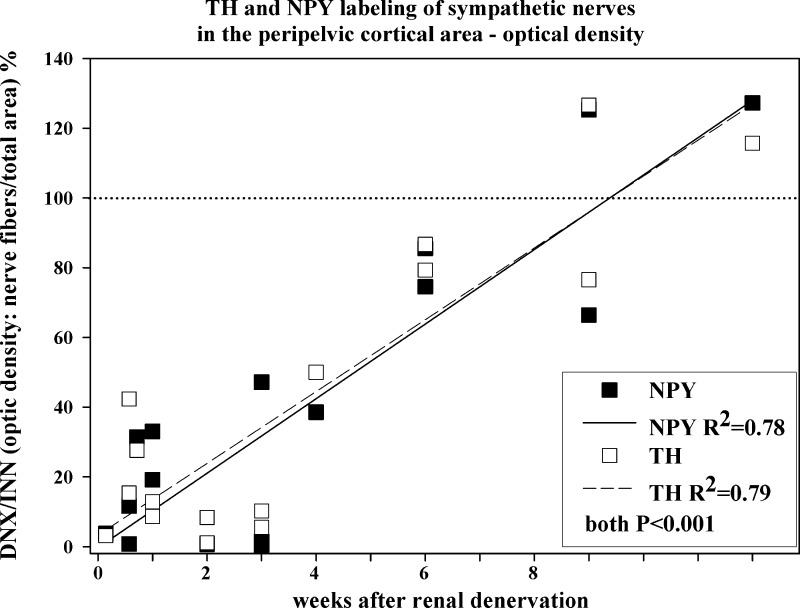
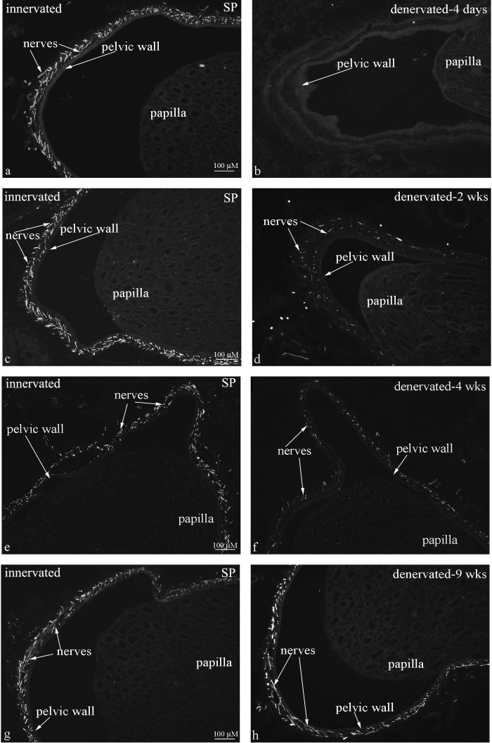
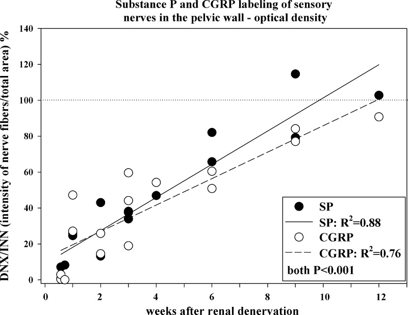
Efferent renal sympathetic nerves reinnervate the kidney after renal denervation in animals and humans. Therefore, the long-term reduction in arterial pressure following renal denervation in drug-resistant hypertensive patients has been attributed to lack of afferent renal sensory reinnervation. However, afferent sensory reinnervation of any organ, including the kidney, is an understudied question. Therefore, we analyzed the time course of sympathetic and sensory reinnervation at multiple time points (1, 4, and 5 days and 1, 2, 3, 4, 6, 9, and 12 wk) after renal denervation in normal Sprague-Dawley rats. Sympathetic and sensory innervation in the innervated and contralateral denervated kidney was determined as optical density (ImageJ) of the sympathetic and sensory nerves identified by immunohistochemistry using antibodies against markers for sympathetic nerves [neuropeptide Y (NPY) and tyrosine hydroxylase (TH)] and sensory nerves [substance P and calcitonin gene-related peptide (CGRP)]. In denervated kidneys, the optical density of NPY-immunoreactive (ir) fibers in the renal cortex and substance P-ir fibers in the pelvic wall was 6, 39, and 100% and 8, 47, and 100%, respectively, of that in the contralateral innervated kidney at 4 days, 4 wk, and 12 wk after denervation. Linear regression analysis of the optical density of the ratio of the denervated/innervated kidney versus time yielded similar intercept and slope values for NPY-ir, TH-ir, substance P-ir, and CGRP-ir fibers (all R(2) > 0.76). In conclusion, in normotensive rats, reinnervation of the renal sensory nerves occurs over the same time course as reinnervation of the renal sympathetic nerves, both being complete at 9 to 12 wk following renal denervation.
Figures
Similar articles
-
Complex reinnervation pattern after unilateral renal denervation in rats.Am J Physiol Regul Integr Comp Physiol. 2016 May 1;310(9):R806-18. doi: 10.1152/ajpregu.00227.2014. Epub 2016 Feb 24. Am J Physiol Regul Integr Comp Physiol. 2016. PMID: 26911463
-
Nerve fibers immunoreactive to calcitonin gene-related peptide, substance P, neuropeptide Y, and dopamine beta-hydroxylase in innervated and denervated oral tissues in ferrets.Acta Odontol Scand. 1998 Aug;56(4):220-8. doi: 10.1080/00016359850142835. Acta Odontol Scand. 1998. PMID: 9765014
-
Reinnervation of renal afferent and efferent nerves at 5.5 and 11 months after catheter-based radiofrequency renal denervation in sheep.Hypertension. 2015 Feb;65(2):393-400. doi: 10.1161/HYPERTENSIONAHA.114.04176. Epub 2014 Nov 17. Hypertension. 2015. PMID: 25403610
-
Reinnervation following catheter-based radio-frequency renal denervation.Exp Physiol. 2015 Apr 20;100(5):485-90. doi: 10.1113/expphysiol.2014.079871. Epub 2015 Jan 22. Exp Physiol. 2015. PMID: 25573386 Review.
-
Role of renal sensory nerves in physiological and pathophysiological conditions.Am J Physiol Regul Integr Comp Physiol. 2015 Jan 15;308(2):R79-95. doi: 10.1152/ajpregu.00351.2014. Epub 2014 Nov 19. Am J Physiol Regul Integr Comp Physiol. 2015. PMID: 25411364 Free PMC article. Review.
Cited by
-
The Effects of Renal Nerve Denervation on Blood Pressure and Target Organs in Different Hypertensive Rat Models.Int J Hypertens. 2021 Apr 4;2021:8615253. doi: 10.1155/2021/8615253. eCollection 2021. Int J Hypertens. 2021. PMID: 33884205 Free PMC article.
-
Renal denervation restores biomechanics of carotid arteries in a rat model of hypertension.Sci Rep. 2024 Jan 4;14(1):495. doi: 10.1038/s41598-023-50816-8. Sci Rep. 2024. PMID: 38177257 Free PMC article.
-
Correlated Sensory and Sympathetic Innervation Between the Acupoint BL23 and Kidney in the Rat.Front Integr Neurosci. 2021 Jan 11;14:616778. doi: 10.3389/fnint.2020.616778. eCollection 2020. Front Integr Neurosci. 2021. PMID: 33505253 Free PMC article.
-
Renal Denervation Influences Angiotensin II Types 1 and 2 Receptors.Int J Nephrol. 2022 Oct 10;2022:8731357. doi: 10.1155/2022/8731357. eCollection 2022. Int J Nephrol. 2022. PMID: 36262553 Free PMC article. Review.
-
Sympathetic Nervous System Contributions to Hypertension: Updates and Therapeutic Relevance.Can J Cardiol. 2020 May;36(5):712-720. doi: 10.1016/j.cjca.2020.03.003. Epub 2020 Mar 6. Can J Cardiol. 2020. PMID: 32389344 Free PMC article. Review.
References
-
- Barajas L, Powers K. Monoaminergic innervation of the rat kidney: a quantitative study. Am J Physiol Renal Fluid Electrolyte Physiol 259: F503–F511, 1990 - PubMed
-
- Barajas L, Powers K, Wang P. Innervation of the renal cortical tubules: a quantitative study. Am J Physiol Renal Fluid Electrolyte Physiol 247: F50–F60, 1984 - PubMed
-
- Calaresu FR, Ciriello J. Renal afferent nerves affect discharge rate of medullary and hypothalamic single units in cat. J Auton Nerv Syst 3: 311–320, 1981 - PubMed
-
- Campese VM, Kogosov E. Renal afferent denervation prevents hypertension in rats with chronic renal failure. Hypertension 25: 878–882, 1995 - PubMed
Publication types
MeSH terms
Substances
Grants and funding
LinkOut - more resources
Full Text Sources
Other Literature Sources
Research Materials
Miscellaneous