Comparative Study
. 2013 Feb 13;5(172):172ra20.
doi: 10.1126/scitranslmed.3004888.

Mucosal imprinting of vaccine-induced CD8⁺ T cells is crucial to inhibit the growth of mucosal tumors

Affiliations
Comparative Study

Mucosal imprinting of vaccine-induced CD8⁺ T cells is crucial to inhibit the growth of mucosal tumors

Federico Sandoval et al. Sci Transl Med. .

Erratum in

  • Sci Transl Med. 2013 Mar 27;5(178):178er2. Suleman, Muhammed [corrected to Suleman, Muhammad]

Abstract

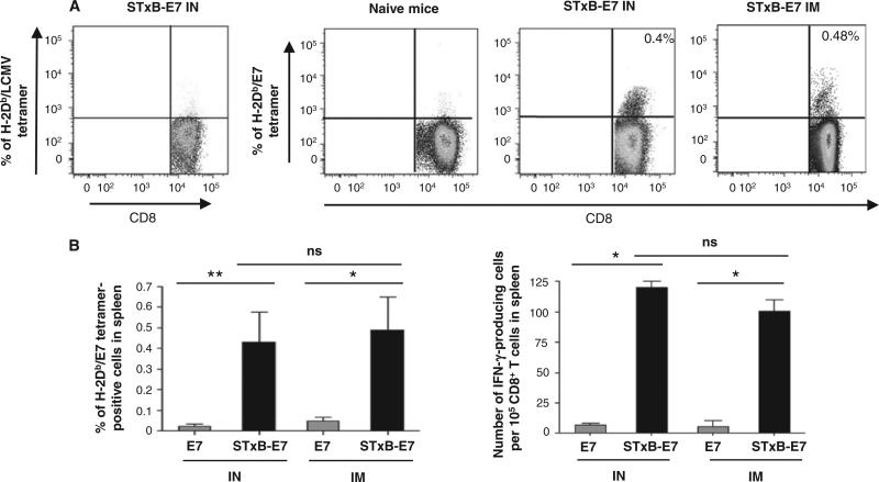
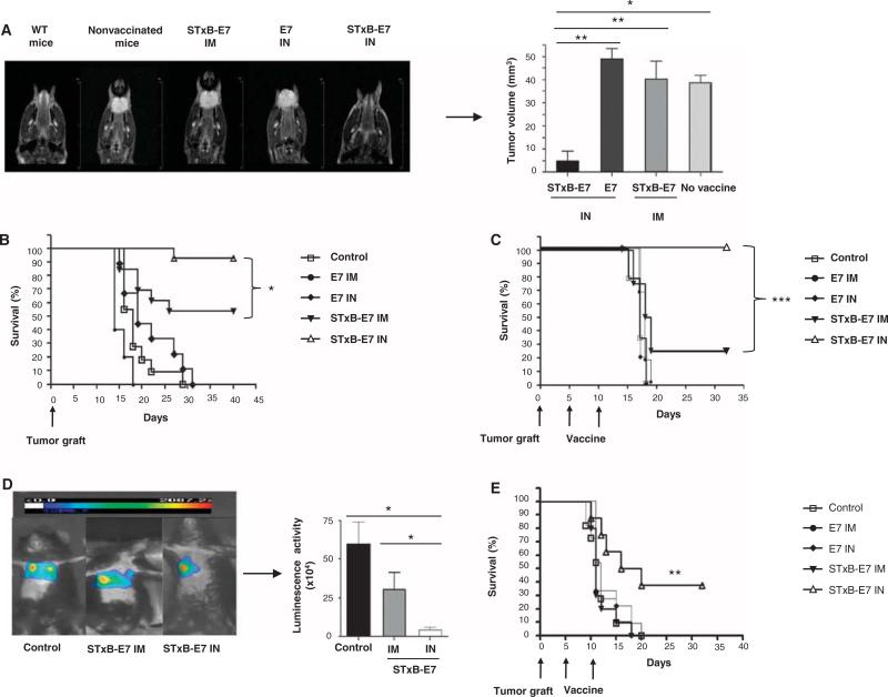
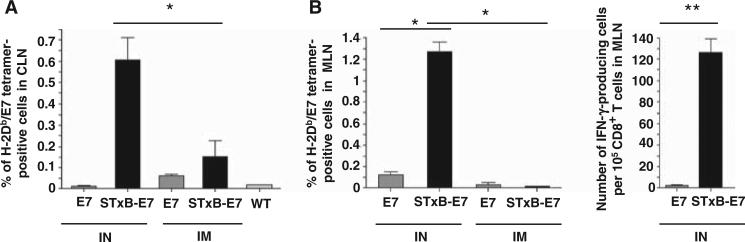
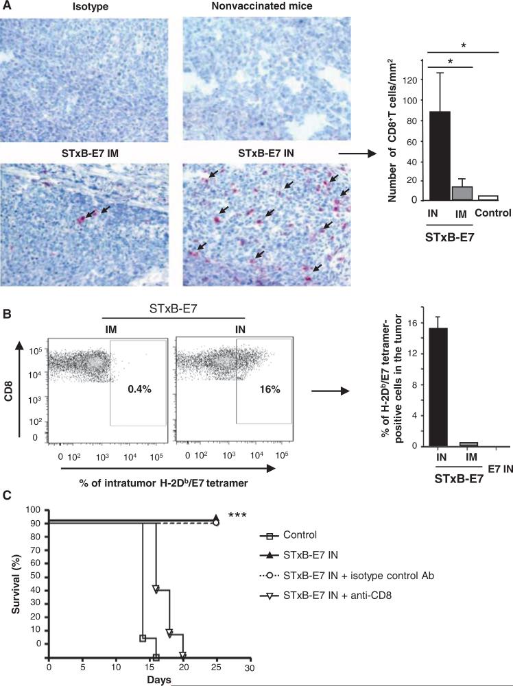
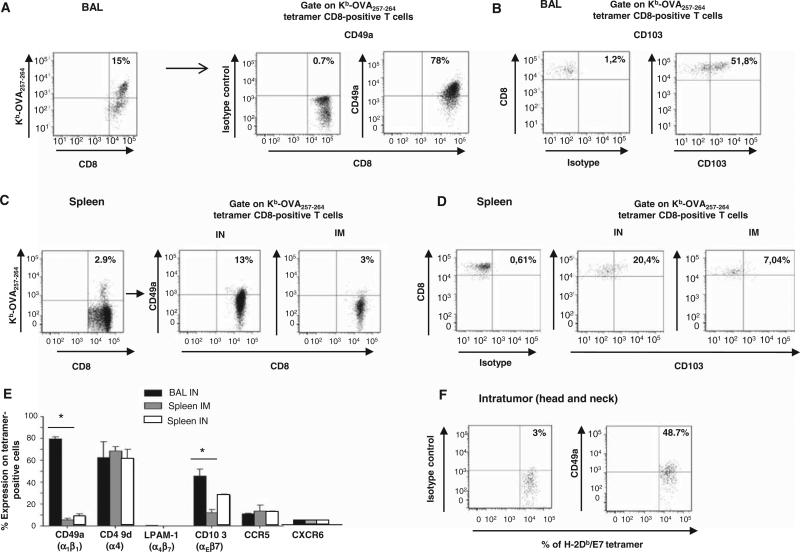
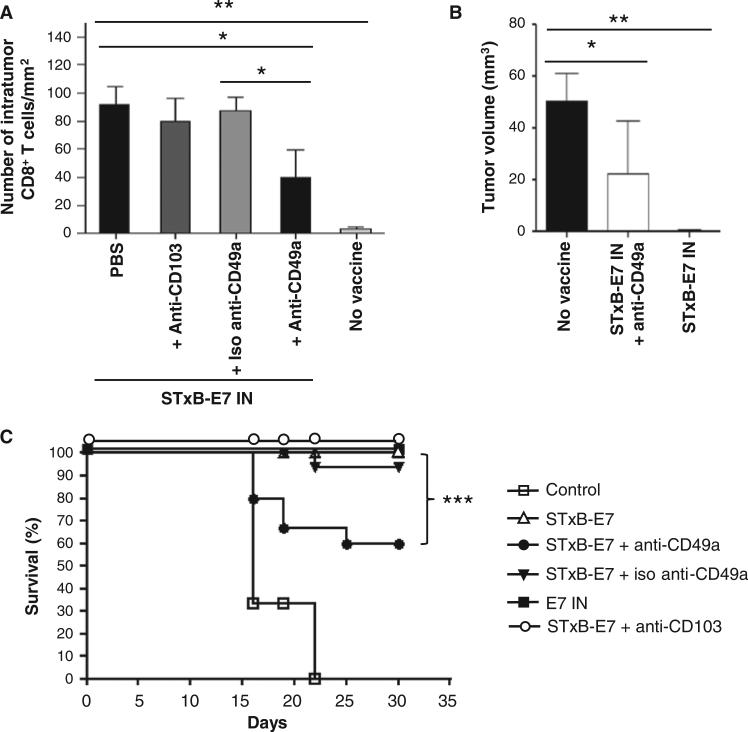
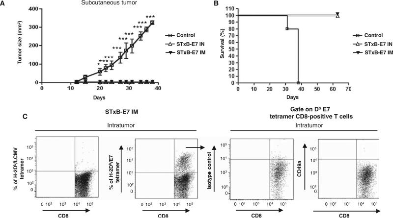
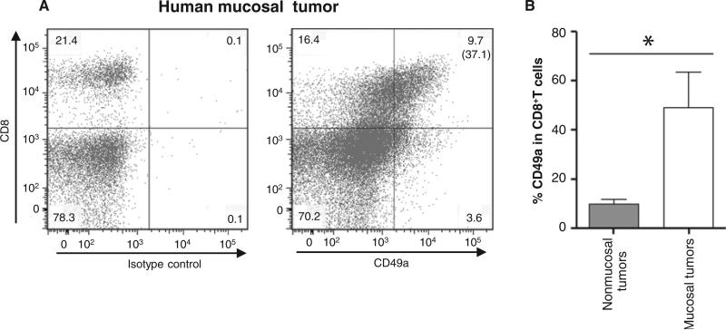
Although many human cancers are located in mucosal sites, most cancer vaccines are tested against subcutaneous tumors in preclinical models. We therefore wondered whether mucosa-specific homing instructions to the immune system might influence mucosal tumor outgrowth. We showed that the growth of orthotopic head and neck or lung cancers was inhibited when a cancer vaccine was delivered by the intranasal mucosal route but not the intramuscular route. This antitumor effect was dependent on CD8⁺ T cells. Indeed, only intranasal vaccination elicited mucosal-specific CD8⁺ T cells expressing the mucosal integrin CD49a. Blockade of CD49a decreased intratumoral CD8⁺ T cell infiltration and the efficacy of cancer vaccine on mucosal tumor. We then showed that after intranasal vaccination, dendritic cells from lung parenchyma, but not those from spleen, induced the expression of CD49a on cocultured specific CD8⁺ T cells. Tumor-infiltrating lymphocytes from human mucosal lung cancer also expressed CD49a, which supports the relevance and possible extrapolation of these results in humans. We thus identified a link between the route of vaccination and the induction of a mucosal homing program on induced CD8⁺ T cells that controlled their trafficking. Immunization route directly affected the efficacy of the cancer vaccine to control mucosal tumors.


Figures

Fig. 1
Fig. 1
Intranasal or intramuscular immunization with STxB-E7 induces E7 antigen-specific CD8+ T cells in spleen. Representative dot plots of spleen E7-specific CD8+ T cells stained by tetramers after two intranasal (IN) or intramuscular (IM) immunizations (days 0 and 14) with STxB-E7 (0.5 nmol = 20 μg). (A) Both vaccines were mixed with αGalCer. (B) E749–57–specific CD8+ T cells from spleen were detected by tetramer assay or IFN-γ ELISpot directly ex vivo. For the tetramer analysis, values shown correspond to means ± SD obtained with specific tetramers. Irrelevant tetramers or tetramer on unimmunized mice gave results <0.05%. For the ELISpot analysis, background obtained with cells not pulsed with the E749–57 peptide was also subtracted (always <10 spots per 105 cells). Spots on unimmunized mice were always <5 spots per 105 cells. Four mice per group were immunized, and these experiments were reproduced three times. *P < 0.05, **P < 0.01. ns, not significant.
Fig. 2
Fig. 2
Intranasal immunization with STxB-E7 inhibits orthotopic head and neck and lung tumors in both prophylactic and therapeutic settings. (A and B) In a prophylactic setting, mice were immunized twice via the intranasal or intramuscular route with STxB-E7 or E7 peptide–based vaccines mixed with the aGalCer adjuvant. Seven days after the second vaccination, mice were grafted with 5 × 10⁴ TC1 cells in the submucosa of the tongue. Representative MRI (coronal T2-weighted image of mouse tongue) (A, left) and tumor volume measured by MRI (A, right) at day 7 after tumor graft in the tongue are shown. (C to E) In the therapeutic setting, mice were immunized 5 and 10 days after grafting TC1 cells in the submucosa of the tongue (C) or 5 and 10 days after grafting TC1 luciferase (105 cells) into the lung (D and E), which was monitored by luminescence. Representative bioluminescence images of tumor-bearing mice in the lung are depicted in (D) (left panel). Luminescence (luciferase activity) in the chest was quantified 4 days after the second immunization with STxB-E7 via the intranasal route (n = 7) or the intramuscular route (n = 4) or in the nonvaccinated control mice (n = 4) (D, right panel). Kaplan-Meier survival curves of prophylactic (B) or therapeutic (C and E) cancer vaccine experiments for head and neck (B and C) and lung tumor (E) are shown. Each experiment was reproduced four times. *P < 0.05, **P < 0.01, ***P < 0.001.
Fig. 3
Fig. 3
Intranasal immunization with STxB-E7 elicits functional E749–57–specific T cells in cervical and mediastinal lymph nodes. Mice were immunized by the intranasal or the intramuscular route with STxB-E7 or the nonvectorized E7 polypeptide. (A and B) E749–57–specific CD8+ T cells from cervical lymph node (CLN) (A) or mediastinal lymph node (MLN) (B) were detected by tetramer assay or IFN-γ ELISpot directly ex vivo. WT, wild type. For the tetramer analysis, means ± SD obtained with specific tetramer after subtracting values from irrelevant tetramer (always <0.1%) are shown. For the ELISpot analysis, background obtained with cells not pulsed with the E749–57 peptide was also subtracted (always <10 spots per 105 cells). These experiments were reproduced three times with four mice per group. *P < 0.05, **P < 0.01.
Fig. 4
Fig. 4
CD8+ T cells are required to control the growth of orthotopic head and neck cancer after intra-nasal immunization with STxB-E7. Eight days after the orthotopic graft of TC1 cells in the submucosa of the tongue, tumors were removed and CD8+ T cells were detected by immunoenzymatic staining. (A) (Left) Arrows show CD8+ T cells infiltrating the tumors in mice previously intranasally or intramuscularly immunized with the STxB-E7 vaccine or nonvaccinated B6 mice (original magnification, ×40). (Right) Histograms show the means ± SEM values from pooled data of three to four mice per group. Intratumoral CD8+ T cells were purified from a pool of three to four tumors of mice vaccinated with various E7 vaccines and stained with H-2Db/E7 tetramer. These experiments were reproduced twice with similar results. (B) (Left) Values shown correspond to means ± SD obtained with specific E7 tetramer after subtracting values from irrelevant tetramer (right). B6 mice were intranasally immunized with STxB-E7 at days 0 and 14. At day 20 and then once a week, mice received anti-CD8 mAb (100 mg, intraperitoneally) or isotype-matched control mAb. (C) Seven days after the second vaccination (day 21), mice were grafted with TC1 cells in the submucosa of the tongue and monitored for survival. All these experiments were reproduced three times. *P < 0.05, ***P < 0.001.
Fig. 5
Fig. 5
Expression of CD49a (α1β1) and CD103 (αEβ7) by antigen-specific CD8+ T cells in the BAL, spleen, and tumor microenvironment after immunization of mice with STxB-based vaccine via the intranasal or intramuscular routes. (A to D) OVA-specific CD8+ T cells were detected in BAL in a pool of six to seven mice (A and B) or in the spleen (C and D) 7 days after the second immunization with STxB-OVA. Expression of CD49a [corresponding to the α1 chain of VLA-1 (α1β1), which associates only with β1] (A and C) and CD103 (B and D) was analyzed after gating on tetramer-positive CD8+ T cells. Isotype controls were included in each experiment. Phenotypic analysis of tetramer-positive OVA-specific CD8+ T cells in the BAL after intramuscular immunization is not shown because this route of immunization did not induce OVA-specific CD8+ T cells in this compartment. (E) Values shown correspond to means ± SD for the expression of CD49a, CD49d, α4β7, CD103, and CCR5 analyzed after gating on tetramer-positive Kb-OVA257–264–positive cells in BAL or spleen using the same protocol described for (A) to (D). Isotype controls were included in each experiment. This experiment was reproduced twice with a pool of five to six mice per experiment. Mice were grafted with TC1 cells in the tongue and then immunized twice (days 5 and 10) by the intranasal route with STxB-E7 mixed with αGalCer. At day 15, tumors (n = 5) were pooled and intratumoral CD8+ T cells were purified from the tumors and stained with H-2Db/E7 tetramer, CD8, and CD49a. (F) Dot plots were generated on gated CD8+ T cells.
Fig. 6
Fig. 6
Blockade of CD49a inhibits CD8+ T cell infiltration and the activity of cancer vaccine on head and neck tumors. Mice were grafted with TC1 cells in the tongue and then immunized two times with STxB-E7 mixed with αGalCer (days 5 and 10). (A to C) They were also treated with either anti-CD49a (250 μg, intraperitoneally) or anti-CD103 (150 μg, intraperitoneally) or isotype-matched control mAb at days 8, 11, and 14. CD8+ T cell infiltration was detected by immunoenzymatic staining (A), and tumor volume was monitored by MRI at day 20 (B). Kaplan-Meier survival curve of mice bearing a head and neck tumor and treated or not with an intranasal cancer vaccine combined or not with anti-CD49a mAb, anti-CD103 (C), or isotype-matched control. Each experiment included five to six mice per group and was reproduced three times. *P < 0.05, **P < 0.01, ***P < 0.001.
Fig. 7
Fig. 7
The intranasal and intramuscular routes are both efficient to control the growth of subcutaneous grafted TC1 cells. B6 mice were immunized via the intranasal or intramuscular routes with STxB-E7 or E7 vaccines mixed with αGalCer. Seven days after the second vaccination, mice were grafted in the right flank with 105 TC1 cells. (A and B) Tumor growth (A) and survival (B) were monitored every 3 days. Five mice per group were included in each experiment, which was reproduced twice. *P < 0.05, ***P < 0.001. (C) Mice were grafted with TC1 cells in the right flank and then immunized two times (days 5 and 10) by the intranasal route with STxBE7 mixed with αGalCer. At day 15, tumors (n = 5) were pooled, and intratumoral CD8+ T cells were purified from the tumors and stained with H-2Db/E7 tetramer, CD8, and CD49a. Dot plots were generated on gated Db-E7 tetramer CD8+ T cells. Tetramer and isotype control were included in each experiments.
Fig. 8
Fig. 8
CD49a expression in TILs from mucosal tumors. TILs were obtained from four enzymatically treated mucosal tumors (three human lung and one head and neck cancers) and eight nonmucosal tumors (eight renal cell carcinoma biopsies). They were stained by anti-CD8 and anti-CD49a mAb. Isotype controls were included in each experiment. (A) Representative dot plots of CD49a expression on head and neck TILs after gating on CD45+ T cells. Values in the rectangle shown in each quadrant correspond to percent of cells, and the value in bracket corresponds to the percent of CD49a in the CD8+ T cells. (B) Values shown on the histogram correspond to means ± SD for the expression of CD49a in CD8+ T cells from mucosal and nonmucosal tumors. *P < 0.05.

Comment in

  • Vaccination route matters for mucosal tumors.
    Nardelli-Haefliger D, Dudda JC, Romero P. Nardelli-Haefliger D, et al. Sci Transl Med. 2013 Feb 13;5(172):172fs4. doi: 10.1126/scitranslmed.3005638. Sci Transl Med. 2013. PMID: 23408051 No abstract available.

References

    1. Boon T, Coulie PG, Van den Eynde BJ, van der Bruggen P. Human T cell responses against melanoma. Annu. Rev. Immunol. 2006;24:175–208. - PubMed
    1. Fridman WH, Galon J, Pagès F, Tartour E, Sautès-Fridman C, Kroemer G. Prognostic and predictive impact of intra- and peritumoral immune infiltrates. Cancer Res. 2011;71:5601–5605. - PubMed
    1. Broussard EK, Disis ML. TNM staging in colorectal cancer: T is for T cell and M is for memory. J. Clin. Oncol. 2011;29:601–603. - PubMed
    1. Kantoff PW, Higano CS, Shore ND, Berger ER, Small EJ, Penson DF, Redfern CH, Ferrari AC, Dreicer R, Sims RB, Xu Y, Frohlich MW, Schellhammer PF, IMPACT Study Investigators Sipuleucel-T immunotherapy for castration-resistant prostate cancer. N. Engl. J. Med. 2010;363:411–422. - PubMed
    1. Schwartzentruber DJ, Lawson DH, Richards JM, Conry RM, Miller DM, Treisman J, Gailani F, Riley L, Conlon K, Pockaj B, Kendra KL, White RL, Gonzalez R, Kuzel TM, Curti B, Leming PD, Whitman ED, Balkissoon J, Reintgen DS, Kaufman H, Marincola FM, Merino MJ, Rosenberg SA, Choyke P, Vena D, Hwu P. gp100 peptide vaccine and interleukin-2 in patients with advanced melanoma. N. Engl. J. Med. 2011;364:2119–2127. - PMC - PubMed

Publication types

MeSH terms