

doi: 10.1007/s11626-013-9588-2.
Epub 2013 Feb 14.
Gastrin-induced proliferation involves MEK partner 1 (MP1)
Affiliations
- PMID: 23408059
- PMCID: PMC3611038
- DOI: 10.1007/s11626-013-9588-2
Item in Clipboard
Gastrin-induced proliferation involves MEK partner 1 (MP1)
In Vitro Cell Dev Biol Anim.
2013 Mar.
Abstract
The peptide hormone gastrin is an important factor for the maintenance and homeostasis of the gastric mucosa. We show that gastrin stimulates proliferation in a dose-dependent manner in the human gastric adenocarcinoma cell line AGS-GR. Furthermore, we demonstrate that the MAPK scaffold protein MEK partner 1 (MP1) is important for gastrin-induced phosphorylation of ERK1 and ERK2 and that MP1 promotes gastrin-induced proliferation of AGS-GR cells. Our results suggest a role of MP1 in gastrin-induced cellular responses involved in proliferation and homeostasis of the gastric mucosa.
Figures
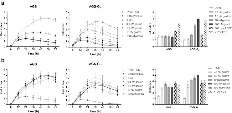
xCELLigence proliferation assay of wild-type AGS and AGS-GR cells in response to gastrin. AGS and AGS-GR cells were seeded in 96-well E-plates for xCELLigence assay monitoring impedance (cell index). The cells were treated with either 10% FCS or 100 ng/ml EGF as controls or gastrin at different concentrations (0.1, 1, 10, or 100 nM) in either absence or presence of serum. (a) Complete growth curves of AGS and AGS-GR cells from 0 to 70 h after seeding. The cells were treated with gastrin and EGF in absence of serum. Panel to the right shows bar graph of cell index at 24 h growth. (b) Complete growth curves of AGS and AGS-GR cells from 0 to 70 h after seeding. The cells were treated with gastrin and EGF in presence of serum. Panel to the right shows bar graph of cell index at 24 h growth. Data is represented as mean ± SD of three independent experiments.
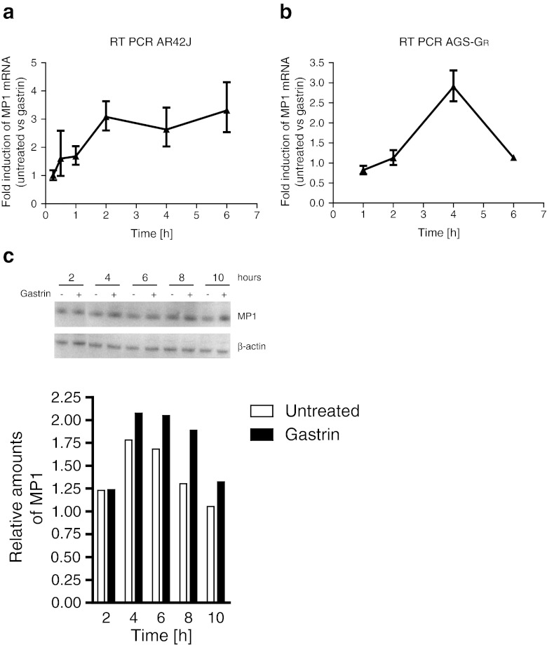
Gastrin-mediated activation of MP1 in AR42J and AGS-GR cells. (a) Verification of gene expression microarray results with quantitative RT-PCR in AR42J. Cells were seeded in 6-well plates and serum starved for 24 h the following day prior to treatment with gastrin (10 nM) as indicated in figure. (b) Quantitative RT-PCR analysis of MP1 expression in AGS-GR. Cells were seeded in 6-well plates and cultivated 24 h prior to gastrin treatment (10 nM) as indicated in figure. Fold induction levels were calculated using the ΔΔCt-method (Livak and Schmittgen 2001), where the expression levels were normalized to the level of β-actin (AR42J) or GAPDH (AGS-GR) expression, and gastrin-treated cells were compared to untreated cells. One representative experiment is shown and data presented as mean ± SD of three technical replicates. (c) Western blot of AGS-GR lysates from cells treated with 10 nM of gastrin 2–10 h before harvesting. The blot was treated with non-commercial antibodies against MP1, and β-actin was used as loading control. Lower panels show relative quantification of band intensities from western blot analysis.
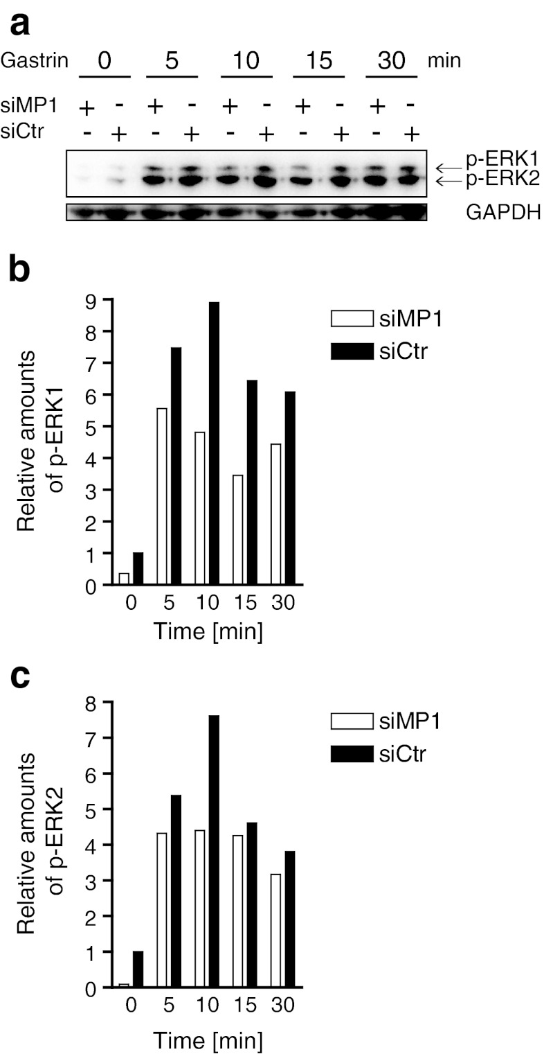
MP1 depletion reduces gastrin-induced phosphorylation of ERK1 and ERK2 (p-ERK1/2). (a) AGS-GR cells were transfected with siMP1 and siCtr for 24 h. Then, the cells were serum starved for 24 h and treated with 10 nM gastrin for 5 to 30 min before harvesting. Protein extracts were subjected to SDS-PAGE and analyzed for p-ERK1 and p-ERK2 in addition to GAPDH (loading control). (b, c) Densiometric analysis of relative amounts of p-ERK1 and p-ERK2, respectively. One representative experiment is shown. Similar results were obtained three times.
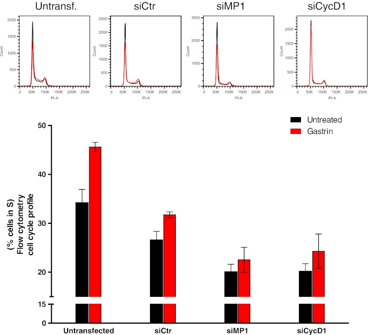
MP1 enhances cell cycle progression. AGS-GR cells were grown and treated with siCtr, siMP1, or siCycD1. Cells were treated with 10 nM gastrin without medium replacement 24 h after transfection and then grown for another 24 h. Finally, the cells were harvested in citrate buffer and treated with buffers A, B, and C according to a slightly modified version of the Vindelov protocol (Vindelov et al. 1983). The proportion of cells in S-phase of the cell cycle in untreated (black) and gastrin-treated (red) bars. DNA histograms were created from 20,000 propidium iodide stained nuclei. Representative histograms are shown, and data is presented as percent cells in S-phase mean ± SD (n = 3). Using qRT-PCR, MP1 depletion was found to be approximately 90% (data not shown).
Similar articles
-
Regulation of protein phosphorylation within the MKK1-ERK2 complex by MP1 and the MP1*P14 heterodimer.Arch Biochem Biophys. 2007 Apr 1;460(1):85-91. doi: 10.1016/j.abb.2006.11.031. Epub 2007 Jan 4. Arch Biochem Biophys. 2007. PMID: 17254543 Free PMC article.
-
BCL2 induced by LAMTOR3/MAPK is a druggable target of chemoradioresistance in mesenchymal lung cancer.Cancer Lett. 2017 Sep 10;403:48-58. doi: 10.1016/j.canlet.2017.05.019. Epub 2017 Jun 10. Cancer Lett. 2017. PMID: 28606806
-
Gastrin stimulates MMP-1 expression in gastric epithelial cells: putative role in gastric epithelial cell migration.Am J Physiol Gastrointest Liver Physiol. 2015 Jul 15;309(2):G78-86. doi: 10.1152/ajpgi.00084.2015. Epub 2015 May 14. Am J Physiol Gastrointest Liver Physiol. 2015. PMID: 25977510 Free PMC article.
-
MEK partner 1 (MP1): regulation of oligomerization in MAP kinase signaling.J Cell Biochem. 2005 Mar 1;94(4):708-19. doi: 10.1002/jcb.20344. J Cell Biochem. 2005. PMID: 15547943
-
ERK1/2 MAP kinases: structure, function, and regulation.Pharmacol Res. 2012 Aug;66(2):105-43. doi: 10.1016/j.phrs.2012.04.005. Epub 2012 Apr 27. Pharmacol Res. 2012. PMID: 22569528 Review.
Cited by
-
The duration of gastrin treatment affects global gene expression and molecular responses involved in ER stress and anti-apoptosis.BMC Genomics. 2013 Jun 28;14:429. doi: 10.1186/1471-2164-14-429. BMC Genomics. 2013. PMID: 23805861 Free PMC article.
-
Optimization of in vitro trophoblast assay for real-time impedimetric sensing of trophoblast-erythrocyte interactions in Plasmodium falciparum malaria.Anal Bioanal Chem. 2020 Jun;412(16):3915-3923. doi: 10.1007/s00216-020-02413-1. Epub 2020 Jan 27. Anal Bioanal Chem. 2020. PMID: 31989195 Free PMC article.
-
Salt-inducible kinase 1 (SIK1) is induced by gastrin and inhibits migration of gastric adenocarcinoma cells.PLoS One. 2014 Nov 10;9(11):e112485. doi: 10.1371/journal.pone.0112485. eCollection 2014. PLoS One. 2014. PMID: 25384047 Free PMC article.
-
Clinical-grade human dental pulp stem cells suppressed the activation of osteoarthritic macrophages and attenuated cartilaginous damage in a rabbit osteoarthritis model.Stem Cell Res Ther. 2021 May 1;12(1):260. doi: 10.1186/s13287-021-02353-2. Stem Cell Res Ther. 2021. PMID: 33933140 Free PMC article.
-
Gastrin Enhances Autophagy and Promotes Gastric Carcinoma Proliferation via Inducing AMPKα.Oncol Res. 2017 Sep 21;25(8):1399-1407. doi: 10.3727/096504016X14823648620870. Epub 2017 Jan 5. Oncol Res. 2017. PMID: 28059052 Free PMC article.
References
-
- Detjen K, Yule D, Tseng MJ, Williams JA, Logsdon CD. CCK-B receptors produce similar signals but have opposite growth effects in CHO and Swiss 3T3 cells. Am J Physiol. 1997b;273:C1449–57. - PubMed
Publication types
MeSH terms
Substances
Associated data
- Actions
LinkOut - more resources
Full Text Sources
Other Literature Sources
Miscellaneous
