Case Reports
. 2012 Nov;9(4):223-6.
doi: 10.5812/iranjradiol.8759. Epub 2012 Nov 20.

Extramedullary plasmacytoma presenting as a solitary mass in the intracranial posterior fossa

Affiliations
Case Reports

Extramedullary plasmacytoma presenting as a solitary mass in the intracranial posterior fossa

Mohammad Hossein Daghighi et al. Iran J Radiol. 2012 Nov.

Abstract

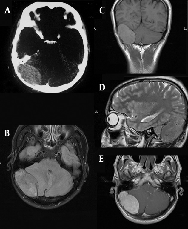
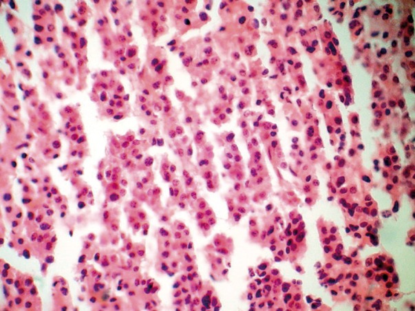
A patient with a 3-month history of headache refractory to pain medication was admitted. The CT scan and MRI showed evidence of a posterior fossa mass. This was pathologically confirmed as an extra medullary plasmacytoma (EMP). He had a pathologic fracture of the left humerus 7 years ago while the radiologist was unaware at the time of diagnosis. A solitary bone plasmacytoma (SBP) was the cause of the pathologic fracture. This report includes the first description of MRI findings in a patient with a rare-incidence intracranial solitary extra medullary plasmacytoma (SEP) in Iran. There is a striking similarity between the features of intracranial SEP and meningiomas. Intracranial SEP, although rare, should be included in the differential diagnosis of brain tumors in areas where meningiomas commonly arise. The MRI findings and differential diagnosis of plasmacytoma are reviewed. Before this case report, only few cases have been reported in the literature. Nonetheless, this is the first report of posterior fossa EMP from Iran.

Keywords: Magnetic Resonance Imaging; Plasmacytoma; Posterior Fossa.


Figures

Figure 1
Figure 1. A 37-year-old man with headache, large extraaxial iso-signal space occupying lesion on the right side of the posterior fossa with erosion of the internal table and compressive effect to the right cerebellar hemisphere and marked enhancement. A, CT image with erosion of the internal table; B, Axial FLAIR MRI with isosignal extra axial SOL at the right side of the posterior fossa; C, Coronal T1 WI without contrast; D, Sagittal T2 WI; E. Axial post-contrast T1 WI.
Figure 2
Figure 2. Diffuse infiltration of medium-sized plasma cells with the characteristic of mild pleomorphic round oval cells, eccentric nuclei and moderate cytoplasm on H&E staining indicating a plasmacytoma.

References

    1. Krumholz A, Weiss HD, Jiji VH, Bakal D, Kirsh MB. Solitary intracranial plasmacytoma: two patients with extended follow-up. Ann Neurol. 1982;11(5):529–32. doi: 10.1002/ana.410110514. - DOI - PubMed
    1. Susnerwala SS, Shanks JH, Banerjee SS, Scarffe JH, Farrington WT, Slevin NJ. Extramedullary plasmacytoma of the head and neck region: clinicopathological correlation in 25 cases. Br J Cancer. 1997;75(6):921–7. doi: 10.1038/bjc.1997.162. - DOI - PMC - PubMed
    1. Straetmans J, Stokroos R. Extramedullary plasmacytomas in the head and neck region. Eur Arch Otorhinolaryngol. 2008;265(11):1417–23. doi: 10.1007/s00405-008-0613-0. - DOI - PubMed
    1. Ching AS, Khoo JB, Chong VF. CT and MR imaging of solitary extramedullary plasmacytoma of the nasal tract. AJNR Am J Neuroradiol. 2002;23(10):1632–6. - PMC - PubMed
    1. Galieni P, Cavo M, Pulsoni A, Avvisati G, Bigazzi C, Neri S, et al. Clinical outcome of extramedullary plasmacytoma. Haematologica. 2000;85(1):47–51. - PubMed

Publication types

LinkOut - more resources