Review
. 2013 May 1;98(2):240-7.
doi: 10.1093/cvr/cvt024. Epub 2013 Feb 12.

'Ryanopathy': causes and manifestations of RyR2 dysfunction in heart failure

Affiliations
Review

'Ryanopathy': causes and manifestations of RyR2 dysfunction in heart failure

Andriy E Belevych et al. Cardiovasc Res. .

Abstract

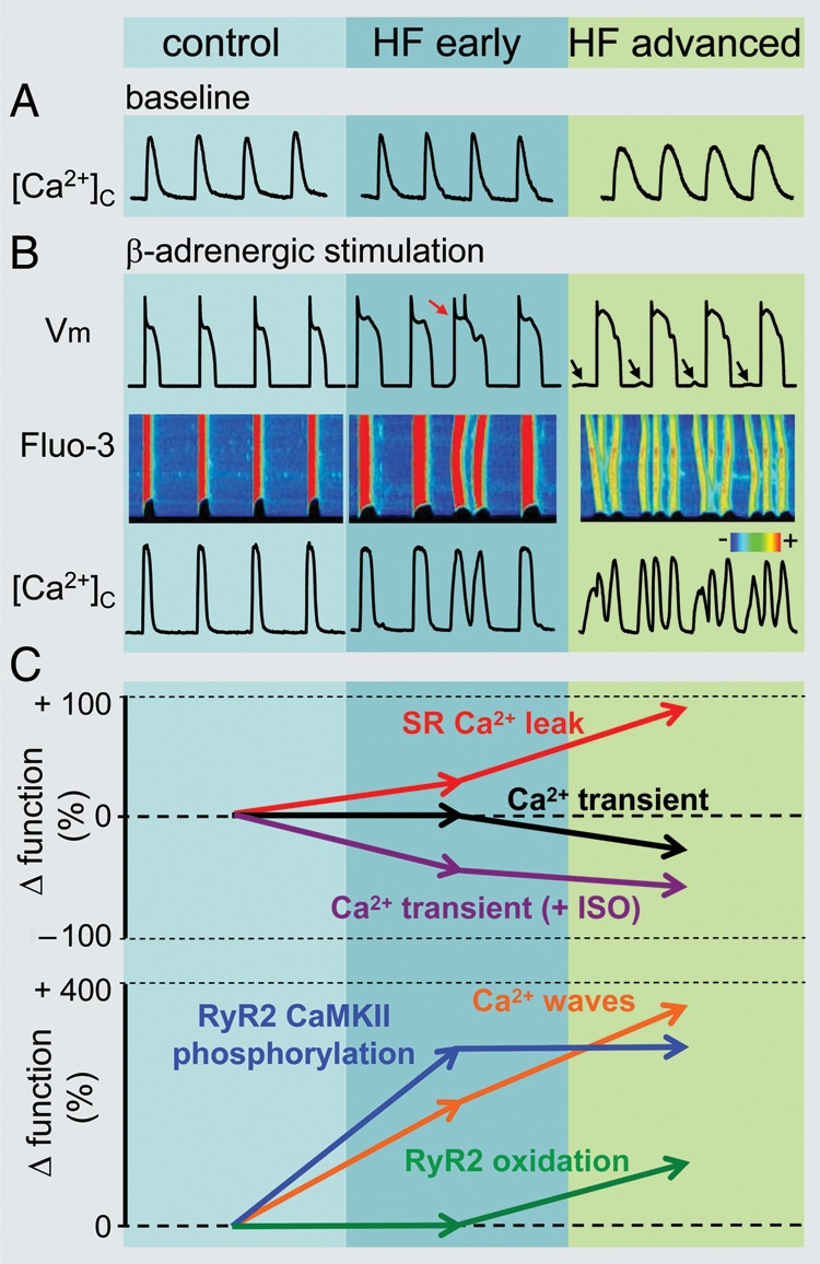
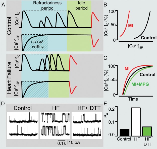
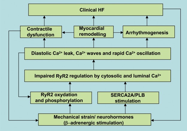
The cardiac ryanodine receptor (RyR2), a Ca(2+) release channel on the membrane of the sarcoplasmic reticulum (SR), plays a key role in determining the strength of the heartbeat by supplying Ca(2+) required for contractile activation. Abnormal RyR2 function is recognized as an important part of the pathophysiology of heart failure (HF). While in the normal heart, the balance between the cytosolic and intra-SR Ca(2+) regulation of RyR2 function maintains the contraction-relaxation cycle, in HF, this behaviour is compromised by excessive post-translational modifications of the RyR2. Such modification of the Ca(2+) release channel impairs the ability of the RyR2 to properly deactivate leading to a spectrum of Ca(2+)-dependent pathologies that include cardiac systolic and diastolic dysfunction, arrhythmias, and structural remodelling. In this article, we present an overview of recent advances in our understanding of the underlying causes and pathological consequences of abnormal RyR2 function in the failing heart. We also discuss the implications of these findings for HF therapy.


Figures

Figure 1.
Figure 1.
Remodelling of intracellular Ca2+cycling during heart failure (HF) progression. (A) Representative Ca2+ transients recorded in control as well as in ventricular myocytes isolated from hearts with early and advanced stages of HF. (B) Representative action potential (AP) traces (top) along with corresponding line scan images of Fluo-3 fluorescence (middle) and Ca2+ transients ([Ca2+]c; bottom) recorded during β-adrenergic receptor stimulation in the three different types of cardiomyocytes. Arrows indicate delayed after-depolarizations. Red arrow points to extrasystolic AP. (C) Summary graphs illustrate that progression of HF is associated with early and progressive increase in the rate of SR Ca2+ leak (red line). The amplitude of Ca2+ transients recorded under baseline conditions (black line) decreases with significant time-delay in respect to elevation of leak during the onset of HF. Such relationships are attributable to the capacity of myocyte Ca2+ handling for autoregulation until SR is depleted by a drastic increase in the SR Ca2+ leak (see text for detailed explanation). In contrast, the frequency of diastolic Ca2+ waves recorded in the presence of β-adrenergic agonist isoproterenol (ISO) (brown line) parallels increases in the SR Ca2+ leak. Ca2+ waves facilitate diastolic SR Ca2+ loss resulting in decrease in Ca2+ transient amplitude (+ISO; purple line). Note that in advanced stages of HF persistent cytosolic Ca2+ oscillations effectively uncouple electrical excitation from mechanical response. Progressive alterations in Ca2+ cycling in HF are coupled to sequential modification of RyR2 by CaMKII-dependent phosphorylation (blue line) and oxidation (green line). Adapted from Belevych et al.
Figure 2.
Figure 2.
Shortened Ca2+ signalling refractoriness in heart disease. (A) Schematic illustration of time-dependent processes occurring between systolic SR Ca2+ depletion and onset of diastolic spontaneous Ca2+ wave (DCW). This latency is composed of a refractory period, reflecting recovery from store-dependent deactivation, and an idle period required for the transition of stochastic release events through functionally recovered RyR2s to regenerating Ca2+ waves. A shorter time delay between systolic Ca2+ release and DCW in diseased myocytes is attributed to reduced refractoriness of the SR Ca2+ release determined by recording the restitution of the Ca2+ transient amplitude measured with a two-pulse protocol. (B) Shortened refractoriness in post-myocardial infarction (MI) myocytes is associated with altered regulation of Ca2+ release by SR luminal Ca2+ resulting in a diminished capability of reduced SR Ca2+ to inhibit RyR2 activity during diastole. Fast restitution of Ca2+ transient (C) and abnormally high activity of RyR2 (D) observed in diseased hearts can be normalized by treatment with reducing agents monopropyonylglycine (MPG; C) and dithiothreitol (DTT; D and E), respectively. Adapted from Belevych et al., and Terentyev et al.
Figure 3.
Figure 3.
Flow diagram summarizing the events involved in time and Ca2+-dependent progression to heart failure (HF). Initial myocardial injury/stress via post-translational modifications of RyR2 and stimulation of SERCA2a activity leads to abnormal regulation of RyR2 by cytosolic and luminal Ca2+. Dysregulated intracellular Ca2+ handling results in myocardial contractile dysfunction, increased arrhythmogenesis, and altered intracellular metabolic and cell survival pathways.

References

    1. Fabiato A. Calcium-induced release of calcium from the cardiac sarcoplasmic reticulum. Am J Physiol. 1983;245:C1–C14. - PubMed
    1. Bers DM. Excitation-Contraction Coupling and Cardiac Contractile Force. Dordrecht, Boston: Kluwer Academic Publishers; 2001.
    1. Cheng H, Lederer WJ. Calcium sparks. Physiol Rev. 2008;88:1491–1545. - PubMed
    1. Li P, Chen SR. Molecular basis of Ca2+ activation of the mouse cardiac Ca2+ release channel (ryanodine receptor) J Gen Physiol. 2001;118:33–44. - PMC - PubMed
    1. Cheng H, Lederer MR, Lederer WJ, Cannell MB. Calcium sparks and [Ca2+]i waves in cardiac myocytes. Am J Physiol. 1996;270:C148–C159. - PubMed

Publication types

MeSH terms

Substances