. 2013 Mar 29;288(13):9396-407.
doi: 10.1074/jbc.M112.395913. Epub 2013 Feb 13.

The oncogene metadherin modulates the apoptotic pathway based on the tumor necrosis factor superfamily member TRAIL (Tumor Necrosis Factor-related Apoptosis-inducing Ligand) in breast cancer

Affiliations

The oncogene metadherin modulates the apoptotic pathway based on the tumor necrosis factor superfamily member TRAIL (Tumor Necrosis Factor-related Apoptosis-inducing Ligand) in breast cancer

Ning Zhang et al. J Biol Chem. .

Abstract

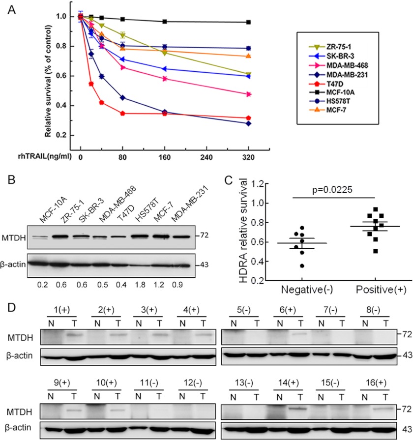
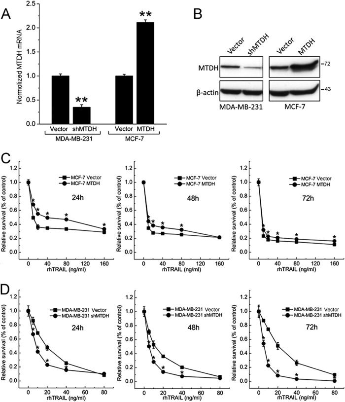
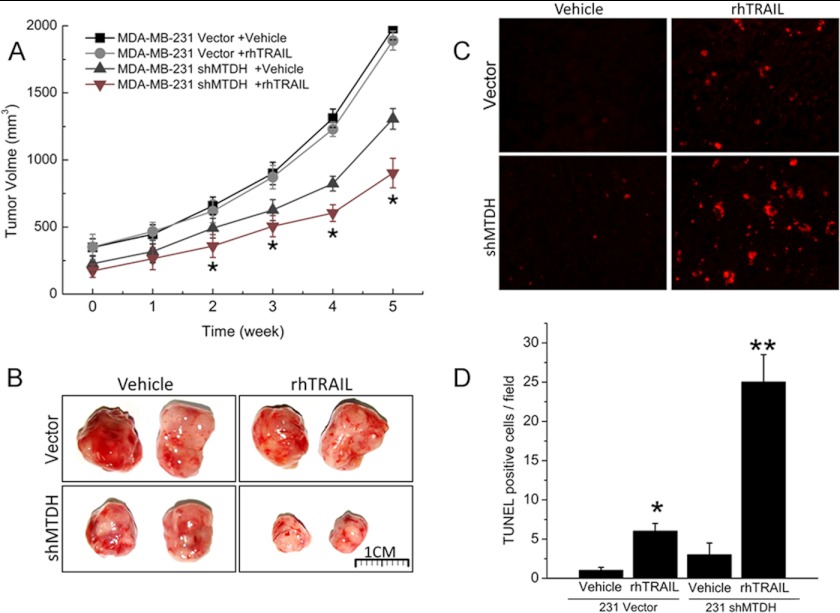
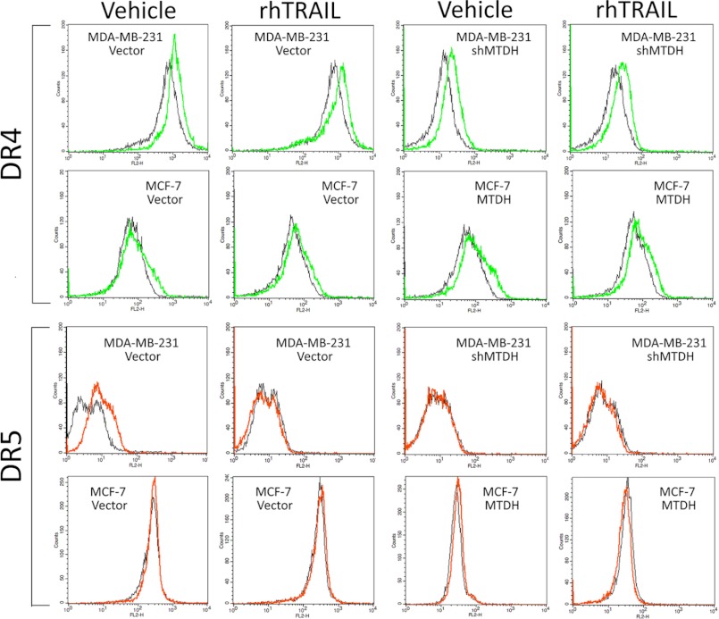
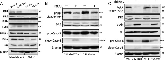
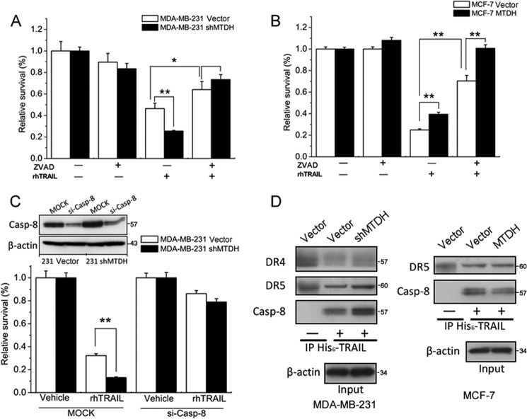
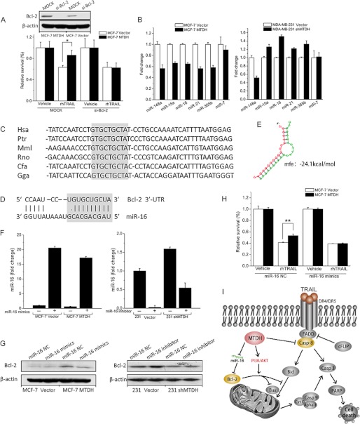
Metadherin (MTDH), the newly discovered gene, is overexpressed in more than 40% of breast cancers. Recent studies have revealed that MTDH favors an oncogenic course and chemoresistance. With a number of breast cancer cell lines and breast tumor samples, we found that the relative expression of MTDH correlated with tumor necrosis factor-related apoptosis-inducing ligand (TRAIL) sensitivity in breast cancer. In this study, we found that knockdown of endogenous MTDH cells sensitized the MDA-MB-231 cells to TRAIL-induced apoptosis both in vitro and in vivo. Conversely, stable overexpression of MTDH in MCF-7 cells enhanced cell survival with TRAIL treatment. Mechanically, MTDH down-regulated caspase-8, decreased caspase-8 recruitment into the TRAIL death-inducing signaling complex, decreased caspase-3 and poly(ADP-ribose) polymerase-2 processing, increased Bcl-2 expression, and stimulated TRAIL-induced Akt phosphorylation, without altering death receptor status. In MDA-MB-231 breast cancer cells, sensitization to TRAIL upon MTDH down-regulation was inhibited by the caspase inhibitor Z-VAD-fmk (benzyloxycarbonyl-VAD-fluoromethyl ketone), suggesting that MTDH depletion stimulates activation of caspases. In MCF-7 breast cancer cells, resistance to TRAIL upon MTDH overexpression was abrogated by depletion of Bcl-2, suggesting that MTDH-induced Bcl-2 expression contributes to TRAIL resistance. We further confirmed that MTDH may control Bcl-2 expression partly by suppressing miR-16. Collectively, our results point to a protective function of MTDH against TRAIL-induced death, whereby it inhibits the intrinsic apoptosis pathway through miR-16-mediated Bcl-2 up-regulation and the extrinsic apoptosis pathway through caspase-8 down-regulation.


Figures

FIGURE 1.
FIGURE 1.
Expression of MTDH correlates with TRAIL resistance in breast cancer cell lines and breast tumor samples. A, relative survival of each breast cell line was tested with MTT assay after a 48-h treatment with rhTRAIL. Points are the average of three independent experiments; error bars represent S.E. B, expression level of MTDH was compared with Western blotting among these cell lines. β-Actin was used as the endogenous control. C, the sensitivity to rhTRAIL studied in the MTDH-negative (−) group was significantly higher than that in the positive (+) group (p = 0.0225). D, the expression of MTDH was determined with Western blotting, and β-actin was used as the loading control. N, paired normal tissue; T, tumor tissues.
FIGURE 2.
FIGURE 2.
Stable overexpression and knockdown of MTDH in breast cancer cell lines and MTDH contributes to TRAIL resistance in vitro. A, stable MDA-MB-231 cells expressing an MTDH-targeting shRNA (shMTDH) or the control pSUPER.retro.puro empty vector (Vector) (left) and stable MCF-7 cells transfected with an MTDH expression plasmid (MTDH) or the control pcDNA3.1 empty vector (Vector) (right) were analyzed for MTDH mRNA levels by real-time PCR after 2 weeks of selection. Columns represent average of three independent experiments; error bars represent S.E.; *, p < 0.05; **, p < 0.01 versus cells transfected with control vector. B, the transfection efficiency was confirmed by Western blot analysis. Cell extracts were prepared for Western blotting of MTDH, and β-actin was used as the endogenous control. The figure shown is representative of three independent experiments. C, MCF-7 vector cells and MCF-7 MTDH cells were treated with TRAIL for the indicated time points. The effect of MTDH on TRAIL-induced apoptosis was measured at different doses of TRAIL by the MTT assay and was plotted as a percentage of change relative to the control. D, sensitization of the MDA-MB-231 cell line by MTDH knockdown (shMTDH) was measured at different doses of TRAIL by the MTT assay. Points are the average of three independent experiments; error bars represent S.E.; *, p < 0.05; **, p < 0.01.
FIGURE 3.
FIGURE 3.
MTDH contributes to TRAIL resistance in vivo. A, MDA-MB-231 vector and MDA-MB-231 shMTDH cells were injected subcutaneously into each flank of BALB/c nu/nu mice. Three weeks later, mice were treated intraperitoneally with the drug vehicle or with rhTRAIL at 7.5 mg/kg, four times per week. Tumor volume was measured weekly by direct caliper. Error bars represent S.E.; *, p < 0.05 versus control vehicle-treated mice. B, representative tumors isolated from mice 5 weeks after implantation with control (Vector) or MTDH-depleted (shMTDH) MDA-MB-231 cells and treated with rhTRAIL or vehicle as described in panel A. C, representative TUNEL staining (red fluorescence) of MDA-MB-231 breast cancer xenografts shown in panel B. D, quantification of terminal deoxynucleotidyl transferase-mediated dUTP nick end labeling-positive cells. Columns represent average of three independent experiments; error bars represent S.E.; *, p < 0.05; **, p < 0.01, significantly different from respective control.
FIGURE 4.
FIGURE 4.
Flow cytometry analysis of surface protein levels of DR4 and DR5 after MTDH overexpression and knockdown. MTDH overexpression or knockdown did not alter surface DR4 (top panel) and DR5 (bottom panel) protein levels in the presence or absence of TRAIL or vehicle, as determined by flow cytometry. IgG was used as the negative control.
FIGURE 5.
FIGURE 5.
MTDH does not regulate DR or c-FLIP protein levels but increases Bcl-2 and decreases caspase-8 expression levels. A, expression of the indicated proteins was examined in MTDH knockdown (shMTDH) or overexpressing (MTDH) cells, and in their respective controls (Vector), by Western blot analysis. The blot for β-actin was performed to confirm equal loading of samples. casp-8, caspase-8. B, MDA-MB-231 vector and shMTDH cells were treated for 12 h with (+) or without (−) TRAIL at 40 ng/ml and then analyzed by Western blotting for the indicated proteins. C, MCF-7 cells stably expressing MTDH (MTDH) and their control (Vector) were treated for 24 h with (+) or without (−) TRAIL at 100 ng/ml and analyzed by Western blotting for the indicated proteins. p-Akt: phospho-Akt.
FIGURE 6.
FIGURE 6.
Inhibition of TRAIL-induced apoptosis depends on the down-regulation of caspase-8 and on decreased recruitment of caspase-8 into the DISC. A, MDA-MB-231 vector cells and MDA-MB-231 shMTDH cells were treated with 100 μm Z-VAD-fmk (ZVAD). One hour later, cells were treated with (+) or without (−) TRAIL at 20 ng/ml for an additional 24 h. Cell viability was quantified with the MTT assay. B, MCF-7 vector and MCF-7 MTDH cells were pretreated for 1 h with vehicle or 100 μm Z-VAD-fmk μm and then exposed to vehicle or TRAIL at 80 ng/ml for an additional 24 h. Cell viability was monitored with the MTT assay. C, MDA-MB-231 vector cells and MDA-MB-231 shMTDH cells were transfected with control (MOCK) or caspase-8 siRNA (si-Casp-8). After 2 days, cells were treated for 24 h with vehicle or TRAIL at 20 ng/ml. The efficiency of caspase-8 (Casp-8) knockdown was assessed by Western blotting using β-actin as a loading control. Cell viability was quantified with the MTT assay. D, MDA-MB-231 vector or shMTDH cells (left panel) and MCF-7 vector or MTDH cells (right panel) were stimulated with His6-tagged TRAIL for 30 min. Cells were lysed, His6-TRAIL was immunoprecipitated (IP) with anti-His mouse mAb, and the co-immunoprecipitated DR4, DR5, or caspase-8 was detected by Western blotting. The negative control (−) was processed identically, except that TRAIL was added to cells after lysis. β-Actin was probed in cell lysates to ensure that similar amounts of lysate were used in the immunoprecipitation. A–C, columns represent average of three independent experiments; error bars represent S.E.; *, p < 0.05,**, p < 0.01 when compared with controls.
FIGURE 7.
FIGURE 7.
MTDH up-regulates Bcl-2 by reducing expression of microRNA-16. A, MCF-7 vector or MTDH cells were transfected with either control (MOCK) or Bcl-2 siRNAs (si-Bcl-2). After 2 days, cells were treated with vehicle or TRAIL at 20 ng/ml, as indicated. The transient knockdown of caspase-8 was validated by Western blotting, using β-actin as loading control. B, the abundance of miR-148a, miR-15a, miR-16, miR-21, miR-365b, and miR-7 was monitored by real-time PCR in MCF-7 vector and MTDH cells and MDA-MB-231 vector and shMTDH cells. Fold change represents the relative quantification of each microRNA versus control cells. C, the alignment of the miR-16 targeting sequences located in the 3′-untranslated region of the Bcl-2 genes from six organisms (Hsa, human; Ptr, chimpanzee; Mml, mouse; Rno, rat; Cfa, dog; Gga, chicken). The evolutionarily conserved nucleotides are highlighted in light gray. D, the predicted miR-16 targeting sequence located in the 3′-untranslated region (3′-UTR) of Bcl-2 mRNA. The seed region is highlighted in light gray. E, structure of predicted duplex. Minimum free energy (mfe) is −24.1 kcal/mol. F, MCF-7 vector and MTDH cells were transfected with 50 nm miR-16 mimics (+) or with a control oligonucleotide (−). MDA-MB-231 vector and shMTDH cells were transiently transfected with 50 nm miR-16 inhibitor (+) or with a control oligonucleotide (−). miR-16 expression levels monitored by real-time PCR were normalized either to expression levels in MCF-7 vector cells or to those in MDA-MB-231 vector cells. G, the MCF-7 and MDA-MB-231 cell lines were treated as described in panel F. Cell lysates were analyzed by Western blotting to detect the effect of miR-16 on the expression of Bcl-2. NC: negative control. H, MCF-7 vector or MTDH cells were transfected with either miR-16 mimic control or miR-16 mimics. After 2 days, cells were treated with vehicle or TRAIL at 40 ng/ml. I, a schematic model of the mechanisms through which MTDH modulates TRAIL-induced apoptosis in breast cancer cells. FADD, Fas-Associated protein with death domain; Casp-8, caspase-8; PARP, poly(ADP-ribose) polymerase-2. In A, B, F, and H, columns represent average of three independent experiments; error bars represent S.E.; *, p < 0.05.

References

    1. Wiley S. R., Schooley K., Smolak P. J., Din W. S., Huang C. P., Nicholl J. K., Sutherland G. R., Smith T. D., Rauch C., Smith C. A., et al. (1995) Identification and characterization of a new member of the TNF family that induces apoptosis. Immunity 3, 673–682 - PubMed
    1. Pitti R. M., Marsters S. A., Ruppert S., Donahue C. J., Moore A., Ashkenazi A. (1996) Induction of apoptosis by Apo-2 ligand, a new member of the tumor necrosis factor cytokine family. J. Biol. Chem. 271, 12687–12690 - PubMed
    1. Ashkenazi A., Pai R. C., Fong S., Leung S., Lawrence D. A., Marsters S. A., Blackie C., Chang L., McMurtrey A. E., Hebert A., DeForge L., Koumenis I. L., Lewis D., Harris L., Bussiere J., Koeppen H., Shahrokh Z., Schwall R. H. (1999) Safety and antitumor activity of recombinant soluble Apo2 ligand. J. Clin. Invest. 104, 155–162 - PMC - PubMed
    1. Walczak H., Miller R. E., Ariail K., Gliniak B., Griffith T. S., Kubin M., Chin W., Jones J., Woodward A., Le T., Smith C., Smolak P., Goodwin R. G., Rauch C. T., Schuh J. C., Lynch D. H. (1999) Tumoricidal activity of tumor necrosis factor-related apoptosis-inducing ligand in vivo. Nat. Med. 5, 157–163 - PubMed
    1. Newsom-Davis T., Prieske S., Walczak H. (2009) Is TRAIL the holy grail of cancer therapy? Apoptosis 14, 607–623 - PubMed

Publication types

MeSH terms