. 2013 Mar 29;288(13):9408-17.
doi: 10.1074/jbc.M112.419861. Epub 2013 Feb 13.

Identification of small molecule inhibitors of Jumonji AT-rich interactive domain 1B (JARID1B) histone demethylase by a sensitive high throughput screen

Affiliations

Identification of small molecule inhibitors of Jumonji AT-rich interactive domain 1B (JARID1B) histone demethylase by a sensitive high throughput screen

Joyce Sayegh et al. J Biol Chem. .

Abstract

JARID1B (also known as KDM5B or PLU1) is a member of the JARID1 family of histone lysine demethylases responsible for the demethylation of trimethylated lysine 27 in histone H3 (H3K4me3), a mark for actively transcribed genes. JARID1B is overexpressed in several cancers, including breast cancer, prostate cancer, and lung cancer. In addition, JARID1B is required for mammary tumor formation in syngeneic or xenograft mouse models. JARID1B-expressing melanoma cells are associated with increased self-renewal character. Therefore, JARID1B represents an attractive target for cancer therapy. Here we characterized JARID1B using a homogeneous luminescence-based demethylase assay. We then conducted a high throughput screen of over 15,000 small molecules to identify inhibitors of JARID1B. From this screen, we identified several known JmjC histone demethylase inhibitors, including 2,4-pyridinedicarboxylic acid and catechols. More importantly, we identified several novel inhibitors, including 2-4(4-methylphenyl)-1,2-benzisothiazol-3(2H)-one (PBIT), which inhibits JARID1B with an IC50 of about 3 μm in vitro. Consistent with this, PBIT treatment inhibited removal of H3K4me3 by JARID1B in cells. Furthermore, this compound inhibited proliferation of cells expressing higher levels of JARID1B. These results suggest that this novel small molecule inhibitor is a lead compound that can be further optimized for cancer therapy.


Figures

FIGURE 1.
FIGURE 1.
Screen setup and hit validation strategy. A, schematic of the demethylase assay used for detection of JARID1B demethylase activity. Upon laser excitation, energy is transferred from the streptavidin (Strep)-coated donor beads to the protein A-coated acceptor beads. A luminescence signal is detected at 520–620 nm. Ab, antibody. B, AlphaScreen optimization for antibody specificity. Bio-H3K4me3/2/1 peptides were titrated in the absence of enzyme and detected by the H3K4me1 antibody/bead mix. Error bars indicate S.E. C, overview of the high throughput screen and validation for JARID1B inhibitors.
FIGURE 2.
FIGURE 2.
Analysis of recombinant FLAG-JARID1B by Coomassie Brilliant Blue staining (A) and Western blot analysis (B). FLAG-JARID1B appears as an ∼170-kDa band. FT, flow-through; WB, Western blot.
FIGURE 3.
FIGURE 3.
Characterization of JARID1B. A, enzymatic activity of FLAG-JARID1B (4 nm) as monitored by AlphaScreen signal in the presence and absence of bio-H3K4me3 peptide substrate (64 nm). Bio-H3K4me2 peptide (64 nm) in the absence of enzyme serves as a positive control for the AlphaScreen assay. B, titration and time course of the FLAG-JARID1B. All assays were carried out in triplicate using 64 nm bio-H3K4me3 peptide and 2, 5, or 7.5 nm FLAG-JARID1B. Reactions were quenched with EDTA at various time points. No signal is seen for bio-H3K4me3 peptide assayed in the absence of FLAG-JARID1B, and bio-H3K4me2 (64 nm) assayed in the absence of enzyme represents a positive control for the AlphaScreen assay. C, demethylase activity of FLAG-JARID1B upon titration of the bio-H3K4me3 peptide for Km determination. D, demethylase activity of FLAG-JARID1B on the bio-H3K4me3 peptide substrate upon titration of α-ketoglutarate for Km determination. Error bars in panels A–D indicate S.E.
FIGURE 4.
FIGURE 4.
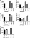
PBIT is selective for JARID1 enzymes. A–C, JARID1B (A), JARID1A (B), and JARID1C (C) were assayed with 64 nm bio-H3K4me3 peptide and PBIT or 2,4-PDCA (10 μm). D and E, UTX (D) and JMJD3 (E) were assayed with 64 nm bio-H3K27me3 peptide and PBIT or 2,4-PDCA (10 μm), and demethylase activity was detected using anti-H3K27me2 antibody. Error bars in panels A–E indicate S.E.
FIGURE 5.
FIGURE 5.
PBIT inhibits H3K4me3 demethylation in vivo. 3×HA-JARID1B was expressed in HeLa cells, and cells were incubated with 0.1% DMSO or 10 or 30 μm PBIT for 24 h. Cell nuclei were identified by DAPI staining (top panel, blue). 3×HA-JARID1B was identified by HA immunofluorescence (second panel, red), and H3K4me3 was visualized by H3K4me3 immunofluorescence (third panel, green). The merged images of HA and H3K4me3 immunofluorescence are shown in the bottom panel. Triangles indicate transfected cells.
FIGURE 6.
FIGURE 6.
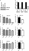
PBIT inhibits cell proliferation in a JARID1B level-dependent manner. A, Western blot analysis of UACC-812, MCF7, and MCF10A cells with the indicated antibodies. B–D, WST-1 cell proliferation assays of UACC-812 (B), MCF7 (C), and MCF10A (D) cells in the presence of PBIT at the indicated concentrations. Shown are the ratio of absorbance at 440 nm of day 3/day 0 (D3/D0) with S.E. E, real time RT-PCR analysis of JARID1B mRNA in stable cell lines with the indicated shRNA hairpins. Shown are mean values with S.E. F–H, WST-1 cell proliferation assays of UACC-812 (F), MCF7 (G), and MCF10A (H) cells with control or JARID1B shRNA hairpins. Shown are the ratio of absorbance at 440 nm of day 3 or 4/day 0 (D3 or D4/D0) with S.E.

References

    1. Blair L. P., Yan Q. (2012) Epigenetic mechanisms in commonly occurring cancers. DNA Cell Biol. 31, Suppl. 1, S49–S61 - PMC - PubMed
    1. Blair L. P., Cao J., Zou M. R., Sayegh J., Yan Q. (2011) Epigenetic regulation by lysine demethylase 5 (KDM5) enzymes in cancer. Cancers 3, 1383–1404 - PMC - PubMed
    1. Klose R. J., Yan Q., Tothova Z., Yamane K., Erdjument-Bromage H., Tempst P., Gilliland D. G., Zhang Y., Kaelin W. G., Jr. (2007) The retinoblastoma binding protein RBP2 is an H3K4 demethylase. Cell 128, 889–900 - PubMed
    1. Lee M. G., Norman J., Shilatifard A., Shiekhattar R. (2007) Physical and functional association of a trimethyl H3K4 demethylase and Ring6a/MBLR, a polycomb-like protein. Cell 128, 877–887 - PubMed
    1. Christensen J., Agger K., Cloos P. A., Pasini D., Rose S., Sennels L., Rappsilber J., Hansen K. H., Salcini A. E., Helin K. (2007) RBP2 belongs to a family of demethylases, specific for tri-and dimethylated lysine 4 on histone 3. Cell 128, 1063–1076 - PubMed

Publication types

MeSH terms