. 2013 Feb 13:7:16.
doi: 10.3389/fncir.2013.00016. eCollection 2013.

Control of breathing by interacting pontine and pulmonary feedback loops

Affiliations

Control of breathing by interacting pontine and pulmonary feedback loops

Yaroslav I Molkov et al. Front Neural Circuits. .

Abstract

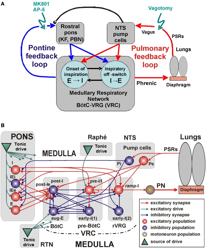
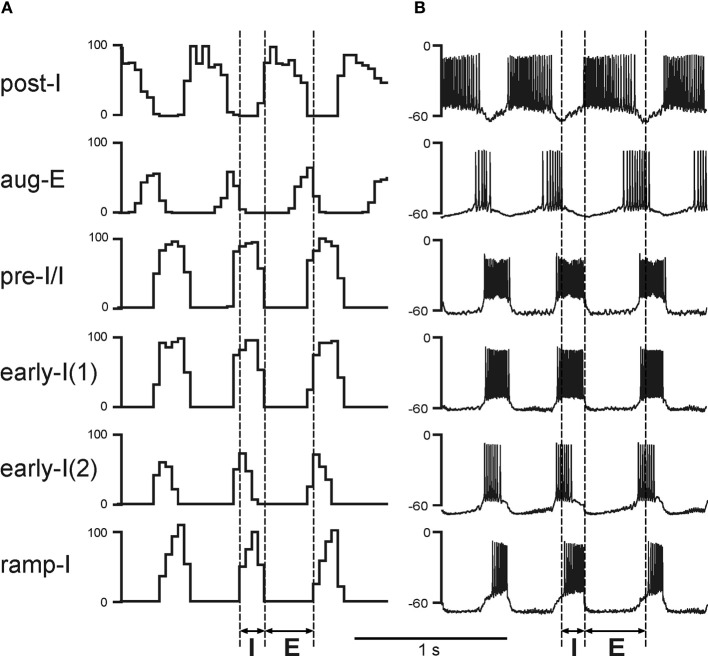
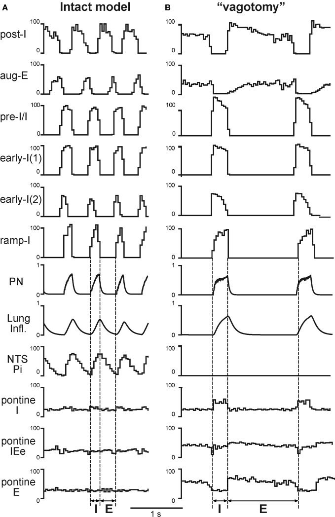
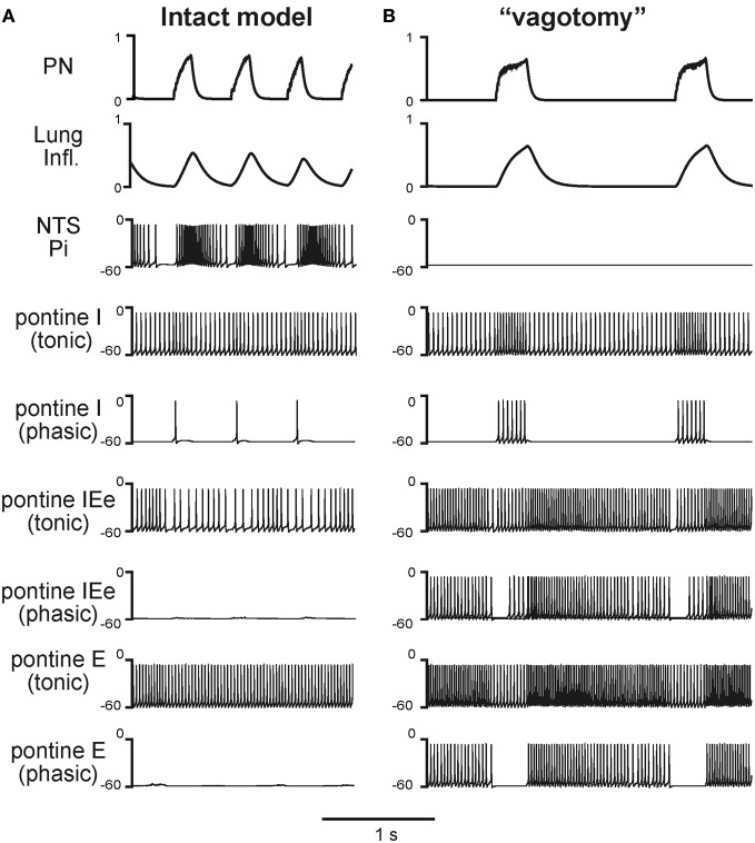
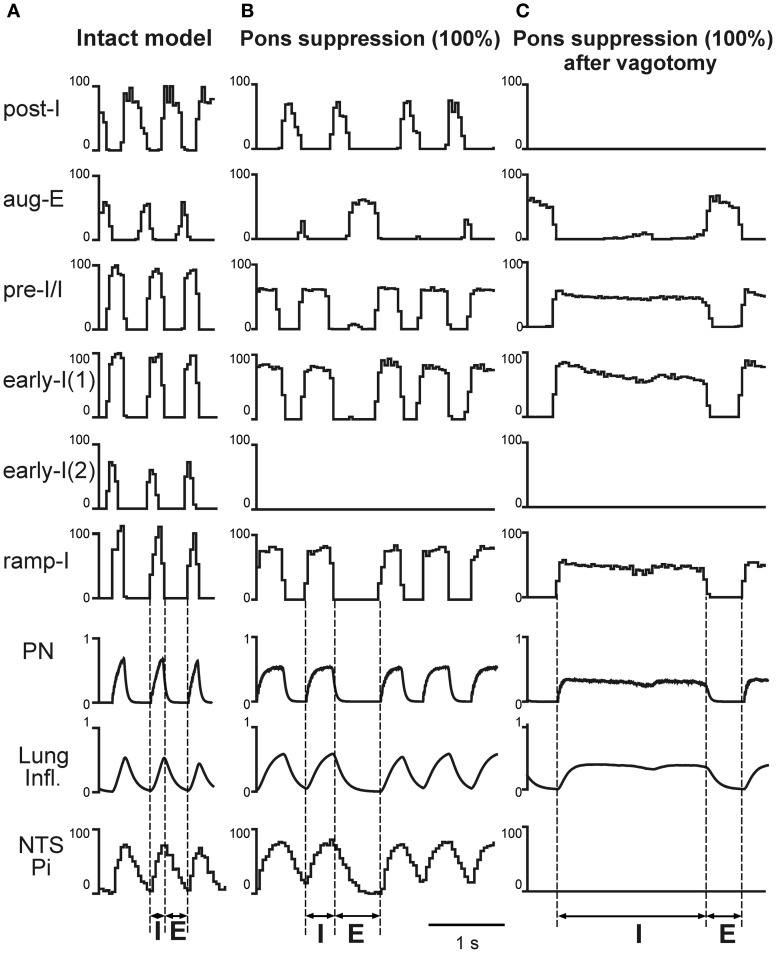
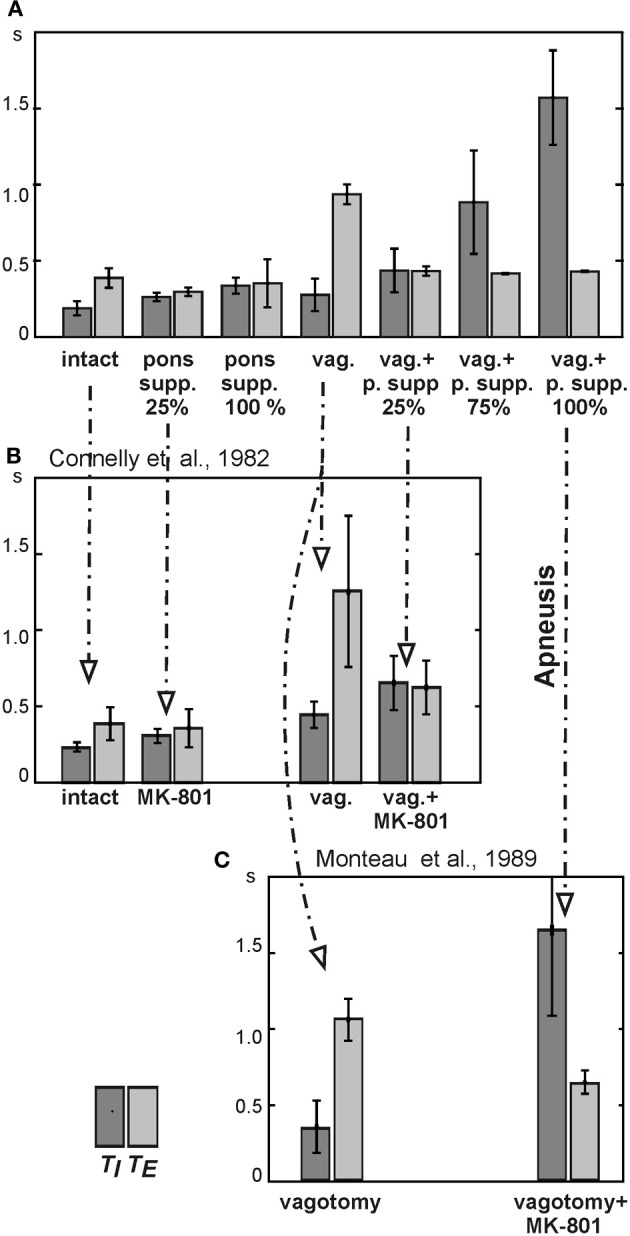
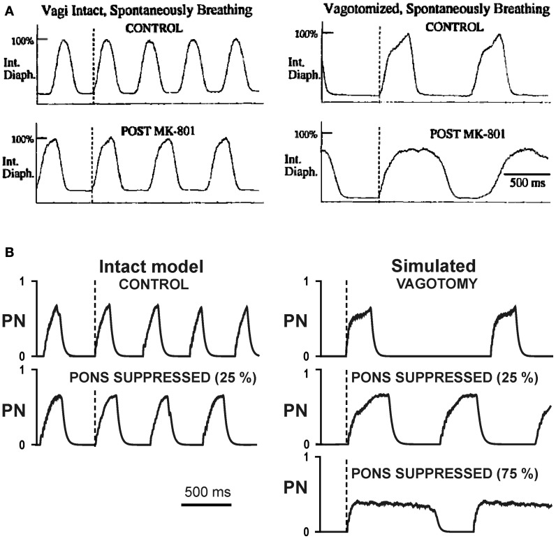
The medullary respiratory network generates respiratory rhythm via sequential phase switching, which in turn is controlled by multiple feedbacks including those from the pons and nucleus tractus solitarii; the latter mediates pulmonary afferent feedback to the medullary circuits. It is hypothesized that both pontine and pulmonary feedback pathways operate via activation of medullary respiratory neurons that are critically involved in phase switching. Moreover, the pontine and pulmonary control loops interact, so that pulmonary afferents control the gain of pontine influence of the respiratory pattern. We used an established computational model of the respiratory network (Smith et al., 2007) and extended it by incorporating pontine circuits and pulmonary feedback. In the extended model, the pontine neurons receive phasic excitatory activation from, and provide feedback to, medullary respiratory neurons responsible for the onset and termination of inspiration. The model was used to study the effects of: (1) "vagotomy" (removal of pulmonary feedback), (2) suppression of pontine activity attenuating pontine feedback, and (3) these perturbations applied together on the respiratory pattern and durations of inspiration (T(I)) and expiration (T(E)). In our model: (a) the simulated vagotomy resulted in increases of both T(I) and T(E), (b) the suppression of pontine-medullary interactions led to the prolongation of T(I) at relatively constant, but variable T(E), and (c) these perturbations applied together resulted in "apneusis," characterized by a significantly prolonged T(I). The results of modeling were compared with, and provided a reasonable explanation for, multiple experimental data. The characteristic changes in T(I) and T(E) demonstrated with the model may represent characteristic changes in the balance between the pontine and pulmonary feedback control mechanisms that may reflect specific cardio-respiratory disorders and diseases.

Keywords: apneusis; brainstem; control of breathing; pontine-medullary interactions; pre-Bötzinger complex; pulmonary feedback; respiratory central pattern generator; ventrolateral respiratory column.


Figures

Figure 1
Figure 1
The medullary respiratory network with pulmonary and pontine feedbacks. (A) A general schematic diagram representing the respiratory network with two interacting feedback. See text for details. (B) The detailing model schematic showing interactions between different populations of respiratory neurons within major brainstem compartments involved in the control of breathing (pons, BötC, pre-BötC, and rVRG) and the organization of pulmonary and pontine feedbacks. Each neural population (shown as a sphere) consists of 50 single-compartment neurons described in the Hodgkin-Huxley style. The model includes 3 sources of tonic excitatory drive located in the pons, RTN, and raphé—all shown as green triangles. These drives, project to multiple neural populations in the model (green arrows; the particular connections to target populations are not shown for simplicity, but are specified in Table A3 in the Appendix). See text for details. Abbreviations: AP-5, amino-5-phosphonovaleric acid, NMDA receptor antagonist; BötC, Bötzinger complex; e, excitatory; E, expiratory or expiration; i, inhibitory; I, inspiratory or inspiration; IE, inspiratory-expiratory; KF, Kölliker-Fuse nucleus; MK801, dizocilpine maleate, NMDA receptor antagonist; NTS, Nucleus Tractus Solitarii; P, pump cells; PBN, ParaBrachial Nucleus; PN, Phrenic Nerve; pre-BötC, pre-Bötzinger Complex; PSRs, pulmonary stretch receptors; RTN, retrotrapezoid nucleus; r, rostral; VRC, ventral respiratory column; VRG, ventral respiratory group.
Figure 2
Figure 2
Performance of the core medullary network under normal conditions (with both feedbacks intact). (A) The activity of main neural populations of the core respiratory network under normal conditions. The shown population activities include (top–down): post-inspiratory (post-I) and augmenting expiratory (aug-E) (both in BötC); pre-inspiratory/ inspiratory (pre-I/I) and early-inspiratory [early-I(1)] (both in pre-BötC); early-inspiratory [early-I(2)] and ramp-inspiratory (ramp-I) (both in rVRG). The activity of each population is represented by the histogram of neuronal firing in the population (spikes/s; bin = 30 ms). (B) Traces of membrane potentials of the corresponding single neurons (randomly selected from each population). Vertical dashed line indicate the inspiratory (I) and expiratory (E) phases.
Figure 3
Figure 3
Simulated vagotomy (removal of the pulmonary feedback). Activity of major VRC (post-I, aug-E, early-I(1), pre-I/I, early-I(1), early-I(2), and ramp-I), NTS (Pi) and pontine (I, IEe, and E) neural populations, lung inflation and PN activity before (A) and after (B) simulated vagotomy. Vertical dashed line indicate the inspiratory (I) and expiratory (E) phases. See text for details.
Figure 4
Figure 4
Respiratory modulation in the activity of pontine neurones before (A) and after (B) simulated vagotomy. The changes of phrenic activity (PN) and the lung inflation are shown at the top. Below these graphs, membrane potentials traces of representative single neurons from the Pi and pontine populations (tonic and phasic subpopulations) are shown. See text for details.
Figure 5
Figure 5
The effects of pontine suppression before and after simulated vagotomy. Activity of major medullary [post-I, aug-E, early-I(1), pre-I/I, early-I(1), early-I(2), and ramp-I], NTS (Pi) and pontine (I, IEe, and E) neural populations, lung inflation and PN activity under control conditions (A) and following the 100% suppression of pontine activity before (B) and after (C) simulated vagotomy. The activity pattern shown in (C) represents typical apneusis. Vertical dashed line indicate the inspiratory (I) and expiratory (E) phases. See text for details.
Figure 6
Figure 6
Changes in the durations of inspiration (TI) and expiration (TE) following pontine suppression and/or vagotomy. (A) Changes in TI and TE following the simulated pontine suppression at different degrees (25%, 75%, and 100%) before and after (vag. +) vagotomy. (B) Changes in TI and TE in the study of Connelly et al. (1992): diagrams are built for spontaneously breathing Wistar rats under control conditions and after administration of NMDA blocker MK-801 before and after vagotomy. (C) Changes in TI and TE in the study of Monteau et al. (1990) performed in anaesthetized vagotomized rats using MK-801 administration.
Figure 7
Figure 7
Changes in the breathing pattern (phrenic activity, PN) following MK-801 application (pontine suppression in the model) before and after vagotomy. (A) Changes in integrated phrenic nerve activity (Int. Diaph.) from spontaneously breathing Wistar rats before (top traces) and after (bottom traces) NMDA channel blockade, before (left diagrams) and after (right diagrams) vagotomy (from Connelly et al., 1992) (B) Changes in integrated phrenic nerve activity (PN) in our simulations before (top traces) and after (bottom traces) simulated pontine suppression, before (left diagrams) and after (right diagrams) simulated vagotomy.

Similar articles

Cited by

References

    1. Abdala A. P., Dutschmann M., Bissonnette J. M., Paton J. F. (2010). Correction of respiratory disorders in a mouse model of Rett syndrome. Proc. Natl. Acad. Sci. U.S.A. 107, 18208–18213 10.1073/pnas.1012104107 - DOI - PMC - PubMed
    1. Abdala A. P., Rybak I. A., Smith J. C., Paton J. F. (2009). Abdominal expiratory activity in the rat brainstem-spinal cord in situ: patterns, origins and implications for respiratory rhythm generation. J. Physiol. 587, 3539–3559 10.1113/jphysiol.2008.167502 - DOI - PMC - PubMed
    1. Anders K., Ohndorf W., Dermietzel R., Richter D. W. (1993). Synapses between slowly adapting lung stretch receptor afferents and inspiratory beta-neurons in the nucleus of the solitary tract of cats: a light and electron microscopic analysis. J. Comp. Neurol. 335, 163–172 10.1002/cne.903350203 - DOI - PubMed
    1. Averill D. B., Cameron W. E., Berger A. J. (1984). Monosynaptic excitation of dorsal medullary respiratory neurons by slowly adapting pulmonary stretch receptors. J. Neurophysiol. 52, 771–785 - PubMed
    1. Backman S. B., Anders C., Ballantyne D., Rohrig N., Camerer H., Mifflin S., et al. (1984). Evidence for a monosynaptic connection between slowly adapting pulmonary stretch receptor afferents and inspiratory beta neurones. Pflugers Arch. 402, 129–136 - PubMed

Publication types

LinkOut - more resources