Review
. 2013 May-Jun;5(3):307-42.
doi: 10.1002/wsbm.1212. Epub 2013 Feb 13.

Lower urinary tract development and disease

Affiliations
Review

Lower urinary tract development and disease

Hila Milo Rasouly et al. Wiley Interdiscip Rev Syst Biol Med. 2013 May-Jun.

Abstract

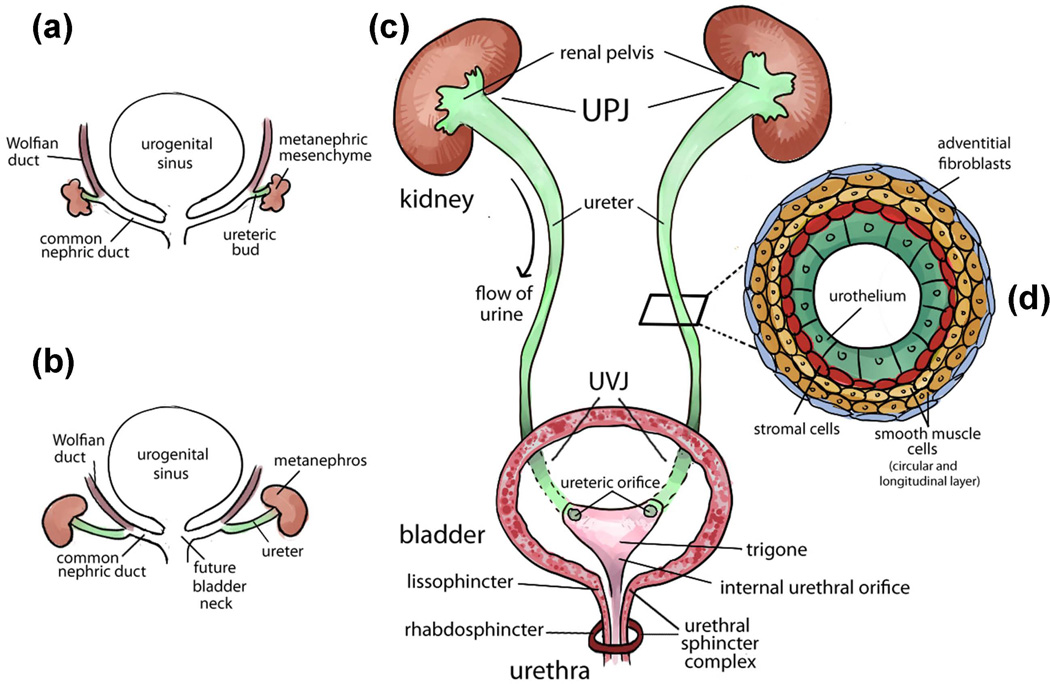
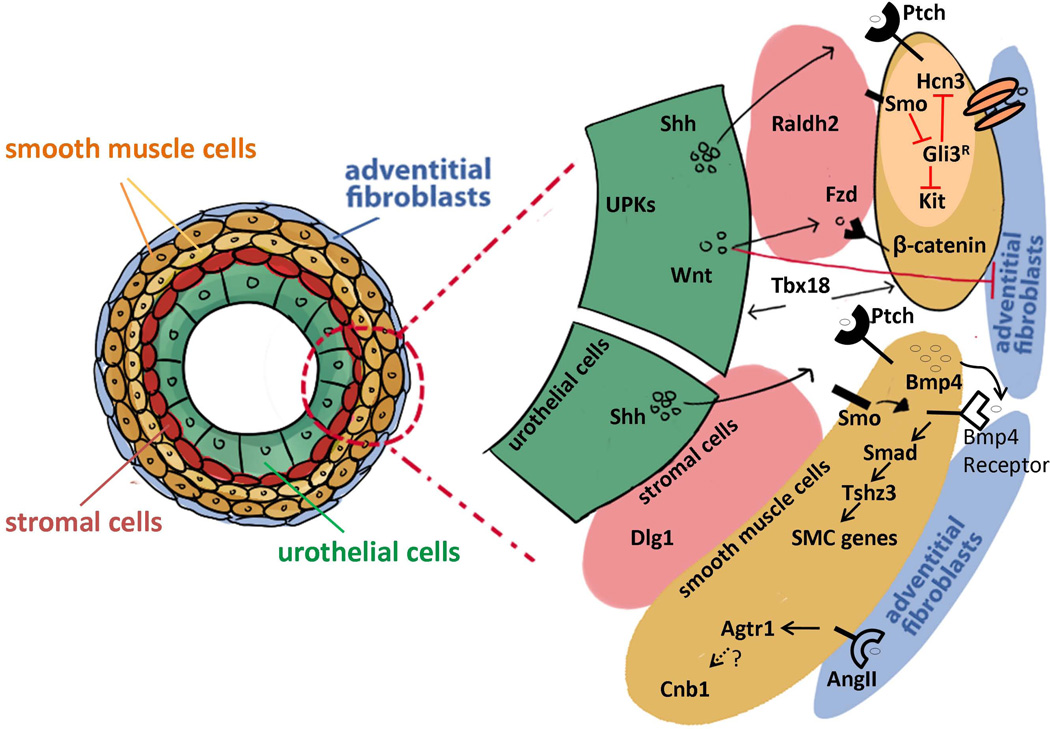
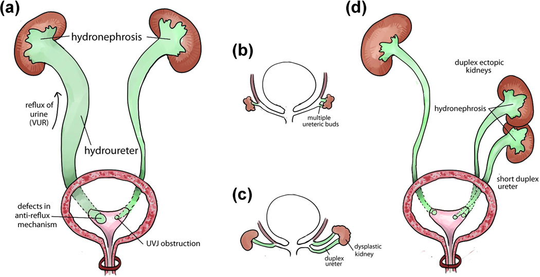
Congenital anomalies of the lower urinary tract (CALUT) are a family of birth defects of the ureter, the bladder, and the urethra. CALUT includes ureteral anomaliesc such as congenital abnormalities of the ureteropelvic junction (UPJ) and ureterovesical junction (UVJ), and birth defects of the bladder and the urethra such as bladder-exstrophy-epispadias complex (BEEC), prune belly syndrome (PBS), and posterior urethral valves (PUVs). CALUT is one of the most common birth defects and is often associated with antenatal hydronephrosis, vesicoureteral reflux (VUR), urinary tract obstruction, urinary tract infections (UTI), chronic kidney disease, and renal failure in children. Here, we discuss the current genetic and molecular knowledge about lower urinary tract development and genetic basis of CALUT in both human and mouse models. We provide an overview of the developmental processes leading to the formation of the ureter, the bladder, and the urethra, and different genes and signaling pathways controlling these developmental processes. Human genetic disorders that affect the ureter, the bladder and the urethra and associated gene mutations are also presented. As we are entering the postgenomic era of personalized medicine, information in this article may provide useful interpretation for the genetic and genomic test results collected from patients with lower urinary tract birth defects. With evidence-based interpretations, clinicians may provide more effective personalized therapies to patients and genetic counseling for their families.


Figures

FIGURE 1
FIGURE 1
Urinary tract development and structure. (a) Early development of the urinary tract (4th week of gestation in human and E10.5 in mice). An epithelial diverticulum called ureteric bud (UB) emanates from the Wolffian duct and grows into an adjacent group of mesenchymal cells (metanephric mesenchyme). (b) Elongation of the ureter and formation of the kidney (metanephros) during development. The common nephric ducts shorten, expand and integrate into the urogenital sinus (the future bladder) close to the region where the future bladder neck is located. (c) Structure of mature urinary tract in human and mice. Urine flows from the renal pelvis in the kidney through the ureter to the bladder for storage and eliminates to the outside through the urethra. The ureter is connected to the kidney at the ureteropelvic junction (UPJ) and is connected to the bladder at the ureterovesical junction (UVJ). Inside the bladder, two ureteric orifices and the internal urethral orifice form the trigone. The urethral sphincter complex includes the lissosphincter which is a continuation of the bladder smooth muscle and the rhabdosphincter which consists of striated muscles. (d) Transverse section of the mature ureter depicts four layers of cells: urothelium, stromal cells, smooth muscle cells and adventitial fibroblasts.
FIGURE 2
FIGURE 2
Genes and signaling pathways involved in ureteric budding. Scheme of signaling pathways between the ureteric buds (green) and the metanephric mesenchyme (yellow-orange). The most important inducer of UB outgrowth is the Receptor Tyrosine Kinase signaling pathway mediated by Gdnf and its receptor Ret. Ret is expressed by the nephric duct (green) and ureteric buds (green). Gdnf is secreted by the metanephric mesenchyme (yellow). The coordination of different signaling pathways in the anterior mesenchyme and the metanephric mesenchyme play a crucial role in the development of a single ureteric bud.
FIGURE 3
FIGURE 3
Genes and signaling pathways involved in ureter development. Scheme of genes and signaling pathways involved in the development of the urothelial cells (green), the stromal cells (pink), the smooth muscle cells (yellow) and the adventitial fibroblasts (blue). The transcription factor Tbx18 is one of the major early genes in ureter differentiation. Tbx18 is expressed in undifferentiated ureter mesenchymal cells and promotes the differentiation of both urothelium and smooth muscle cells. Uroplakins (UPKs) are expressed on the apical surface of urothelial cells which also express Shh (sonic hedgehog) and Wnt molecules. Dlg1 plays an essential role in the differentiation of the stromal cells that express the marker gene Raldh2. The Hedgehog signaling (Shh and its receptor Ptch) plays a major role in the ureteric smooth muscle maturation through molecules in the TGF-β signaling pathway such as Bmp4 and Tshz3. Shh is also necessary for the differentiation of ureteric pacemaker cells by suppressing the Gli3 repressor (Gli3R) through Smo (Smoothened) which in turn activates the expression of Kit and Hcn3. The canonical Wnt signaling is necessary for the differentiation of smooth muscle cells and the repression of the adventitial fibroblast cell differentiation. The smooth muscle cells express the Wnt receptors Frizzled (Fzd) which activates β-catenin (Ctnnb1). Calcineurin b1 (Cnb1) is required in the mesenchyme and smooth muscle cells for the development of pyeloureteral peristaltic machinery. The Angiotensin pathway may activate Cnb1.
FIGURE 4
FIGURE 4
Common congenital anomalies of the ureter. (a) Two main causes of hydronephrosis and hydroureter: vesicoureteral reflux (VUR) caused by defects of anti-reflux mechanism is on the left side and urinary obstruction caused by abnormal structure of the ureterovesical junction (UVJ) is on the right side. (b–d) Early abnormal ureteric budding can lead to congenital anomalies of the ureter: Abnormal multiple ureteric buds formation from the right Wolffian duct (b) can lead to abnormal phenotypes including duplex ureter, dysplastic kidney (c), ectopic kidney, duplex kidney, duplex ureter, short ureter, which are often associated with hydronephrosis phenotype on the right side (d), compared to the normal ureteric buds development on the left side. Each of these malformations can be found separately or coexist with other types of anomalies.
FIGURE 5
FIGURE 5
Chromosome map of genomic loci associated with congenital anomalies of the lower urinary tract. Each arrow indicates the physical mapping position of a single locus. Different malformations are represented by different colors as following: hydronephrosis (Red), vesicoureteral reflux (Green), bladder anomalies (Brown), urethra anomalies (Purple). See Table 1–4 for details about gene names, chromosome locations, and associated phenotypes, etc.

References

    1. Christianson A, Howson CP, Modell B. March of Dimes global report on birth defects. 2006 Pages Retrieved from http://www.marchofdimes.com/downloads/Birth_Defects_Report-PF.pdf.
    1. CDC. Update on overall prevalence of major birth defects--Atlanta, Georgia, 1978–2005. MMWR Morb Mortal Wkly Rep. 2008;57:1–5. - PubMed
    1. Yoon PW, Olney RS, Khoury MJ, Sappenfield WM, Chavez GF, Taylor D. Contribution of birth defects and genetic diseases to pediatric hospitalizations. A population-based study. Arch Pediatr Adolesc Med. 1997;151:1096–1103. - PubMed
    1. Pope JCt, Brock JW, 3rd, Adams MC, Stephens FD, Ichikawa I. How they begin and how they end: classic and new theories for the development and deterioration of congenital anomalies of the kidney and urinary tract, CAKUT. J Am Soc Nephrol. 1999;10:2018–2028. - PubMed
    1. Miyazaki Y, Ichikawa I. Ontogeny of congenital anomalies of the kidney and urinary tract, CAKUT. Pediatr Int. 2003;45:598–604. - PubMed

Publication types

MeSH terms

Supplementary concepts