. 2013 Apr;87(8):4545-57.
doi: 10.1128/JVI.03083-12. Epub 2013 Feb 13.

Nuclear localization of dengue virus nonstructural protein 5 does not strictly correlate with efficient viral RNA replication and inhibition of type I interferon signaling

Affiliations

Nuclear localization of dengue virus nonstructural protein 5 does not strictly correlate with efficient viral RNA replication and inhibition of type I interferon signaling

Anil Kumar et al. J Virol. 2013 Apr.

Abstract

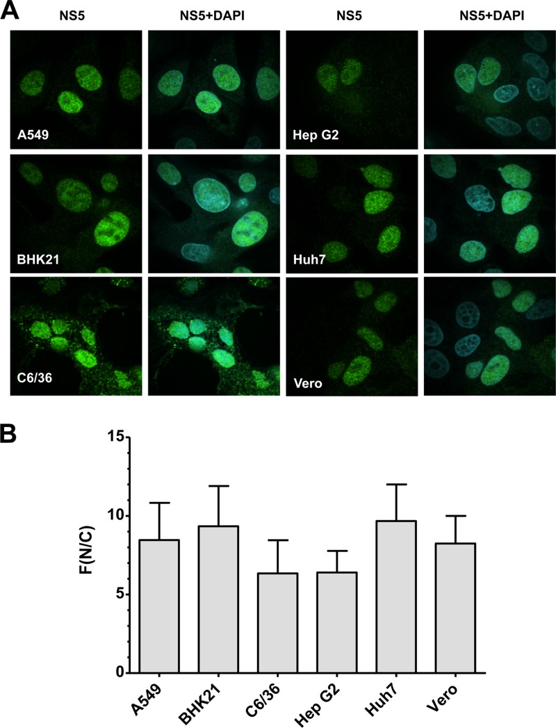
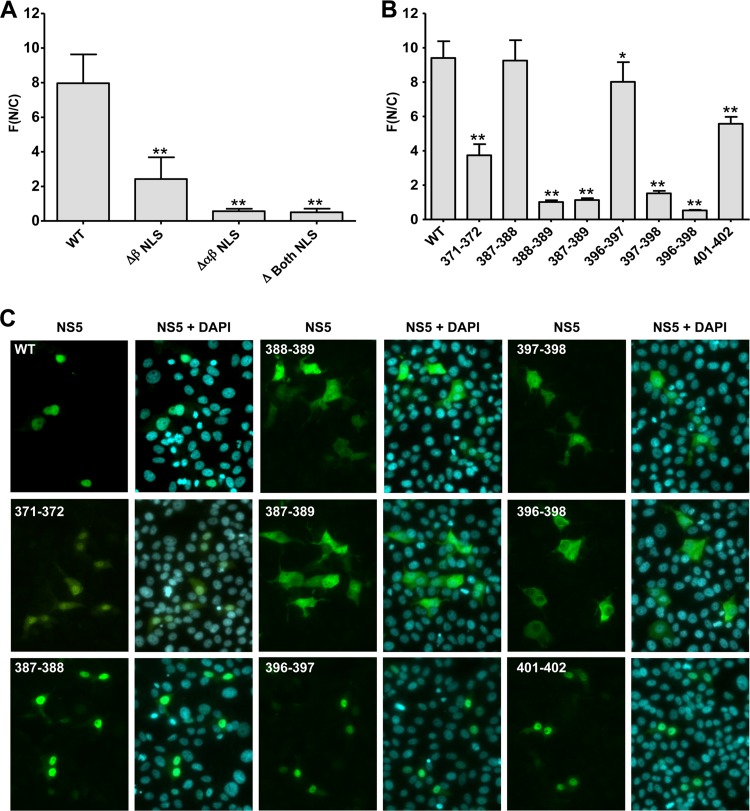
Dengue virus (DENV) is an important human pathogen, especially in the tropical and subtropical parts of the world, causing considerable morbidity and mortality. DENV replication occurs in the cytoplasm; however, a high proportion of nonstructural protein 5 (NS5), containing methyltransferase (MTase) and RNA-dependent RNA polymerase (RdRp) activities, accumulates in the nuclei of infected cells. The present study investigates the impact of nuclear localization of NS5 on its known functions, including viral RNA replication and subversion of the type I interferon response. By using a mutation analysis approach, we identified the most critical residues within the αβ nuclear localization signal (αβNLS), which are essential for the nuclear accumulation of this protein. Although we observed an overall correlation between reduced nuclear accumulation of NS5 and impaired RNA replication, we identified one mutant with drastically reduced amounts of nuclear NS5 and virtually unaffected RNA replication, arguing that nuclear localization of NS5 does not correlate strictly with DENV replication, at least in cell culture. Because NS5 plays an important role in blocking interferon signaling via STAT-2 (signal transducer and activator of transcription 2) degradation, the abilities of the NLS mutants to block this pathway were investigated. All mutants were able to degrade STAT-2, with accordingly similar type I interferon resistance phenotypes. Since the NLS is contained within the RdRp domain, the MTase and RdRp activities of the mutants were determined by using recombinant full-length NS5. We found that the C-terminal region of the αβNLS is a critical functional element of the RdRp domain required for polymerase activity. These results indicate that efficient DENV RNA replication requires only minimal, if any, nuclear NS5, and they identify the αβNLS as a structural element required for proper RdRp activity.


Figures

Fig 1
Fig 1
Nuclear localization of NS5 is independent of the cell line used. (A) The indicated cell lines were infected with DENV-2 (strain NGC; multiplicity of infection, 1), and cells were fixed 24 h after infection. Subcellular localization of NS5 was determined by immunofluorescence microscopy using an NS5-specific antibody. Images were captured by confocal laser scanning microscopy using a 100× objective. (B) Quantification of NS5-specific immunofluorescence by use of the ImageJ software package. The extent of nuclear localization of NS5 was determined by quantifying the NS5 signals detected in the nucleus (N) and the cytoplasm (C) and calculating the ratio of mean fluorescence (F) in these two compartments. Results are means ± standard deviations (n, ≥30).
Fig 2
Fig 2
Overview of constructs and mutants used in this study. (A) (Top) Diagram of the full-length DENV genome. The 5′ and 3′ UTRs are shown with their putative secondary structures (9). Polyprotein cleavage products are separated by vertical lines and are labeled as specified in the introduction. (Center) Genomic organization of the selectable subgenomic replicon. The gene encoding hygromycin resistance is fused via the ubiquitin coding sequence (Ubi) to NS1. (Bottom) Structure of the DENV reporter genome. The gene encoding firefly luciferase (Ffluc) is fused via Ubi to the N terminus of the polyprotein. All constructs are derived from the DENV-2 (DV2) NGC isolate (26). (B) (Top) Schematic diagrams of wild-type (WT) and mutant DENV-2 NS5. Wild-type NS5 is composed of an N-terminal methyltransferase domain and a C-terminal RNA-dependent RNA polymerase domain (shaded). The latter contains the βNLS (amino acids 321 to 368) and the αβNLS (amino acids 369 to 405). The positions of the amino acids deleted are given above each NLS deletion mutant. (Bottom) Amino acid sequences of the wild-type αβNLS and mutants derived from it. The minimal αβNLS according to reference (amino acids 369 to 393) is shaded in the wild-type sequence. For each mutant, the double or triple alanine substitutions are indicated by AA or AAA, and they are given below the corresponding positions of the wild type.
Fig 3
Fig 3
Subcellular localization of NS5 NLS mutants. Huh7 cells were transfected with plasmids directing the expression of NS5 bearing NLS deletions or the indicated amino acid substitutions under the control of the cytomegalovirus promoter, and cells were fixed 16 h later. The subcellular localization of NS5 was determined by immunofluorescence using an NS5-specific antibody. Images were captured with a 20× objective using a confocal laser scanning microscope. NS5-specific immunofluorescence was quantified by using the ImageJ software package. (A and B) The extent of the nuclear localization of NLS mutants was determined by quantifying the NS5 signals inside the nucleus (N) and the cytoplasm (C) and calculating the ratio of the mean fluorescence (F) in the nucleus to that in the cytoplasm. Results are means ± standard deviations (n, ≥50) for two independent experiments. Asterisks indicate significant differences (*, P < 0.05; **, P < 0.001) from the wild type. (A) Nuclear accumulation of NLS deletion mutants lacking amino acids 321 to 368 (ΔβNLS), amino acids 369 to 405 (ΔαβNLS), or amino acids 321 to 405 (Δ Both NLS). (B) Nuclear accumulation of αβNLS mutants. (C) Representative immunofluorescence images of NS5 mutants.
Fig 4
Fig 4
Replication competence of NLS mutants in BHK-21 cells. (A) Capped RNAs generated by in vitro transcription of cloned DENV reporter genomes were transfected by electroporation into BHK-21 cells. Lysates of cells prepared at the time points given above the graph were used to determine luciferase activity. Values were normalized to the 4-h value, which reflects transfection efficiency, and are expressed as means ± standard deviations for three independent experiments. (B) Titers of infectious virus released into culture supernatants of cells that had been transfected with the indicated DENV reporter genomes. Supernatants of the cells used for the experiment for which results are shown in panel A were harvested 96 h postelectroporation and were used to infect naïve BHK-21 cells. Seventy-two hours later, DENV replication was determined by a luciferase reporter assay reflecting the titers of infectious virus released from transfected cells. Values are means ± standard deviations for three independent experiments and are expressed relative to wild-type (WT) titers, which were set at 100%. Asterisks indicate significant differences (**, P < 0.001) from the wild type. (C) Replication of nonreporter DENV genomes carrying NLS mutations. BHK-21 cells were electroporated with the indicated DENV genomes. RNA replication was analyzed 72 h postelectroporation by determining the number of viral RNA copies by use of quantitative real-time RT-PCR. Values are means ± standard deviations for two independent experiments with three replicates each. Asterisks indicate significant differences (*, P < 0.05; **, P < 0.001) from the wild type. (D) Quantification of nuclear NS5 as described in the legend to Fig. 1B. Results are means ± standard errors of the means (n, ≥50) from two independent experiments. (E) Localization of NS5 in nonreporter DENV genomes carrying NLS mutations. The subcellular localization of selected mutants was determined by immunofluorescence using an NS5-specific polyclonal antibody 72 h postelectroporation. Nuclear DNA was visualized by DAPI staining. Images were captured with a 60× objective using a confocal laser scanning microscope.
Fig 5
Fig 5
Mutations in the αβNLS affecting viral replication cannot be rescued by trans-complementation. (A) Schematic of the experimental strategy. A Huh7-derived cell line containing a stably replicating subgenomic NGC replicon (as shown in Fig. 2A) was transfected with full-length genomes containing mutations in the αβNLS. Cells were seeded into multiwell culture dishes and were harvested 4, 24, 48, 72, and 96 h after transfection. The replication of the transfected reporter virus genome was determined by a luciferase assay. Note that the subgenomic helper replicon contains the selectable marker but lacks a luciferase reporter gene. p.e., postelectroporation. (B) The αβNLS mutants (x axis) were transfected into Huh7 replicon cells. The replication of the NLS mutants was quantified by a luciferase assay using cell lysates prepared at the indicated time points. The NS1 deletion mutant (ΔNS1) was used as a positive control. The replication of the wild-type DENV reporter genome and of the ΔNS1 mutant, each transfected into naïve Huh7 cells that lack the subgenomic helper replicon, served as references. (C) NS5 expression in DENV replicon cells was determined by immunofluorescence using an NS5-specific polyclonal antibody. Nuclear DNA was visualized by DAPI staining. Images were captured with a 100× objective using a confocal laser scanning microscope.
Fig 6
Fig 6
STAT-2 degradation is not affected by mutations in the αβNLS. (A) Huh7 cells were transfected either with the wild-type NGC genome (WT) or with the indicated αβNLS mutants. Seventy-two hours later, the cells were treated with 100 U of IFN-α/ml. After 2 h, the cells were fixed, and E protein and STAT-2 were detected by indirect immunofluorescence using E- and STAT-2-specific antisera. The images were acquired with a 100× objective using a confocal laser scanning microscope. Note that in cells expressing DENV E-protein, STAT-2 is absent, as manifested by the “empty” nucleus. (B) The amount of STAT-2 was determined by measuring the average STAT-2 signal inside the nuclei of infected cells. The values, expressed as percentages of the amount of STAT-2 in uninfected control cells, are means ± standard deviations (n, ≥30) for two independent experiments.
Fig 7
Fig 7
Expression of singly or doubly tagged full-length NS5 in E. coli. (A) Schematic representation of the expression constructs encoding full-length NS5 fused either to a C-terminal hexahistidine tag alone (construct 5His) or to an N-terminal HA tag and a C-terminal hexahistidine tag (construct 5HAHis). Mutations abrogating the methyltransferase (MTase) activity (S56A) or the RdRp activity (D663N), used as controls in the biochemical assays, are indicated. (B) Analysis of full-length NS5 proteins expressed in E. coli. Protein samples were analyzed by SDS-PAGE and Coomassie blue staining of the gel (top) or by Western blotting (WB) using an NS5-specific polyclonal antiserum (bottom). For each lane, 1 μg protein, as determined by the Bradford assay, was loaded onto the gel. SM, size marker; UI, uninduced; I, induced; S, soluble fraction; IS, insoluble fraction; FT, flowthrough; E, eluate fraction. Because the amount of protein in the wash fraction (W) was below the detection limit, a 1:700 dilution of the total wash fraction was loaded onto the gel.
Fig 8
Fig 8
Biochemical characterization of MTase and RdRp activities of full-length NS5 containing mutations in the αβNLS. (A) The tagged full-length NS5 proteins given above the gel were expressed in E. coli as described in Materials and Methods and were extracted from cell lysates by affinity purification using Ni-NTA affinity chromatography. Proteins were eluted with imidazole, and 5 μg protein was analyzed for purity and integrity by SDS-PAGE and Coomassie blue staining. Numbers on the left are the sizes of molecular mass standards (Marker). (B) A 2′O-methyltransferase assay of NLS mutants was carried out using a capped DENV-2 genomic RNA template (corresponding to nucleotides 1 to 175 of the genome), 100 ng purified NS5, and 2 μCi of 3H-radiolabeled S-adenosylmethionine. 3H incorporation was determined by liquid scintillation counting. (C) RdRp assay of NLS mutants using in vitro-transcribed DENV-2 genomic RNA as the template and 100 ng purified NS5. The amount of 32P-radiolabeled GMP incorporated was measured by liquid scintillation counting. The 2′O-MTase and RdRp activities are expressed as percentages of activities for wild-type (WT) NS5, with means and standard deviations estimated from three independent measurements.
Fig 9
Fig 9
Structural model of the DENV-2 RNA polymerase domain and localization of the NLS mutations. (A) Model of the DENV-2 RdRp domain derived from the structure of the DENV-3 polymerase domain (Protein Data Bank entry 2J7W). Shown is the classical closed “right-hand” structure of the RdRp domain, with the active site provided mainly by elements from the palm subdomain, situated in the center. The finger subdomain contributes the RNA template tunnel, and the thumb subdomain contributes the priming loop, which is essential for de novo initiation. The αβNLS consists of two helices, which reside on the surface of the finger subdomain. The N-terminal helix harbors residues K371-K372 at its N terminus and residues K387-K388-K389 at its C terminus. The C-terminal helix bears residues R396-E397-E398 at its N terminus. The C terminus of this helix contributes to the RNA template tunnel. Whereas residue R401 is at the surface of the domain, K402 is orientated toward the tunnel. (B) Model of the DENV-2 RdRp domain in complex with the 3′ end of the genome (CU-3′, represented as sticks color coded according to the atom type: white, C; red, O; blue, N; orange, P), as well as the first and second nucleotides used to initiate synthesis of the negative-strand RNA: ATP (represented as sticks and color coded according to the atom type: yellow, C; red, O; blue, N; orange, P) and GTP (represented as sticks color coded according to the atom type: light blue, C; red, O; blue, N; orange, P). Green spheres represent the two catalytic Mg2+ ions. R401 resides on the surface of the domain and is not engaged in an interaction. In contrast, K402 is engaged in a salt bridge to residue E491 (red dotted lines) and is situated in close proximity to partially negatively charged groups of the RNA template (O atoms of the phosphodiester and ribose backbone) to allow possible interactions (black dotted line). The models were generated as described elsewhere (36). PyMol was used to create the images.

References

    1. Kyle JL, Harris E. 2008. Global spread and persistence of dengue. Annu. Rev. Microbiol. 62:71–92 - PubMed
    1. Halstead SB.2007. Dengue. Lancet 370:1644–1652 - PubMed
    1. Gubler DJ. 2012. The economic burden of dengue. Am. J. Trop. Med. Hyg. 86:743–744 - PMC - PubMed
    1. Guy B, Almond JW. 2008. Towards a dengue vaccine: progress to date and remaining challenges. Comp. Immunol. Microbiol. Infect. Dis. 31:239–252 - PubMed
    1. Guy B, Guirakhoo F, Barban V, Higgs S, Monath TP, Lang J. 2010. Preclinical and clinical development of YFV 17D-based chimeric vaccines against dengue, West Nile and Japanese encephalitis viruses. Vaccine 28:632–649 - PubMed

Publication types

LinkOut - more resources