. 2013 Apr;87(8):4609-22.
doi: 10.1128/JVI.02424-12. Epub 2013 Feb 13.

Membrane topology and function of dengue virus NS2A protein

Affiliations

Membrane topology and function of dengue virus NS2A protein

Xuping Xie et al. J Virol. 2013 Apr.

Abstract

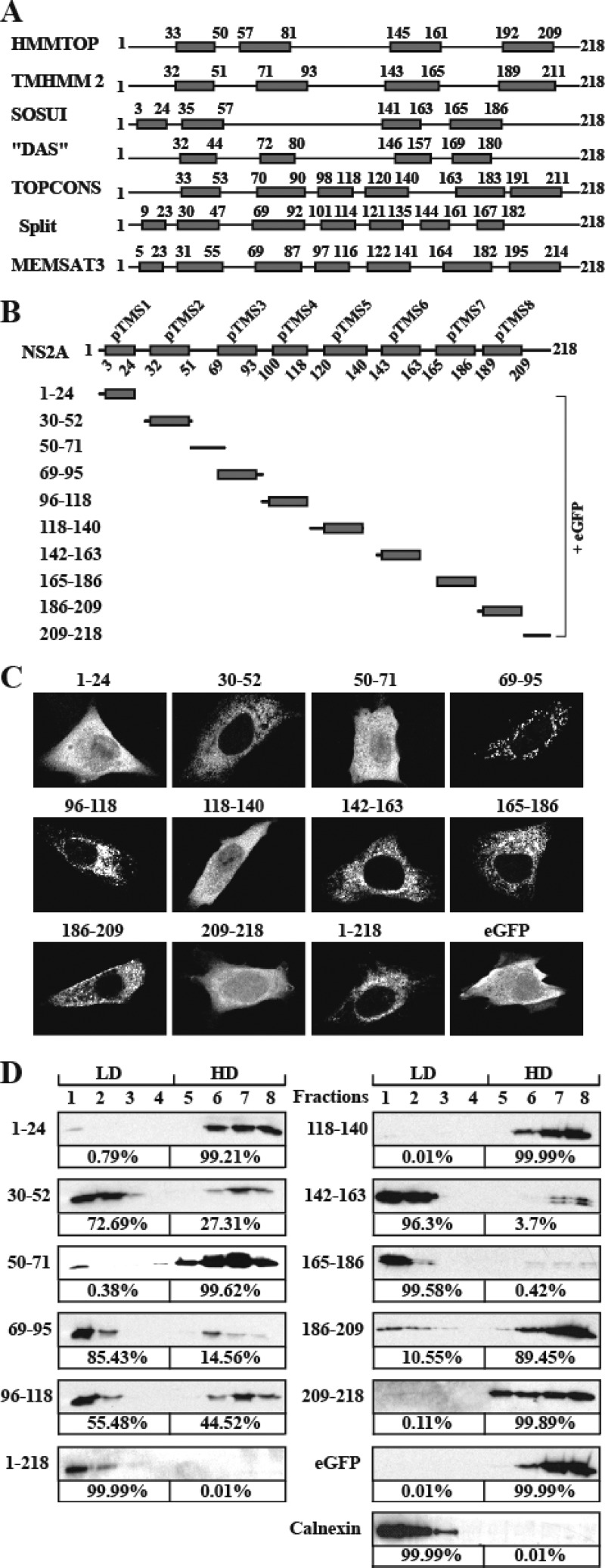
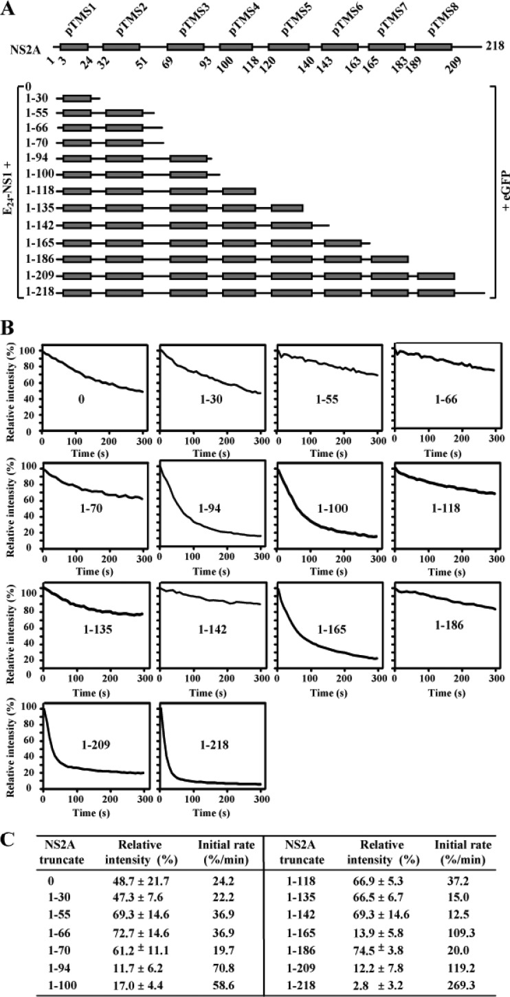
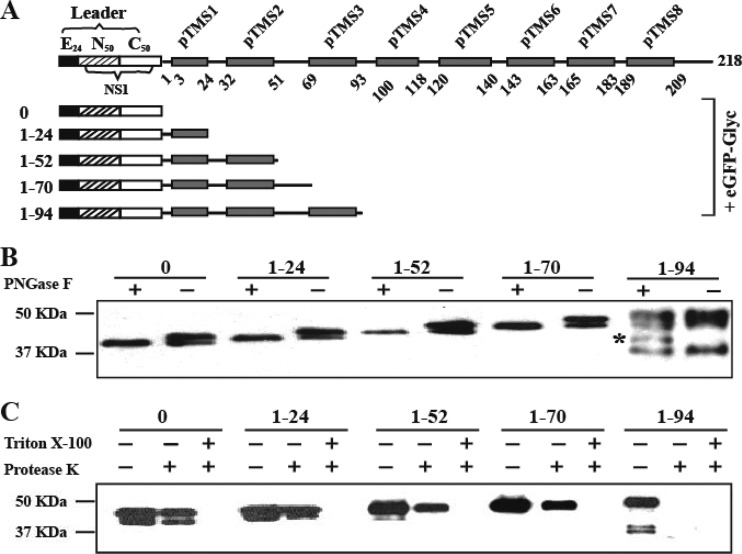
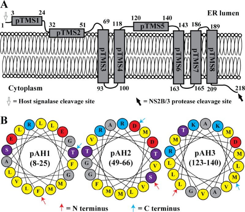
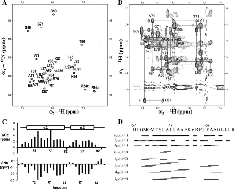
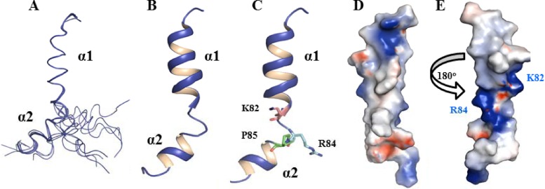
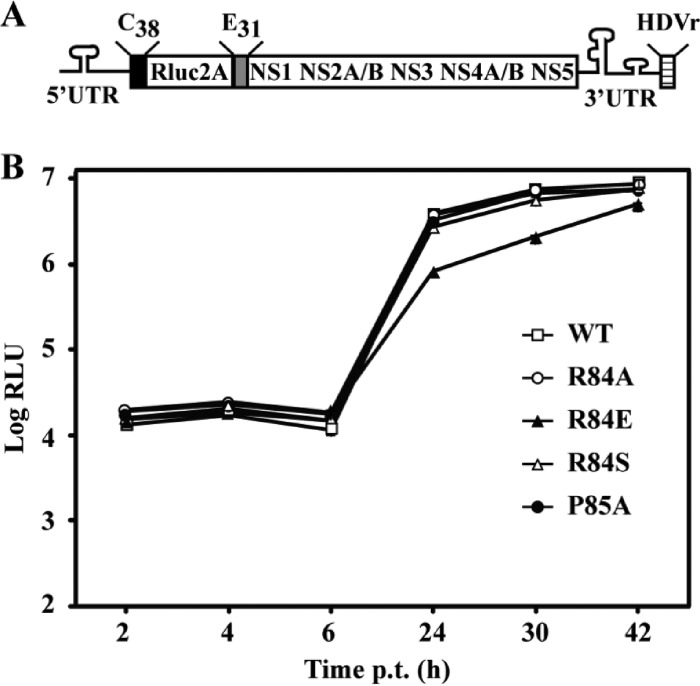
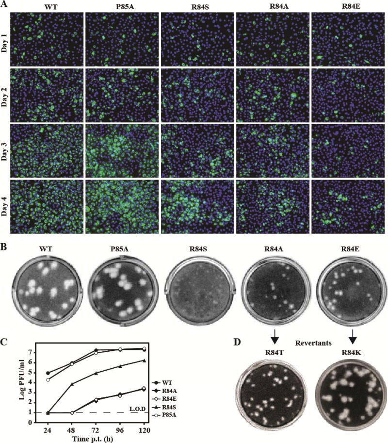
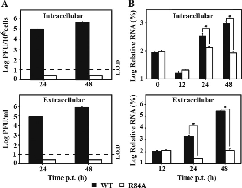
Flavivirus nonstructural protein 2A (NS2A) is a component of the viral replication complex that functions in virion assembly and antagonizes the host immune response. Although flavivirus NS2A is known to associate with the endoplasmic reticulum (ER) membrane, the detailed topology of this protein has not been determined. Here we report the first topology model of flavivirus NS2A on the ER membrane. Using dengue virus (DENV) NS2A as a model, we show that (i) the N-terminal 68 amino acids are located in the ER lumen, with one segment (amino acids 30 to 52) that interacts with ER membrane without traversing the lipid bilayer; (ii) amino acids 69 to 209 form five transmembrane segments, each of which integrally spans the ER membrane; and (iii) the C-terminal tail (amino acids 210 to 218) is located in the cytosol. Nuclear magnetic resonance (NMR) structural analysis showed that the first membrane-spanning segment (amino acids 69 to 93) consists of two helices separated by a "helix breaker." The helix breaker is formed by amino acid P85 and one positively charged residue, R84. Functional analysis using replicon and genome-length RNAs of DENV-2 indicates that P85 is not important for viral replication. However, when R84 was replaced with E, the mutation attenuated both viral RNA synthesis and virus production. Remarkably, an R84A mutation did not affect viral RNA synthesis but blocked intracellular formation of infectious virions. Collectively, the mutagenesis results demonstrate that NS2A functions in both DENV RNA synthesis and virion assembly/maturation. The topology model of DENV NS2A provides a good starting point for studying how flavivirus NS2A modulates viral replication and evasion of host immune response.


Figures

Fig 1
Fig 1
Prediction of membrane topology and analysis of membrane-associated activity of DENV-2 NS2A. (A) Schematic representation of DENV-2 NS2A transmembrane segments predicted by HMMTOP, TMHMM2, SOSUI, DAS, TOPCONS, Split, and MEMSAT3. The gray boxes indicate predicted transmembrane segments (pTMS). The positions of the first and last amino acid of pTMS are indicated. (B) A reference model of DENV-2 NS2A topology. Different fragments covering the entire NS2A were C-terminally fused with eGFP. The amino acid positions of each NS2A fragment are indicated on the left. (C) IFA analysis of BHK-21 cells transfected with various NS2A-eGFP constructs. At 24 h p.t., the expression of eGFP was monitored by a mouse monoclonal antibody against eGFP and a goat anti-mouse IgG conjugated with Alexa Fluor 488. The eGFP signal is in white. (D) Membrane flotation analysis of 293T cells transfected with plasmids expressing NS2A fragment-eGFP fusion proteins. NS2A fragment-eGFP proteins in each fraction were detected using an antibody against eGFP. Calnexin, probed with rabbit IgG against calnexin (Sigma), was used as an integral membrane protein control. The percentages of signals detected in the low-density (LD) fractions (1 to 4) and high-density (HD) fractions (5 to 8) were calculated by ImageJ software and are indicated below the panels.
Fig 2
Fig 2
In situ fluorescence protease protection assay. (A) E24-NS1-NS2A (truncated)-eGFP fusion constructs. Each NS2A fragment was N-terminally fused with E24-NS1 (representing a signal peptide derived from the last 24 residues of E protein [E24] and NS1 protein), and C-terminally fused with eGFP. pTMS are shown as gray boxes. (B) In situ fluorescence protease protection assay. BHK-21 cells were transfected with the indicated NS2A constructs. At 24 h p.t., the cells were permeabilized with digitonin followed by protease K treatment. Once the protease K was added, the fluorescence intensities were continuously quantified for 300 s (see Materials and Methods). Relative intensities were calculated using the fluorescence intensity at every 8 s divided by that at time zero when protease K was added. The means of relative intensities derived from 5 to 7 fields (each field containing 4 to 6 positive cells) are presented. The positions of NS2A truncates are indicated in the corresponding panels. (C) Summary of relative fluorescence intensity and initial rate of fluorescence degradation. Average results from at least two independent experiments are presented. See Materials and Methods for calculations.
Fig 3
Fig 3
Deglycosylation and in vitro protease protection assays. (A) Leader-NS2A truncate-eGFP-Glyc constructs. The leader sequence contains a signal peptide derived from the last 24 residues of E protein (E24; black box), the first 50 residues of NS1 (striped box), and the last 50 residues of NS1 (white box). pTMS are shown as gray boxes. Each NS2A fragment was N-terminally fused with the leader sequence and C-terminally fused with eGFP-Glyc (a glycosylation sequence). The amino acid positions of each NS2A constructs are indicated on the left. (B) In vitro deglycosylation assay. HEK293T cells were transfected with various constructs depicted in (A). An extra band of unknown identity is indicated with an asterisk. At 24 h p.t., the expressed fusion proteins were extracted and treated with PNGase F (+) or PBS (−). A mouse monoclonal antibody against GFP was used to probe eGFP-tagged proteins (see Materials and Methods). Molecular mass markers are shown on the left. (C) In vitro protease protection assay. Cell membranes of the transfected cells as described for panel B were extracted and digested with protease K in the presence or absence of 0.5% Trion X-100. The samples were analyzed by Western blotting as described for panel B.
Fig 4
Fig 4
Membrane topology of DENV NS2A. (A) A topology model of DENV-2 NS2A on the ER membrane. The N-terminal 68 residues are located in the ER lumen; pTMS1 does not associate with the membrane, whereas pTMS2 is likely to peripherally associate with ER membrane. pTMS3, pTMS6, and pTMS8 span the membrane from lumen to cytosol, whereas pTMS4 and pTMS7 span the membrane from cytosol to lumen. The C-terminal 9 residues are located in cytosol. It should be noted that the topology of pTMS4-8 is supported only by results from Fig. 1 and 2; due to technical difficulties (see details in the text), we were not able to validate the topology of pTMS4-8 by the deglycosylation and in vitro protease protection assays (Fig. 3). (B) Three amphipathic helices (pAH1, pAH2, and pAH3) predicted by HeliQuest. Each pAH construct is an α-helix composed of 18 amino acids. The numbers represent amino acid positions of NS2A for each pAH. Red, blue, purple, gray, and yellow circles represent negatively charged, positively charged, polar, glycine/alanine, and hydrophobic residues, respectively.
Fig 5
Fig 5
Resonance assignment of pTMS3. (A) 1H-15N-HSQC of pTMS3 in 15N natural abundance. The assigned cross-peaks are labeled with amino acid and residue position. (B) Assignment of the 2D NOESY spectrum of pTMS3 in the Hα region. Connections between residues are shown with lines. (C) Secondary structure analysis of pTMS3. Deviations of Cα (top) and Hα (bottom) from random coil values are shown. (D) Summary of NOE connectivity. The NOE plot was made from CYANA.
Fig 6
Fig 6
Structures of NS2A pTMS3. (A) Stereo view of the ensemble of 15 NMR structures from CYANA. Only the Cα trace is shown with PyMOL. (B) Cartoon representation of the pTMS3 structure. Two α-helices are shown with labels. (C) Positions of K82, R84, and P85 in pTMS3. In the cartoon representation, the side chains of K82, R84, and P85 are shown in stick mode. (D) Surface representation of pTMS3. The charge analysis was conducted in Pymol. The positively charged residues, negatively charged residues, and hydrophobic residues are shown in blue, red, and white, respectively. (E) Surface representation of pTMS3 with a 180° rotation from panel D. All images were prepared using Pymol (www.pymol.org).
Fig 7
Fig 7
Replicon analysis of NS2A R84 and P85. (A) Schematic diagram of a luciferase replicon of DENV-2. Rluc2A, Renilla luciferase gene followed by the foot-and-mouth disease virus 2A peptide; C38, nucleotides encoding the first 38 amino acids of C protein; E31, nucleotides encoding the last 31 amino acids of E protein; HDVr, hepatitis delta virus ribozyme sequence. (B) Transient replicon assay. Equal amounts of replicon RNA (WT or mutant R84A, R84E, R84S, or P85A) were electroporated into BHK-21 cells. At the indicated time points, the transfected cells were lysed and assayed for luciferase activities. The y axis shows the log10 value of Renilla luciferase activity (RLU). Each data point is the average for three replicates, and error bars show the standard deviations.
Fig 8
Fig 8
Analysis of NS2A R84 and P85 in DENV-2 genome-length RNA. (A) IFA analysis. Equal amounts of WT or mutant genome-length RNA were electroporated into BHK-21 cells. The transfected cells were monitored for E protein expression at indicated time points. An anti-E monoclonal antibody 4G2 and a goat anti-mouse IgG conjugated with Alexa Fluor 488 were used as primary and secondary antibodies, respectively. Green and blue indicate E protein and nucleus staining, respectively. (B) Plaque morphology of WT and mutant viruses. The viruses were derived from the media collected on day 5 posttransfection. (C) Virus production. Culture media from cells transfected with genome-length RNAs from (A) were collected at the indicated time points; viral titers were quantified by plaque assay. Average results with standard deviations are presented. The dashed line indicates the limit of detection (L.O.D; 10 PFU/ml). (D) Plaque morphology of revertant viruses after 5 rounds of passaging on Vero cells. Amino acid substitutions identified at position 84 of NS2A are indicated.
Fig 9
Fig 9
Comparison of intra- and extracellular infectious viral particles and viral RNA between the WT and the R84A mutant. BHK-21 cells were transfected with equal amounts of WT and mutant genome-length RNA. At the indicated time points, intra- and extracellular infectious viral particles were quantified by plaque assay (A); viral RNAs were measured by quantitative RT-PCR (B) (for details, see Materials and Methods). The intracellular viral particles were normalized to the cell number and presented as PFU per 106 cells. The relative RNA was calculated using formula 100% × 2(CTi − CTo), where CTi is the CT value for the individual virus sample and CTo is the CT value derived from the intracellular WT viral RNA collected at 0 h p.t. for intracellular RNA calculation or at 12 h p.t. for extracellular RNA calculation. The intracellular viral RNAs were normalized to the CT values derived from GAPDH mRNA. An asterisk indicates statistical significance based on Student's t test (P < 0.05). L.O.D, limit of detection. Each data point is an average; error bars indicate the standard deviations derived from three independent experiments (n = 3).

References

    1. Lindenbach BD, Thiel H-J, Rice CM. 2007. Flaviviridae: the viruses and their replication, p 1101–1152 In Knipe DM, Howley PM. (ed), Fields virology, 5th ed, vol 1 Lippincott William & Wilkins, Philadelphia, PA
    1. Cleaves GR, Dubin DT. 1979. Methylation status of intracellular dengue type 2 40 S RNA. Virology 96:159–165 - PubMed
    1. Mackenzie JM, Khromykh AA, Jones MK, Westaway EG. 1998. Subcellular localization and some biochemical properties of the flavivirus Kunjin nonstructural proteins NS2A and NS4A. Virology 245:203–215 - PubMed
    1. Lindenbach BD, Rice CM. 1997. Trans-complementation of yellow fever virus NS1 reveals a role in early RNA replication. J. Virol. 71:9608–9617 - PMC - PubMed
    1. Avirutnan P, Punyadee N, Noisakran S, Komoltri C, Thiemmeca S, et al. 2006. Vascular leakage in severe dengue virus infections: a potential role for the nonstructural viral protein NS1 and complement. J. Infect. Dis. 193:1078–1088 - PubMed

Publication types

Substances