

Cephalometric comparison of obstructive sleep apnea patients and healthy controls
- PMID: 23408768
- PMCID: PMC3571509
Cephalometric comparison of obstructive sleep apnea patients and healthy controls
Abstract
Objective: This study aimed to compare the cephalometric characteristics of obstructive sleep apnea (OSA) patients with those of healthy subjects and to determine possible relationships between cephalometric measurements of OSA patients and control subjects.
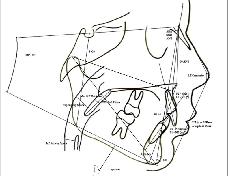
Methods: Standardized lateral cephalograms of 16 OSA patients and 16 healthy controls were obtained. Airway dimensions and dentofacial parameters were measured using a cephalometric analysis program (Dolphin Imaging Cephalometric and Tracing Software, Chatsworth, CA, USA). All statistical analyses were conducted using SPSS version 17.0.0 (SPSS Inc., Chicago, IL, USA). Descriptive statistics were calculated for all measurements, and the Mann-Whitney U test was used to evaluate intergroup differences.
Results: Midface length was significantly shorter and upper lip E-plane length was significantly longer in the OSA group than in the controls (P<.05). SNA, SNB, and mandibular plane angles (GoGn-SN), anterior and posterior facial heights, and posteroanterior face height ratio were similar in both groups. Maxillary length was slightly longer in the OSA group, whereas the mandibular length showed a slight increase in the control group (P<.05). The axial inclination of the lower incisor to its respective plane was normal, whereas the upper incisor was significantly protrusive (P<.05) in the OSA group. Distance between the hyoid and mandible was significantly greater in the OSA group than in the controls, indicating that the hyoid bone was positioned more downward in the OSA group (P<.05).
Conclusions: In this study, the patients with OSA demonstrated significant differences in several craniofacial measurements. OSA patients showed reduced midface length and inferiorly placed hyoid bone and tended to have smaller airway dimensions.
Keywords: Cephalometry; airway; obsrtructive sleep apnea.
Figures
References
-
- Kim J, In K, Kim J, You S, Kang K, Shim J, Lee S, Lee J, Lee S, Park C, Shin C. Prevalence of sleep-disordered breathing in middle-aged Korean men and women. Am J Respir Crit Care Med. 2004;170:1108–1113. - PubMed
-
- Young T, Palta M, Dempsey J, Skatrud J, Weber S, Badr S. The occurrence of sleep-disordered breathing among middle-aged adults. N Engl J Med. 1993;328:1230–1235. - PubMed
-
- Sunitha C, Kumar SA. Obstructive sleep apnea and its management. Indian J Dent Res. 2010;21:119–124. - PubMed
-
- Sunitha C, Aravindkumar S. Obstructive sleep apnea: clinical and diagnostic features. Indian J Dent Res. 2009;20:487–491. - PubMed
-
- Meyer JB, Jr, Knudson RC. The sleep apnea syndrome. Part I: Diagnosis. J Prosthet Dent. 1989;62:675–679. - PubMed
LinkOut - more resources
Full Text Sources
Research Materials