. 2013 Feb;24(4):495-509.
doi: 10.1091/mbc.E12-11-0843.

Direct binding of the Kex2p cytosolic tail to the VHS domain of yeast Gga2p facilitates TGN to prevacuolar compartment transport and is regulated by phosphorylation

Affiliations

Direct binding of the Kex2p cytosolic tail to the VHS domain of yeast Gga2p facilitates TGN to prevacuolar compartment transport and is regulated by phosphorylation

Mithu De et al. Mol Biol Cell. 2013 Feb.

Abstract

Human Golgi-localized, γ-ear-containing, ADP-ribosylation factor-binding proteins (Ggas) bind directly to acidic dileucine sorting motifs in the cytosolic tails (C-tails) of intracellular receptors. Despite evidence for a role in recruiting ubiquitinated cargo, it remains unclear whether yeast Ggas also function by binding peptide-sorting signals directly. Two-hybrid analysis shows that the Gga1p and Gga2p Vps27, Hrs, Stam (VHS) domains both bind a site in the Kex2p C-tail and that the Gga2p VHS domain binds a site in the Vps10p C-tail. Binding requires deletion of an apparently autoinhibitory sequence in the Gga2p hinge. Ser(780) in the Kex2p C-tail is crucial for binding: an Ala substitution blocks but an Asp substitution permits binding. Biochemical assays using purified Gga2p VHS-GGA and TOM1 (GAT) and glutathione S-transferase-Kex2p C-tail fusions show that Gga2p binds directly to the Kex2p C-tail, with relative affinities Asp(780) > Ser(780) > Ala(780). Affinity-purified antibody against a peptide containing phospho-Ser-(780) recognizes wild-type Kex2p but not S(780)A Kex2p, showing that Ser(780) is phosphorylated in vivo; phosphorylation of Ser(780) is up-regulated by cell wall-damaging drugs. Finally, mutation of Ser(780) alters trafficking of Kex2p both in vivo and in cell-free trans-Golgi network (TGN)-prevacuolar compartment (PVC) transport. Thus yeast Gga adaptors facilitate TGN-PVC transport by direct binding of noncanonical phosphoregulated Gga-binding sites in cargo molecules.


Figures
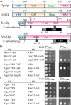
FIGURE 1:
FIGURE 1:
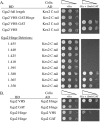
Yeast Gga1p and Gga2p VHS domains interact with the C-tail of Kex2p. (A) Shown are 1) operational domain boundaries of Gga1p and Gga2p used in two-hybrid analyses, 2) positions of TLS1, TLS2, and the GBS (Gga-binding site) in the Kex2p C-tail, and 3) positions of the N-terminal boundary (40–45) of the internal inhibitory region and of the GBS in the Vps10p C-tail. (B) Two-hybrid constructs in the LexA-BD (binding domain) and VP16-AD (activation domain) plasmids are indicated by BD and AD, respectively. The regions of Kex2p and Vps10p C-tails within each construct are indicated either by C-tail residue number (1–115, corresponding to residues 700–814 in Kex2p, and 1–164, corresponding to residues 1416–1579 in Vps10p) or, in the case of Tyr713 and Ser780 in Kex2p, by the position within the overall Kex2p sequence. Full-length Gga2p (1–585) and Gga2p-VHS-GAT (1–336) were used. (C) Gga2p-VHS (1–169), Gga2p-GAT (170–336), and Gga1p-VHS (1x165) two-hybrid constructs in the LexA-BD plasmid were tested for interaction with the full-length Kex2p C-tail (700–814) in the VP16-AD plasmid. (B, C) Strains were spotted at decreasing cell densities on plates that allowed growth without (left, +His) or only with (right, –His + 3 AT) interaction.
FIGURE 2:
FIGURE 2:
Gga2p contains an internal autoinhibitory site that binds its VHS domain. (A) Full-length Gga2p (residues 1–585), Gga2p-VHS-GAT-hinge (residues 1–472), Gga2-VHS-GAT (residues 1–336), and Gga2p-VHS (residues 1–169) in the LexA-BD plasmid and Kex2p C-tail (residues 1–115) in the VP16-AD plasmid were used. C-terminal truncations of Gga2p-VHS-GAT-hinge are indicated by Gga2p residue numbers and were tested in the LexA-BD plasmid for interaction with the Kex2p C-tail (residues 1–115) in the VP16-AD plasmid. (B) Gga2-hinge (residues 337–472) alone binds Gga2-VHS (residues 1–169) and not Gga2-GAT (170–336). Strains were spotted as in Figure 1.
FIGURE 3:
FIGURE 3:
Delimiting the Gga-binding site of Kex2p by mutational analysis. (A) C-terminal and (B) N-terminal truncation mutants of Kex2p (C-tail) in the VP16-AD plasmid were tested for interaction with Gga2-VHS-GAT (residues 1–336) in the LexA-BD plasmid. (C) Kex2p C-tail construct expressing residues 45–90 (i.e., Kex2p residues 746-791) in the VP16-AD plasmid was tested for interaction with Gga2-VHS-GAT (residues 1–336) in the LexA-BD plasmid. (D) Ala-scanning mutations in the full-length Kex2p C-tail in the VP16-AD two-hybrid plasmid were tested for interaction with Gga2-VHS-GAT (residues 1–336) in the LexA-BD plasmid. (E) Single-Ala mutations of F779 and S780, double-Ala mutations of F779S780, and the S780D mutation, all in the full-length Kex2p C-tail in the VP16-AD plasmid, were tested for interaction with Gga2-VHS-GAT (residues 1–336) in the LexA-BD plasmid. The regions of Kex2p C-tail within each construct are indicated by C-tail residue number. Strains were spotted as in Figure 1.
FIGURE 4:
FIGURE 4:
The C-tail of Vps10p contains a GBS that interacts with the Gga2p VHS domain. Two-hybrid constructs in the LexA-BD plasmid and the VP16-AD plasmid were as indicated. The regions of Vps10p C-tail within each construct are indicated by C-tail residue number. Gga2-VHS-GAT (residues 1–336) and Gga2-VHS (residues 1–169) were used. (A) A GBS in the Vps10p tail is revealed after deletion of an inhibitory sequence. (B) Fine deletion analysis of the N-terminal end of the inhibitory sequence. (C) Minimal GBS in Vps10p C-tail. (D) The Vps10p GBS binds the Gga2p VHS domain. Strains were spotted as in Figure 1.
FIGURE 5:
FIGURE 5:
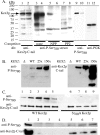
Kex2p Ser780 is phosphorylated in vivo, and phosphorylation is increased under conditions of cell wall damage. (A) WT Kex2p is phosphorylated at S780 in vivo. After glass bead lysis of CBO17 cells containing pG5-KX22 (WT Kex2p, odd-numbered lanes) or pG5-KX22-S780A (S780A-Kex2p, even-numbered lanes), cell extracts were analyzed by immunoblotting with anti-Kex2p antibody (lanes 1 and 2), anti–P-Ser780 serum (lanes 3–8), anti–P-Ser780 antibody (lanes 9 and 10), or anti–yeast phosphoglycerate kinase antibody (lanes 11 and 12). Lanes 3 and 4 contained no competing peptide, lanes 5 and 6 contained competing NPP, and lanes 7 and 8 contained competing PP2. Measurement of membrane-associated Kex2 proteolytic activity (Brenner and Fuller, 1992) confirmed the presence of equivalent amounts of WT and Ser780A Kex2p. (B) Kex2p expressed at its WT level is phosphorylated at Ser780. Extracts of kex2Δ strain CBO16 (Δ), KEX2 WT strain CBO18, or kex2Δ strain CBO17 containing YCpKX22 (25×) or pG5-KX22 (150×) were analyzed by immunoblotting with anti–P-Ser780 antibody (left) or anti-Kex2p antibody (right). (C) CBO17 containing YCpKX22 (lanes 1–5) or YCpKX22-S­780A (lanes 6–9) was grown to log phase (lane 1 and 6) and then grown for 4 h in the presence of calcofluor white (lanes 2 and 7), caffeine (lanes 3 and 8), Congo red (lanes 4 and 9), or no drug (lane 5). Lysates were analyzed by immunoblotting with anti–P-Ser780 antibody (top) or anti-Kex2p antibody (bottom). The slight reduction of S780A Kex2p seen here in stressed cells was not a reproducible result. (D) kex2Δ strain CBO16 (lanes 2–4) and KEX2 WT strain CBO18 (lanes 5–7) were grown to log phase (lanes 2 and 5) and treated with caffeine for 4 h (lanes 3 and 6) or grown overnight to high density (lanes 4 and 7). Equal amounts of extracted protein were analyzed by immunoblotting with anti–P-Ser780 antibody (top) or anti-Kex2p antibody (bottom). Lane 1 contains extract of CBO17 containing YCpKX22 to provide a marker for the Kex2p band. See Materials and Methods for further details.
FIGURE 6:
FIGURE 6:
Direct binding of yeast Gga2p VHS-GAT-His6 to the GBS in the Kex2p C-tail. Binding assays were performed as described in Materials and Methods. (A) Binding of His6-Gga2p-VHS-GAT (12.5, 25, 50 μg/ml) to the WT GST-Kex2p C-tail fusion protein (100 μg/ml) in the absence or presence of indicated amounts of untagged VHS-GAT was performed at 4°C. The GST fusion protein was visualized by Coomassie blue staining after SDS–PAGE, and bound His6-VHS-GAT was detected by immunoblotting with antibody to His6. (B) Binding of His6-VHS-GAT (50 μg/ml) with GST (100 μg/ml), a GST fusion protein bearing the C-tail of Kex2p (100 μg/ml), or a GST fusion protein bearing Kex2p C-tail residues 45–90. Bound His6-VHS-GAT was detected by immunoblotting with antibody to His6. Binding was performed at 4°C. (C) Binding of His6-VHS-GAT to WT, S780A, and S780D GST-Kex2p C-tail fusion proteins. Binding was performed at RT. (D) Preincubation of GST-Kex2p C-tail fusions with anti–P-Ser780 antibody inhibits binding of Gga2p VHS-GAT-His6. Preincubations with antibody and binding reactions were both performed at RT. (E) Effects of preincubating Gga2p VHS-GAT-His6 with phosphorylated peptide (PP2) and nonphosphorylated peptide (NPP) on binding to the WT and S780D GST-Kex2p C-tail fusions. Preincubations with peptides and binding assays were both performed at RT. (F) Dissociation of Gga2p VHS-GAT-His6 from WT, S780A, and S780D GST-Kex2p C-tail fusions, a representative experiment. Incubations were performed at RT. (G) Quantification of dissociation rates. Data represent the average of two independent assays, including the one shown in F. Error bars, SE of the mean. The asterisk indicates that the difference between S780A Kex2p and either WT or S­780D Kex2p at 30 min is statistically significant (p < 0.05, unpaired t test). The data are fitted to a first-order decay curve (Prism; GraphPad Software, La Jolla, CA) with t1/2 values of 160 min (S­780D), 97 min (WT), and 40 min (S780A) and R2 values, respectively, of 0.76, 0.83, and 0.97.
FIGURE 7:
FIGURE 7:
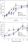
Effects of FS779,780AA and S780D mutations on Kex2p localization in vivo. (A) The FS779,780AA mutation suppresses and the S780D mutation enhances the accelerated loss of retrieval-defective Y713A-Kex2p from the pro–α-factor processing compartment. Strain MAY15-8B harboring WT and mutant derivatives of KEX2 under the control of the GAL1 promoter were grown on galactose for several generations. The strains were then shifted to glucose-containing media to repress expression of the wild-type and mutant KEX2 constructs. At the indicated time points after the shift to glucose the ability of the cells to mate was measured to generate an index of mating efficiency. Data points represent mean values of duplicates, and the error bars represent the SE. The experiment was performed twice with comparable results. (B) Rates of turnover of WT and Y713A, Y713A, FS779,780AA, and Y713A, S780D mutant forms of Kex2p. Turnover experiments were performed as described in Materials and Methods. Samples were processed in triplicate at the times indicated. The entire experiment was performed three times with comparable results. Half-lives, calculated by fitting the data in Figure 7B to first-order decay curves using Kaliedagraph (Synergy Software, Reading, PA), were 330 min (WT), 20 min (Y713A), 26 min (Y713A, FS779,780AA), and <7.1 min (Y713A, S780D).
FIGURE 8:
FIGURE 8:
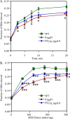
The FS779,780AA mutation suppresses and the S780D mutation enhances cell free TGN–PVC transport. Cell-free TGN–PVC transport reactions were conducted using donor MSS from cells expressing WT, FS779,780AA, or S780D Kex2p and acceptor MSS from cells expressing the chimeric Kex2p substrate PSHA, which is localized to the PVC. The assay measures the cleavage of PSHA in acceptor PVC membranes by Kex2p delivered from TGN donor membranes. For details see Materials and Methods. (A) Time course. (B) Donor MSS titration. Data points represent mean values of triplicates; error bars represent the SD of the mean. Entire experiments were performed three times with comparable results. Asterisks indicate that the difference between the indicated points and points on the other curves were significant (p < 0.05, unpaired t test).
FIGURE 9:
FIGURE 9:
Mutation of Ser780 reduces Kex2p activity available for TGN homotypic fusion. Cell-free TGN-homotypic fusion reactions were conducted using donor MSS from cells expressing WT, FS779,780AA, or S780D Kex2p and acceptor MSS from cells expressing the chimeric Kex2p substrate SHA, which is localized to the TGN. The assay measures the cleavage of SHA in TGN membranes after fusion with TGN membranes containing Kex2p. For details see Materials and Methods. (A) Time course. (B) Donor MSS titration. Data points represent mean values of triplicates; error bars represent the SD of the mean. Entire experiments were performed three times with comparable results. The significance (unpaired t test) of the difference between the indicated points and WT points was as follows: *p < 0.05; **p < 0.01; ***p < 0.001. In B, the difference between values for the two mutants for points from 60 to 240 μg was significant (p < 0.01 for 60 and 120 μg and p < 0.05 for 180 and 240 μg).

Similar articles

Cited by

References

    1. Abazeed ME, Blanchette JM, Fuller RS. Cell-free transport from the trans-Golgi network to late endosome requires factors involved in formation and consumption of clathrin-coated vesicles. J Biol Chem. 2005;280:4442–4450. - PubMed
    1. Abazeed ME, Fuller RS. Yeast Golgi-localized, gamma-Ear-containing, ADP-ribosylation factor-binding proteins are but adaptor protein-1 is not required for cell-free transport of membrane proteins from the trans-Golgi network to the prevacuolar compartment. Mol Biol Cell. 2008;19:4826–4836. - PMC - PubMed
    1. Bilodeau PS, Winistorfer SC, Allaman MM, Surendhran K, Kearney WR, Robertson AD, Piper RC. The GAT domains of clathrin-associated GGA proteins have two ubiquitin binding motifs. J Biol Chem. 2004;279:54808–54816. - PMC - PubMed
    1. Blanchette JM, Abazeed ME, Fuller RS. Cell-free reconstitution of transport from the trans-Golgi network to the late endosome/prevacuolar compartment. J Biol Chem. 2004;279:48767–48773. - PubMed
    1. Bonifacino JS. The GGA proteins: adaptors on the move. Nat Rev Mol Cell Biol. 2004;5:23–32. - PubMed

Publication types

MeSH terms

Substances