

Cutting edge: innate memory CD8+ T cells are distinct from homeostatic expanded CD8+ T cells and rapidly respond to primary antigenic stimuli
- PMID: 23408840
- PMCID: PMC4033301
- DOI: 10.4049/jimmunol.1202988
Cutting edge: innate memory CD8+ T cells are distinct from homeostatic expanded CD8+ T cells and rapidly respond to primary antigenic stimuli
Abstract
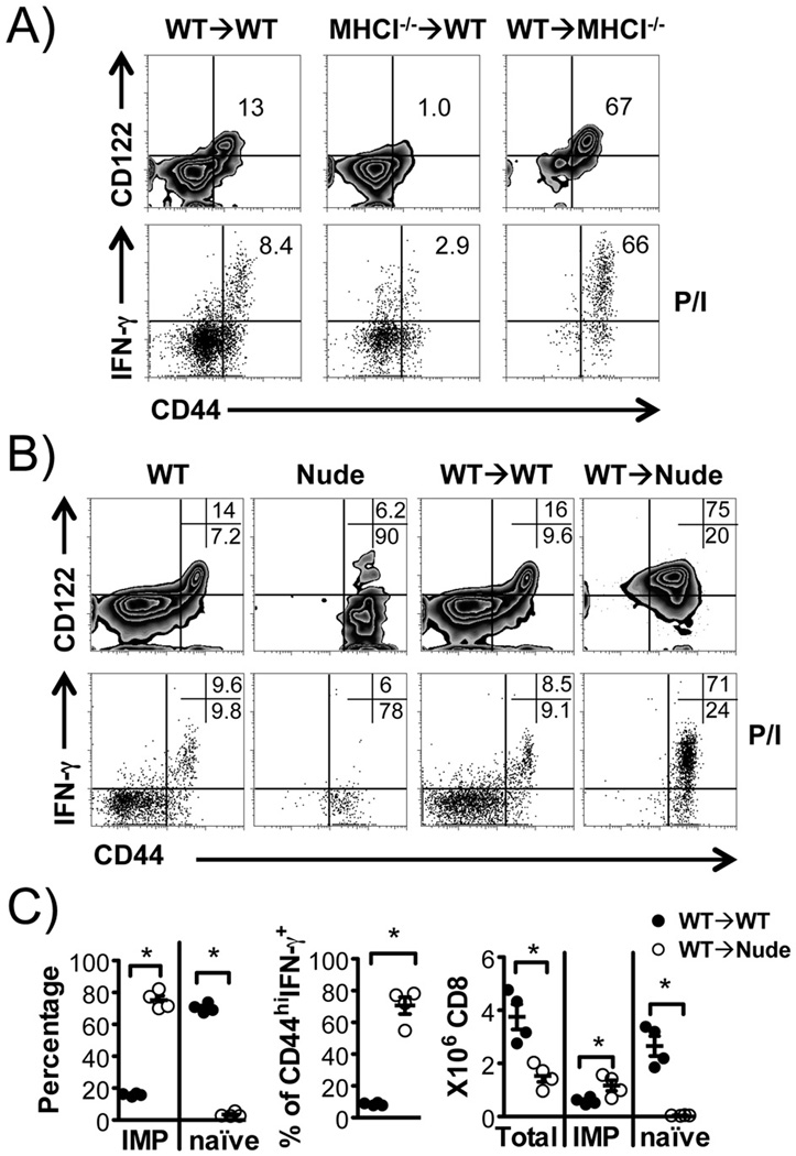
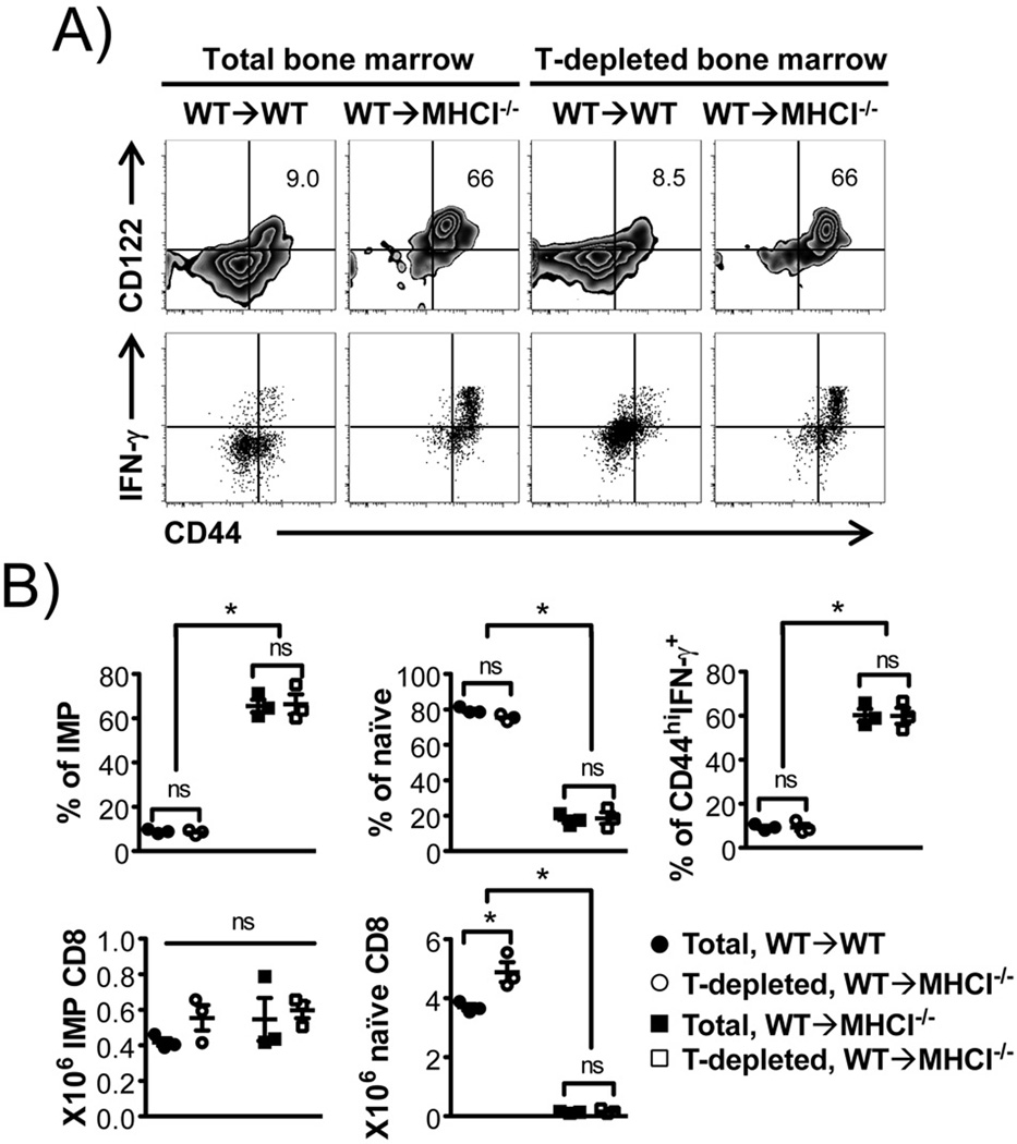
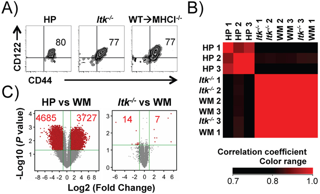
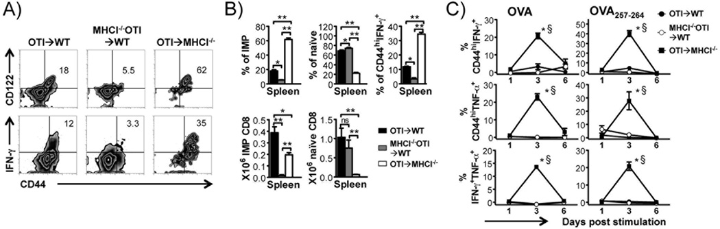
Innate memory phenotype (IMP) CD8(+) T cells are nonconventional αβ T cells exhibiting features of innate immune cells and are significantly increased in the absence of ITK. Their developmental path and function are not clear. In this study, we show hematopoietic MHC class I (MHCI)-dependent generation of Ag-specific IMP CD8(+) T cells using bone marrow chimeras. Wild-type bone marrow gives rise to IMP CD8(+) T cells in MHCI(-/-) recipients, resembling those in Itk(-/-) mice, but distinct from those derived via homeostatic proliferation, and independent of recipient thymus. In contrast, MHCI(-/-) bone marrow does not lead to IMP CD8(+) T cells in wild-type recipients. OTI IMP CD8(+) T cells generated via this method exhibited enhanced early response to Ag without prior primary stimulation. Our findings suggest a method to generate Ag-specific "naive" CD8(+) IMP T cells, as well as demonstrate that they are not homeostatic proliferation cells and can respond promptly in an Ag-specific fashion.
Conflict of interest statement
The authors have no financial conflicts of interest.
Figures
Similar articles
-
Memory phenotype of CD8+ T cells in MHC class Ia-deficient mice.J Immunol. 2003 Jun 1;170(11):5414-20. doi: 10.4049/jimmunol.170.11.5414. J Immunol. 2003. PMID: 12759416
-
Homeostatic proliferation of a Qa-1b-restricted T cell: a distinction between the ligands required for positive selection and for proliferation in lymphopenic hosts.J Immunol. 2004 Nov 15;173(10):6065-71. doi: 10.4049/jimmunol.173.10.6065. J Immunol. 2004. PMID: 15528342
-
Cutting edge: Ly9 (CD229), a SLAM family receptor, negatively regulates the development of thymic innate memory-like CD8+ T and invariant NKT cells.J Immunol. 2013 Jan 1;190(1):21-6. doi: 10.4049/jimmunol.1202435. Epub 2012 Dec 7. J Immunol. 2013. PMID: 23225888 Free PMC article.
-
Cytokine synergy in antigen-independent activation and priming of naive CD8+ T lymphocytes.Crit Rev Immunol. 2009;29(3):219-39. doi: 10.1615/critrevimmunol.v29.i3.30. Crit Rev Immunol. 2009. PMID: 19538136 Review.
-
From the thymus to longevity in the periphery.Curr Opin Immunol. 2010 Jun;22(3):274-8. doi: 10.1016/j.coi.2010.03.003. Epub 2010 Apr 6. Curr Opin Immunol. 2010. PMID: 20378321 Free PMC article. Review.
Cited by
-
Targeting Interleukin-2-Inducible T-Cell Kinase (ITK) Differentiates GVL and GVHD in Allo-HSCT.Front Immunol. 2020 Nov 26;11:593863. doi: 10.3389/fimmu.2020.593863. eCollection 2020. Front Immunol. 2020. PMID: 33324410 Free PMC article.
-
T-Bet independent development of IFNγ secreting natural T helper 1 cell population in the absence of Itk.Sci Rep. 2017 Apr 13;7:45935. doi: 10.1038/srep45935. Sci Rep. 2017. PMID: 28406139 Free PMC article.
-
ITK tunes IL-4-induced development of innate memory CD8+ T cells in a γδ T and invariant NKT cell-independent manner.J Leukoc Biol. 2014 Jul;96(1):55-63. doi: 10.1189/jlb.1AB0913-484RR. Epub 2014 Mar 11. J Leukoc Biol. 2014. PMID: 24620029 Free PMC article.
-
The signaling symphony: T cell receptor tunes cytokine-mediated T cell differentiation.J Leukoc Biol. 2015 Mar;97(3):477-85. doi: 10.1189/jlb.1RI0614-293R. Epub 2014 Dec 18. J Leukoc Biol. 2015. PMID: 25525115 Free PMC article. Review.
-
NFAT2 Regulates Generation of Innate-Like CD8+ T Lymphocytes and CD8+ T Lymphocytes Responses.Front Immunol. 2016 Oct 6;7:411. doi: 10.3389/fimmu.2016.00411. eCollection 2016. Front Immunol. 2016. PMID: 27766099 Free PMC article.
References
-
- Berg LJ. Signalling through TEC kinases regulates conventional versus innate CD8(+) T-cell development. Nat Rev Immunol. 2007;7:479–485. - PubMed
-
- Behar SM, Porcelli SA. CD1-restricted T cells in host defense to infectious diseases. Curr Top Microbiol Immunol. 2007;314:215–250. - PubMed
-
- Born WK, Reardon CL, O'Brien RL. The function of gammadelta T cells in innate immunity. Curr Opin Immunol. 2006;18:31–38. - PubMed
Publication types
MeSH terms
Substances
Associated data
- Actions
Grants and funding
LinkOut - more resources
Full Text Sources
Other Literature Sources
Molecular Biology Databases
Research Materials
