. 2013 May 15;207(10):1525-34.
doi: 10.1093/infdis/jit049. Epub 2013 Feb 13.

Rv1894c is a novel hypoxia-induced nitronate monooxygenase required for Mycobacterium tuberculosis virulence

Affiliations

Rv1894c is a novel hypoxia-induced nitronate monooxygenase required for Mycobacterium tuberculosis virulence

Lee G Klinkenberg et al. J Infect Dis. .

Abstract

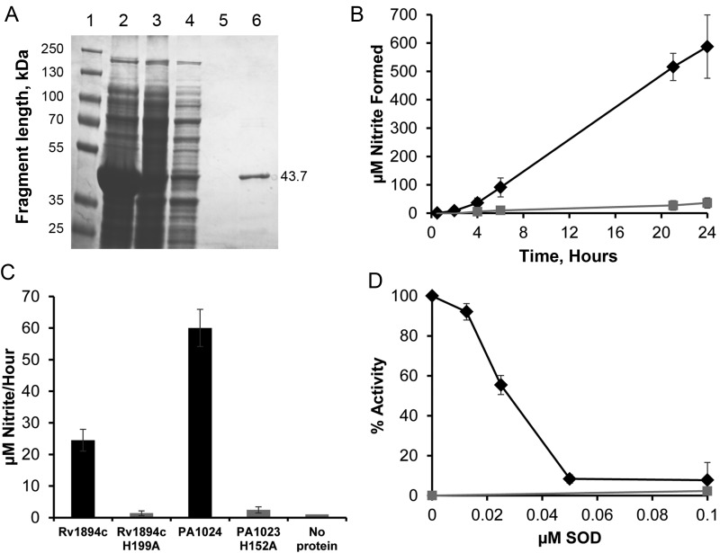
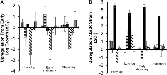
Tuberculosis is difficult to cure, requiring a minimum of 6 months of treatment with multiple antibiotics. Small numbers of organisms are able to tolerate the antibiotics and persist in the lungs of infected humans, but they still require some metabolic activity to survive. We studied the role of the hypoxia-induced Rv1894c gene in Mycobacterium tuberculosis virulence in guinea pigs, which develop hypoxic, necrotic granulomas histologically resembling those in humans and found this gene to be necessary for full bacillary growth and survival. We characterized the function of the encoded enzyme as a nitronate monooxygenase, which is needed to prevent the buildup of toxic products during hypoxic metabolism and is negatively regulated by the transcriptional repressor KstR. Future studies will focus on developing small-molecule inhibitors that target Rv1894c and its homologs, with the goal of killing persistent bacteria, thereby shortening the time needed to treat tuberculosis.


Figures

Figure 1.
Figure 1.
Complementation of Rv1894c::Tn. A, Polymerase chain reaction analysis of genomic DNA. Lanes 1 and 6 contain wild-type genomic template (WT), lanes 2 and 7 contain Rv1894c::Tn genomic template (Tn), lanes 3 and 8 contain the first Rv1894c::Tn Comp candidate (C1), and lanes 4 and 9 contain the second Rv1894c::Tn Comp candidate (C2). Lanes 2–4 show the presence of the kanamycin cassette marking the himar1 insertion in the Rv1894c::Tn strain and both candidate complement strains. Lanes 8 and 9 show the presence of a region of the hygromycin gene marking the integration of the Rv1894c::Tn Comp plasmid into both candidate strains. Lane 5 is the molecular marker, showing 100-bp increments, beginning with 100 bp at the bottom. B, Diagram of expected sizes of genomic fragments binding to the probe-recognizing region of the Rv1894c coding sequence. C, Southern blot showing Rv1894c as a single-copy gene present in the correct fragment sizes in both wild-type (6.2 kb) and Rv1894c::Tn (8.3 kb) strains (lanes 2 and 3, respectively). In both of the complement candidate strains (lanes 4 and 5), the mutant allele (8.3 kb) and the complement allele (3.9 kb) are present. D, Map of genomic region surrounding Rv1894c.
Figure 2.
Figure 2.
Reduced initial growth and long-term survival of the Rv1894c::Tn strain in the lungs of guinea pigs after aerosol infection. Black diamonds refer to wild-type Mycobacterium tuberculosis infection. Black and white crosshatched boxes refer to Rv1894c::Tn Comp infection. Grey boxes refer to Rv1894c::Tn infection. The asterisks indicate significant differences between colony-forming units (CFUs) recovered from wild-type– and mutant–infected lungs (P < .0006 for day 14, P < .003 for day 28, and P < .002 for day 56). The CFUs recovered from wild-type– and complement-infected lungs were not significantly different at any time point.
Figure 3.
Figure 3.
Effect of Rv1894c deficiency on guinea pig pathology. A, Guinea pig lung weights of lungs infected with Mycobacterium tuberculosis. B, Guinea pig spleen weights, normalized to mean body weight on day 1 divided by body weight of individual animal at time of sacrifice. Black bars represent wild type; grey bars represent Rv1894c::Tn; and white bars represent Rv1894c::Tn Comp.
Figure 4.
Figure 4.
The proposed nitronate monooxygenase family shows amino acid sequence and structural similarity. A, Clustal X multiple alignment of PA1024 from Pseudomonas aeruginosa with 5 nitronate monooxygenase (NMO) proteins from Mycobacterium tuberculosis and with NCU04803 from Neurospora crassa. Segment of whole protein alignment showing one area of conservation near the proposed catalytic histidine residue (shown in red). B, Overlaid alignment of alignment of PA1024 and Rv1894c, using FATCAT, predicting strong structural similarity. The color scheme shows each amino acid as a different color, as defined by Vector-NTI 3D Molecule Viewer software. The histidine in the catalytic cleft is shown in space-fill. C, View of the PA1024 structure, showing conserved residues from Clustal X paired alignment with Rv1894c in space-fill. Also shown in this image is the space-fill view in yellow of the flavin cofactor and the 2-nitropropane substrate, both near the center of the image. D, Enzymatic reaction, from left to right, catalyzed by NMO.
Figure 5.
Figure 5.
Recombinant Rv1894c protein functions as a nitronate monooxygenase (NMO). A, Coomassie-stained sodium dodecyl sulfate polyacrylamide gel electrophoresis showing purification of Rv1894c from Escherichia coli. Lane 1 is a size standard. Lane 2 is a crude extract from lysed E. coli. Lane 3 is the cleared lysate soluble fraction. Lane 4 is the column flow through. Lane 5 is the final wash of the column. Lane 6 is the eluted protein of the correct size, 43.7 kDa. B, Enzyme assay of NMO activity of Rv1894c, using 2-nitropropane as substrate. Black diamonds represent reactions containing Rv1894c. Grey squares represent a control reaction containing no protein. Conversion from absorbance at 540 nM to micromoles of nitrite was performed using a standard curve of known nitrite concentration. C, Enzyme assay of NMO activity of Rv1894c and PA1024 containing site-directed mutations of the proposed catalytic histidine residues (H199A and H152A, respectively) compared to the native proteins. Each bar represents the rate of nitrite conversion from 3 independent purifications of each protein, and the error bars are SDs. D, Inhibition of NMO activity by superoxide dismutase (SOD). Rv1894c protein (black diamonds) and a no-protein control (grey boxes) were incubated with substrate and increasing concentrations of SOD. Nitrite production was measured after several hours of incubation. Each time point was performed in triplicate with independent protein purifications. Error bars represent SDs.
Figure 6.
Figure 6.
Expression of Rv1894c in Mycobacterium tuberculosis is regulated by KstR but is growth-phase independent. A, Gene expression of the 5 nitronate monooxygenase (NMO) homologs as well as the expression level of the repressor kstR (Rv3574) in wild-type M. tuberculosis grown in 7H9 were compared to normalized sigA expression levels at different growth stages. Each value is shown relative to the levels of expression measured at the early log phase of growth. B, Cycle threshold (CT) values of the 5 NMO homolog expression levels were compared between wild type and a kstR::Tn strain at different growth stages. Rv0021c values are represented by a bar with vertical lines. Rv1533 values are represented by a bar with horizontal lines. Rv1894c values are represented by a solid black bar. Rv2781c values are represented by a bar with diagonal lines. Rv3553 values are represented by a bar with a checkerboard pattern. Rv3574 (kstR) values are represented by a solid grey bar. Each experimental time point was performed in triplicate. Error bars represent SDs.

Similar articles

Cited by

References

    1. Flynn JL, Chan J. Tuberculosis: latency and reactivation. Infect Immun. 2001;69:4195–201. - PMC - PubMed
    1. Kaplan G, Post FA, Moreira AL, et al. Mycobacterium tuberculosis growth at the cavity surface: a microenvironment with failed immunity. Infect Immun. 2003;71:7099–108. - PMC - PubMed
    1. Grosset J. Mycobacterium tuberculosis in the extracellular compartment: an underestimated adversary. Antimicrob Agents Chemother. 2003;47:833–6. - PMC - PubMed
    1. Haapanen JH, Kass I, Gensini G, Middlebrook G. Studies on the gaseous content of tuberculous cavities. Am Rev Respir Dis. 1959;80:1–5. - PubMed
    1. Wayne LG, Hayes LG. An in vitro model for sequential study of shiftdown of Mycobacterium tuberculosis through two stages of nonreplicating persistence. Infect Immun. 1996;64:2062–9. - PMC - PubMed

Publication types

MeSH terms

Substances