. 2013 Apr;41(7):e83.
doi: 10.1093/nar/gkt080. Epub 2013 Feb 13.

Site- and strand-specific nicking of DNA by fusion proteins derived from MutH and I-SceI or TALE repeats

Affiliations

Site- and strand-specific nicking of DNA by fusion proteins derived from MutH and I-SceI or TALE repeats

Lilia Gabsalilow et al. Nucleic Acids Res. 2013 Apr.

Abstract

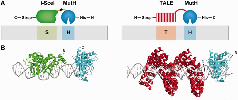
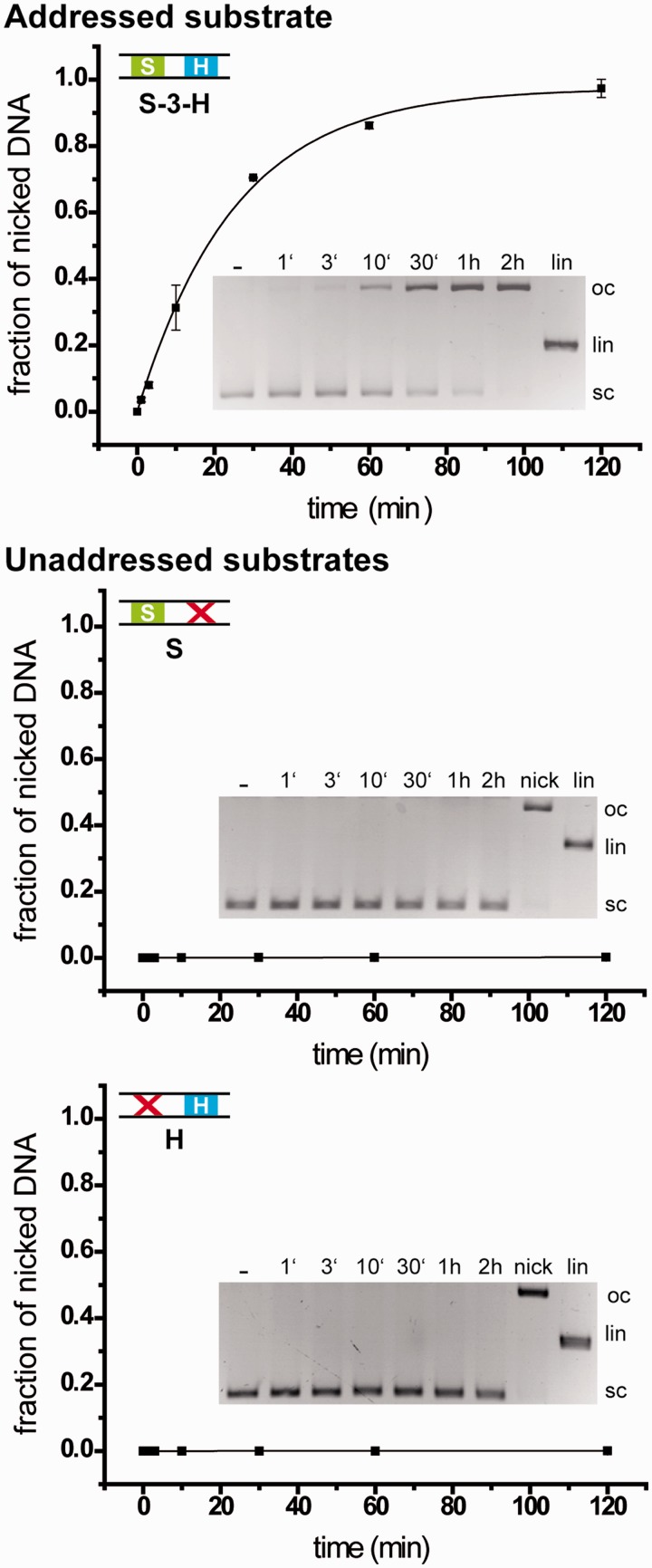
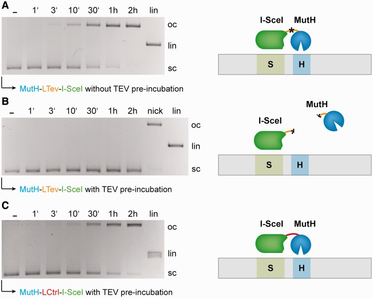
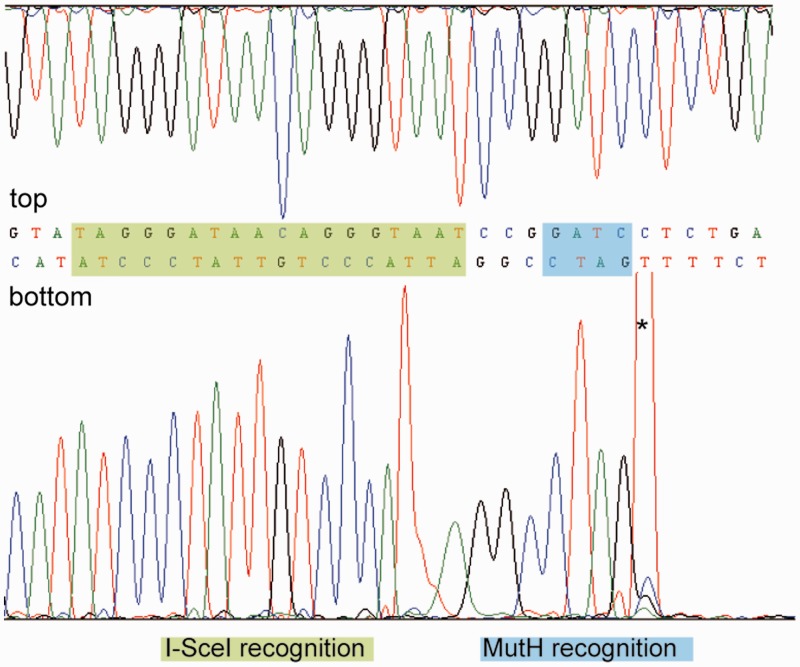
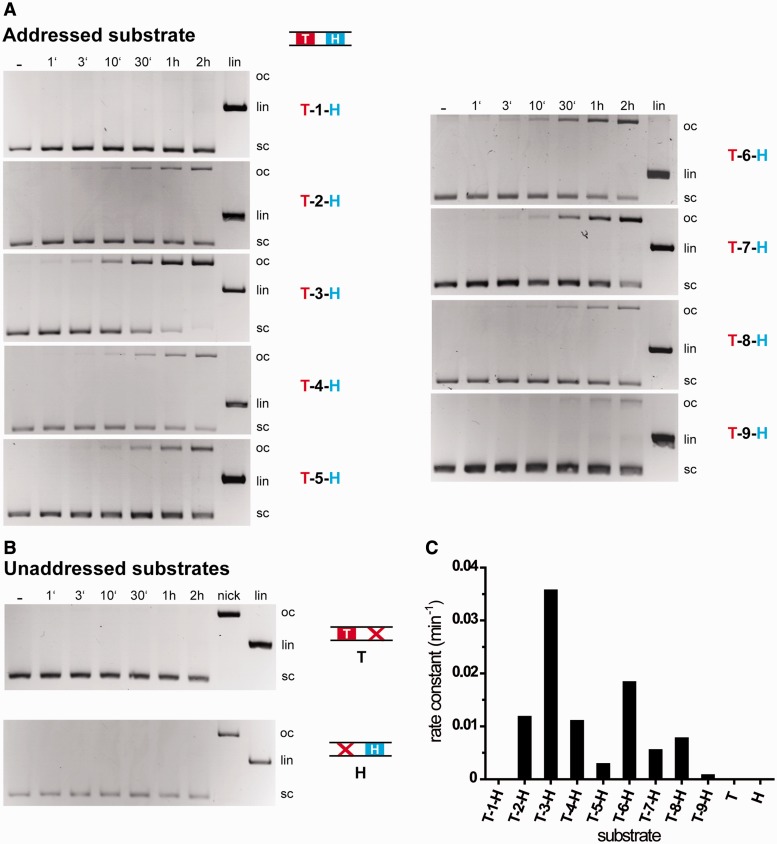
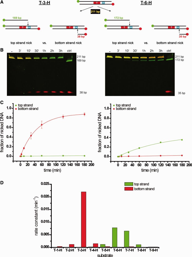
Targeted genome engineering requires nucleases that introduce a highly specific double-strand break in the genome that is either processed by homology-directed repair in the presence of a homologous repair template or by non-homologous end-joining (NHEJ) that usually results in insertions or deletions. The error-prone NHEJ can be efficiently suppressed by 'nickases' that produce a single-strand break rather than a double-strand break. Highly specific nickases have been produced by engineering of homing endonucleases and more recently by modifying zinc finger nucleases (ZFNs) composed of a zinc finger array and the catalytic domain of the restriction endonuclease FokI. These ZF-nickases work as heterodimers in which one subunit has a catalytically inactive FokI domain. We present two different approaches to engineer highly specific nickases; both rely on the sequence-specific nicking activity of the DNA mismatch repair endonuclease MutH which we fused to a DNA-binding module, either a catalytically inactive variant of the homing endonuclease I-SceI or the DNA-binding domain of the TALE protein AvrBs4. The fusion proteins nick strand specifically a bipartite recognition sequence consisting of the MutH and the I-SceI or TALE recognition sequences, respectively, with a more than 1000-fold preference over a stand-alone MutH site. TALE-MutH is a programmable nickase.


Figures

Figure 1.
Figure 1.
(A) Scheme of the fusion constructs MutH–I-SceI and TALE-MutH binding to their respective recognition sites (5′→3′). The highly specific interaction is mediated mainly by the binding module, I-SceI or the DNA-binding domain of the TALE protein AvrBs4, respectively. The monomeric cleavage module MutH is recruited by the binding module to nick the GATC site in close proximity to the I-SceI or TALE site. MutH is fused to I-SceI via a 10-amino-acid linker (ASENLYFQGG, shown as an orange line; the star indicates a TEV protease recognition site). In the case of the TALE–MutH construct, no additional linker was introduced; the fusion was done 28 amino acids (shown as red line) after the last half-repeat. (B) Model of the designed fusion constructs bound to specific DNA, but without the oligopeptide linking the DNA-binding and DNA-cleavage module. The colour code can be deduced from the scheme, I-SceI in green, the TALE protein in red (only the 23 repeats of the 3UGM structure are shown) and MutH in blue. Modelling was done using the crystal structures 1R7M (I-SceI), 3UGM (TALE) and 2AOQ (MutH). The model was generated with PyMOL and 3D-DART (56).
Figure 2.
Figure 2.
Kinetics of DNA nicking by the MutH–I-SceI fusion construct of addressed and unaddressed substrates analysed with plasmid cleavage assays using a 4-fold excess of enzyme over substrate (16 nM enzyme, 4 nM substrate). The addressed substrate plasmid contains an I-SceI and a MutH recognition site separated by 3 bp (S-3-H). The unaddressed substrate plasmids (controls) contain either an I-SceI recognition site without a nearby MutH recognition site (S) or no I-SceI recognition site (H). Each of the substrates has 19 additional GATC sites distributed over the plasmid. The position of the bands representing supercoiled (sc), open circular (oc) and linear (lin) forms of the plasmid is indicated. The first lane shows the reaction without MgCl2 (‘−’). ‘Nick’ and ‘lin’ represent the controls for nicking and cleavage activity, respectively, and correspond to the open circular and linear forms of the plasmid, respectively. The electrophoretic analysis shows that the addressed substrate plasmid is nicked by the fusion construct MutH–I-SceI, whereas the unaddressed substrate plasmids are not nicked or cleaved. The calculated apparent cleavage rate constant for the addressed plasmid substrate is 4.0 × 10⁻² min−1, whereas the apparent cleavage rate constants for both unaddressed substrates are below the detection limit of approximately 4 × 10⁻⁵ min−1.
Figure 3.
Figure 3.
Effect of separating the I-SceI and the MutH module on addressed substrate nicking. The kinetics of DNA-nicking by the MutH–I-SceI fusion construct of the addressed substrate was analysed with a plasmid cleavage assay (16 nM enzyme, 4 nM substrate). The addressed substrate plasmid contains an I-SceI and a MutH recognition site separated by 3 bp (S-3-H). The substrate has 19 additional GATC sites distributed over the plasmid. (A) Assay performed with MutH–I-SceI that had not been pre-incubated with TEV protease. (B) Assay performed with MutH–I-SceI that had been pre-incubated with TEV protease. (C) For control, we have also incubated the fusion protein, in which the linker did not contain a TEV protease cleavage site, with the TEV protease. The position of the bands representing supercoiled (sc), open circular (oc) and linear (lin) forms of the plasmid is indicated. The first lane shows the reaction without MgCl2 (‘−’). ‘Nick’ and ‘lin’ are the controls for cleavage and nicking activity and correspond to the open circular and linear forms of the plasmid, respectively. Whereas in (A) and (C), nicking is observed, in (B), it is not.
Figure 4.
Figure 4.
Determination of the strand specificity of MutH–I-SceI by sequence analysis of the nicked addressed plasmid after cleavage with the fusion construct MutH–I-SceI. The cleavage product was gel-purified and sequenced (top strand and bottom strand) over the specific site (S-3-H). For the purpose of illustration, the sequence is shown in the reverse and complementary orientation. The recognition sites of I-SceI and MutH are highlighted by green and blue bars, respectively. The sequencing results indicate that the bottom strand is cleaved 5′ of the addressed GATC site. *Taq polymerase artefact.
Figure 5.
Figure 5.
Kinetics of DNA-cleavage by TALE–MutH fusion constructs of addressed and unaddressed substrates analysed with plasmid cleavage assays using a 4-fold excess of enzyme over substrate (16 nM enzyme, 4 nM substrate). (A) Catalytic activity of TALE–MutH with a plasmid substrate containing an AvrBs4 and a MutH recognition site separated by × base pairs (T-x-H; x = 1–9 bp). (B) Catalytic activity on the unaddressed substrate plasmids (controls) containing either an AvrBs4 recognition site without a nearby MutH recognition site (T) or no AvrBs4 recognition site (H). Each of the substrates has 18 additional GATC sites distributed over the plasmid. The position of the bands representing supercoiled (sc), open circular (oc) and linear (lin) forms of the plasmid is indicated. The first lane shows the reaction without MgCl2 (‘−’). ‘Nick’ and ‘lin’ represent controls for nicking and cleavage activity, respectively, and correspond to the open circular and linear forms of the plasmid, respectively. (C) Calculated rate constants for TALE–MutH nicking the above-mentioned substrates. The electrophoretic analysis shows that the addressed substrate plasmid is nicked by the fusion construct TALE–MutH, whereas the unaddressed substrate plasmids remain uncleaved. The graph shows the quantitative evaluation of the experiments shown in (A) and (B). Note that the enzyme exhibits the best activity on the addressed substrate with the 3 bp spacer and no activity with the stand-alone TALE (T) or MutH (H) recognition sites. The quantitative analyses reveal the following apparent cleavage rate constants: 1.3 × 10⁻⁴ min−1 (T-1-H), 1.2 × 10⁻² min−1 (T-2-H), 3.6 × 10⁻² min−1 (T-3-H), 1.1 × 10⁻² min−1 (T-4-H), 0.3 × 10⁻² min−1 (T-5-H), 1.8 × 10⁻² min−1 (T-6-H), 0.6 × 10⁻² min−1 (T-7-H), 0.8 × 10⁻² min−1 (T-8-H) and 8.9 × 10⁻⁴ min−1 (T-9-H); the nicking rate for T and H was below the detection limit of approximately 4 × 10⁻⁵ min−1.
Figure 6.
Figure 6.
Determination of the strand specificity of TALE–MutH. (A) Scheme of the reaction: the 211 bp polynucleotide substrate harbours the AvrBs4 recognition site (T) and the MutH (H) recognition site separated by 3 (T-3-H; left) or 6 (T-6-H; right) bp. The substrates are 5′-labelled at the top strand with Atto 488 (green) and with Atto 467 N (red) at the bottom strand. The expected nicking products have a length of 169 (172) and 38 (35) bp for top and bottom strand nicking of the T-3-H substrate (T-6-H substrate). Which strand is attacked preferentially can be determined in the electrophoretic analysis by the characteristic fluorescence of the products. (B) DNA nicking kinetics of the addressed polynucleotide substrates T-3-H (left) and T-6-H (right) by TALE-MutH. The nicking products for top and bottom strand of the T-3-H (T-6-H) substrate are indicated with 169 (172) and 38 (35) bp, respectively. The first lane shows the control reaction without MgCl2 (‘−’). The lane designated as ‘ctrl’ shows the result of the cleavage of the PCR substrate by BamHI which overlaps the MutH site and is shown here to indicate the size of the nicking products. (C) The graphs show the quantitative evaluation of the experiments shown in (B). The calculated cleavage rate constants for T-3-H (T-6-H) top strand nicking is 6.0 × 10⁻⁵ min−1 (7.8 × 10⁻³ min−1) and for bottom strand nicking 2.2 × 10⁻² min−1 (1.3 × 10⁻⁴ min−1). The analysis of the strand specificity shows that the addressed PCR substrate with the 3 bp spacer is preferentially nicked in the bottom strand by the fusion construct TALE–MutH, whereas the substrate with the 6 bp spacer is preferentially nicked in the top strand. In (D), the rate constants for nicking are shown for all substrates investigated: T-1-H to T-9-H.

Similar articles

Cited by

References

    1. Marcaida MJ, Munoz IG, Blanco FJ, Prieto J, Montoya G. Homing endonucleases: from basics to therapeutic applications. Cell. Mol. Life Sci. 2010;67:727–748. - PMC - PubMed
    1. Silva G, Poirot L, Galetto R, Smith J, Montoya G, Duchateau P, Paques F. Meganucleases and other tools for targeted genome engineering: perspectives and challenges for gene therapy. Curr. Gene Ther. 2011;11:11–27. - PMC - PubMed
    1. Urnov FD, Rebar EJ, Holmes MC, Zhang HS, Gregory PD. Genome editing with engineered zinc finger nucleases. Nat. Rev. Genet. 2010;11:636–646. - PubMed
    1. Ramalingam S, Kandavelou K, Rajenderan R, Chandrasegaran S. Creating designed zinc-finger nucleases with minimal cytotoxicity. J. Mol. Biol. 2011;405:630–641. - PMC - PubMed
    1. Rahman SH, Maeder ML, Joung JK, Cathomen T. Zinc-finger nucleases for somatic gene therapy: the next frontier. Hum. Gene. Ther. 2011;22:925–933. - PMC - PubMed

Publication types

MeSH terms

Substances