Case Reports
. 2013 Mar 12;80(11):1033-40.
doi: 10.1212/WNL.0b013e31828726a7. Epub 2013 Feb 13.

CSF1R mutations link POLD and HDLS as a single disease entity

Affiliations
Case Reports

CSF1R mutations link POLD and HDLS as a single disease entity

Alexandra M Nicholson et al. Neurology. .

Abstract

Objective: Pigmented orthochromatic leukodystrophy (POLD) and hereditary diffuse leukoencephalopathy with axonal spheroids (HDLS) are rare neurodegenerative disorders characterized by cerebral white matter abnormalities, myelin loss, and axonal swellings. The striking overlap of clinical and pathologic features of these disorders suggested a common pathogenesis; however, no genetic or mechanistic link between POLD and HDLS has been established. Recently, we reported that mutations in the colony-stimulating factor 1 receptor (CSF1R) gene cause HDLS. In this study, we determined whether CSF1R mutations are also a cause of POLD.

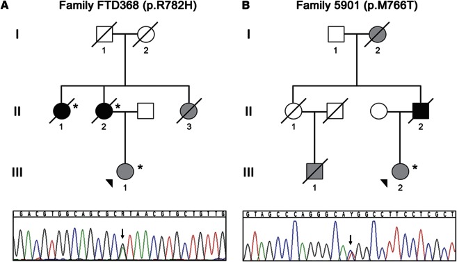
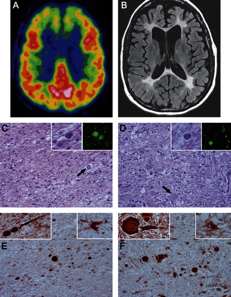
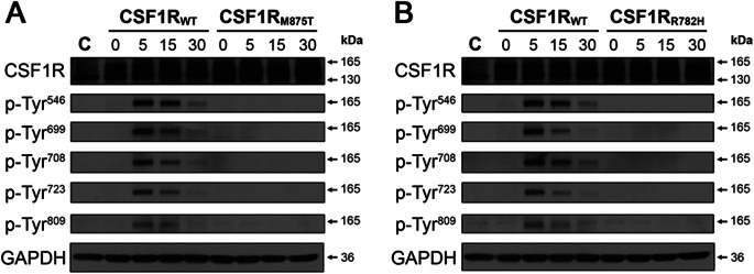
Methods: We performed sequencing of CSF1R in 2 pathologically confirmed POLD families. For the largest family (FTD368), a detailed case report was provided and brain samples from 2 affected family members previously diagnosed with POLD were re-evaluated to determine whether they had HDLS features. In vitro functional characterization of wild-type and mutant CSF1R was also performed.

Results: We identified CSF1R mutations in both POLD families: in family 5901, we found c.2297T>C (p.M766T), previously reported by us in HDLS family CA1, and in family FTD368, we identified c.2345G>A (p.R782H), recently reported in a biopsy-proven HDLS case. Immunohistochemical examination in family FTD368 showed the typical neuronal and glial findings of HDLS. Functional analyses of CSF1R mutant p.R782H (identified in this study) and p.M875T (previously observed in HDLS), showed a similar loss of CSF1R autophosphorylation of selected tyrosine residues in the kinase domain for both mutations when compared with wild-type CSF1R.

Conclusions: We provide the first genetic and mechanistic evidence that POLD and HDLS are a single clinicopathologic entity.


Figures

Figure 1
Figure 1. Families with POLD and CSF1R mutations
Shown are abbreviated pedigrees of 2 families, family FTD368 (A) and family 5901 (B) affected by pigmented orthochromatic leukodystrophy (POLD) included in this study. Gray-filled symbols represent clinically affected individuals, whereas black-filled symbols represent individuals with pathologically confirmed POLD. The proband of each family is indicated with an arrowhead. Cases for which DNA was available for genetic studies are noted with an asterisk. DNA sequence traces observed in a sample from the proband from each family are shown below each pedigree. The single base substitution in each trace is indicated with an arrow.
Figure 2
Figure 2. Neuroimaging and neuropathologic findings in pigmented orthochromatic leukodystrophy
(A) 18-Fluorodeoxyglucose PET scan performed in 2006 from FTD368 II-1 shows hypometabolism in both frontal lobes. (B) MRI scan performed in 2007 shows atrophy of the frontal lobes, white matter hyperintensities in the white matter of the frontal lobes and similar changes in the parietooccipital white matter, enlargement of the frontal horns of the lateral ventricles, and milder diffuse cerebral atrophy. (C–F) Microscopic images of white matter from FTD368 II-2 (C and E) compared with typical hereditary diffuse leukoencephalopathy with axonal spheroids (HDLS) (D and F). On hematoxylin & eosin stains, the white matter has pallor because of myelinated fiber loss and gliosis, with axonal spheroids (arrows in C and D). Within the affected white matter are macrophages with basophilic to brown fine granules (left insets in C and D) that have autofluorescent pigment (right insets in C and D) when unstained sections are viewed with a fluorescent microscope. Immunohistochemistry for amyloid precursor protein (panels E and F) shows many axonal spheroids in affected white matter, which are confirmed to be axonal in origin by immunoreactivity with phosphorylated neurofilament (left insets in E and F). A characteristic feature of HDLS is the presence of bizarre hypertrophic astrocytes demonstrated with immunohistochemistry for αB-crystallin (right insets in E and F). C–F: original magnification ×200; all insets: original magnification ×400.
Figure 3
Figure 3. Functional effects of CSFR1 mutations in cell culture
CSF-1 treatment leads to autophosphorylation of wild-type but not mutant CSF1R. HeLa cells were transfected with either a tGFP control plasmid, or a plasmid containing wild-type (CSF1RWT), the previously characterized mutant (CSF1RM875T) (A), or uncharacterized mutant (CSF1RR782H) (B) CSF1R. Cells were treated with CSF-1 for 5, 15, or 30 minutes. Each blot was probed with antibodies against total CSF1R and with antibodies that detect CSF1R autophosphorylation at several tyrosine residues (p-Tyr). Note that CSF-1 treatment only causes CSF1R autophosphorylation in cells expressing CSF1RWT. GAPDH (glyceraldehyde 3-phosphate dehydrogenase) was used as a loading control.

Similar articles

Cited by

References

    1. Freeman SH, Hyman BT, Sims KB, et al. Adult onset leukodystrophy with neuroaxonal spheroids: clinical, neuroimaging and neuropathologic observations. Brain Pathol 2009;19:39–47 - PMC - PubMed
    1. Van Gerpen JA, Wider C, Broderick DF, Dickson DW, Brown LA, Wszolek ZK. Insights into the dynamics of hereditary diffuse leukoencephalopathy with axonal spheroids. Neurology 2008;71:925–929 - PMC - PubMed
    1. Marotti JD, Tobias S, Fratkin JD, Powers JM, Rhodes CH. Adult onset leukodystrophy with neuroaxonal spheroids and pigmented glia: report of a family, historical perspective, and review of the literature. Acta Neuropathol 2004;107:481–488 - PubMed
    1. Hanaya R, Arita K, Itoh Y, Kiura Y, Iida K, Kurisu K. Zygomatic osteotomy for resection of medial temporal cavernous angioma in dominant hemisphere after subdural grid electroencephalographic study. Hiroshima J Med Sci 2006;55:39–43 - PubMed
    1. Wider C, Van Gerpen JA, DeArmond S, Shuster EA, Dickson DW, Wszolek ZK. Leukoencephalopathy with spheroids (HDLS) and pigmentary leukodystrophy (POLD): a single entity? Neurology 2009;72:1953–1959 - PMC - PubMed

Publication types

MeSH terms

Substances