. 2013 Feb;9(2):e1003137.
doi: 10.1371/journal.ppat.1003137. Epub 2013 Feb 7.

P53-mediated rapid induction of apoptosis conveys resistance to viral infection in Drosophila melanogaster

Affiliations

P53-mediated rapid induction of apoptosis conveys resistance to viral infection in Drosophila melanogaster

Bo Liu et al. PLoS Pathog. 2013 Feb.

Abstract

Arthropod-borne pathogens account for millions of deaths each year. Understanding the genetic mechanisms controlling vector susceptibility to pathogens has profound implications for developing novel strategies for controlling insect-transmitted infectious diseases. The fact that many viruses carry genes that have anti-apoptotic activity has long led to the hypothesis that induction of apoptosis could be a fundamental innate immune response. However, the cellular mechanisms mediating the induction of apoptosis following viral infection remained enigmatic, which has prevented experimental verification of the functional significance of apoptosis in limiting viral infection in insects. In addition, studies with cultured insect cells have shown that there is sometimes a lack of apoptosis, or the pro-apoptotic response happens relatively late, thus casting doubt on the functional significance of apoptosis as an innate immunity. Using in vivo mosquito models and the native route of infection, we found that there is a rapid induction of reaper-like pro-apoptotic genes within a few hours following exposure to DNA or RNA viruses. Recapitulating a similar response in Drosophila, we found that this rapid induction of apoptosis requires the function of P53 and is mediated by a stress-responsive regulatory region upstream of reaper. More importantly, we showed that the rapid induction of apoptosis is responsible for preventing the expression of viral genes and blocking the infection. Genetic changes influencing this rapid induction of reaper-like pro-apoptotic genes led to significant differences in susceptibility to viral infection.


Conflict of interest statement

The authors have declared that no competing interests exist.

Figures

Figure 1
Figure 1. A simplified diagram of apoptosis regulation in Drosophila.
The RHG genes are clustered in a chromosomal region of about 350 kb. Their transcription is coordinated by highly conserved intergenic regulatory regions such as IRER (irradiation responsive enhancer region). Cytotoxic stress such as DNA damage can activate P53, which in turn induces the expression of RHG genes. RHG proteins function as IAP-antagonists to relieve the caspase from inhibition by DIAP (Drosophila Inhibitor of Apoptosis). The relative human orthologs are in parenthesis. Arrows indicate activation and red bars indicate suppression. Dotted lines represent indirect relationship. The genes that were analyzed in this work for their role in mediating the rapid induction of apoptosis following viral infection are underlined.
Figure 2
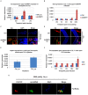
Figure 2. Rapid induction of reaper and hid following viral infection of Drosophila larvae or adults.
A. At 1 hr post AcMNPV injection, hid mRNA level in injected larvae was induced ∼2 fold. By 2 hr p.i., the level of hid went back to normal. B. AcMNPV infection in cultured DL-1 cells (MOI = 20) did not induce reaper/hid until a relative late stage (24 hr p.i.). Data are shown as mean± SD of two to three independent experiments. C. hid was induced in fat body cells as revealed by FISH using digoxin-labeled cRNA probe following AcMNPV injection. D. A few cells in the gut also had elevated hid expression following AcMNPV injection. E. At 1 hr post FHV injection, reaper mRNA level in infected adults was induced ∼1.6 fold. F. FHV infection in DL-1 cells (MOI = 40) did not induce reaper/hid expression until 36 hr p.i.. Data are shown as mean± SD. G. Cells expressing high levels of reaper mRNA (green, FISH) in the fat body were cells were also positive for the FHV capsid protein (red, arrows). DAPI was used to visualize the nuclei. The animal shown was of genotype Lsp-Gal4, UAS-diap1. Photos are representative of at least 2 independent experiments.
Figure 3
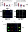
Figure 3. Virus -induced reaper/hid expression and apoptosis requires P53 and IRER.
A. The induction of hid following AcMNPV injection was absent in larvae homozygous for a P53 null mutation (p53−/−) or a deficiency removing the regulatory region IRER (Df(IRER)). B. Likewise, the induction of reaper following FHV infection also required p53 and IRER. Flies of different genotypes were injected in parallel and sacrificed for total RNA extraction at 1 hr p.i,. C & D. Activated caspases were detected in the fat body cells of D. melanogaster larvae (C) and adults (D) following the infection of AcMNPV or FHV, respectively. The percentage of cells positive for activated caspases in different genotypes is listed in the table below corresponding panel. The induction of apoptotic cells (with activated caspases) was significantly blocked in P53−/− or IRER deficient flies (p<0.01 for both). E. Fat body cells in P53−/− animals became necrotic at 4–7 days p.i.. PI was injected to either control-injected or FHV-injected (2000 PFU/animal) animals at 7 days p.i. and the animals were sacrificed and fixed 20 min later. The presence of FHV was visualized with an antibody against the FHV capsid protein. The presence of PI staining indicated that the integrity of the cell membrane was compromised in fat body cells infected by FHV. Photos are representative of at least 2 independent experiments.
Figure 4
Figure 4. Rapid induction of apoptosis functions to limit viral gene expression and proliferation.
A. The mRNA levels of AcMNPV immediate early gene ie0 and ie1 were examined at 6 hr post virus injection by Q-PCR in Drosophila larvae. The data indicate that in both p53−/− and IRER deficient strains that lack rapid induction of apoptosis, ie0 and ie1 expression levels were dramatically higher than in the wild type strain. Data are shown as mean± SD of two to three independent experiments. B. FHV RNA1 and RNA2 levels were examined at 24 hr post injection in wild type, p53−/− and Df(IRER) adults by Q-PCR. Note the significantly higher levels of RNA1 and RNA2 in p53−/− and Df(IRER) flies than in wild type flies. C. RNA1 and RNA2 levels were also higher when apoptosis was suppressed by knocking down the upstream caspase Dronc (Dronc KD) in the fat body cells (genotype LSP2-Gal4;UAS-dronc_RNAi) (*, p<0.05, **, p<0.01).
Figure 5
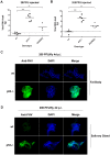
Figure 5. Rapid induction of apoptosis prevents viral proliferation.
A. When a low dose of FHV (20 PFU/animal) was injected, no viral RNA amplification was observed at 4 days p.i. in wild type Drosophila. In contrast, significant amplification of FHV RNA was observed in p53−/− or Df(IRER) flies (**, p<0.01, ***, p<0.001). The dotted line indicates the amount of virus injected into each animal. Total RNA extraction and Q-PCR were performed for individual flies to estimate the copy numbers of RNA1 and RNA2 (Materials and Methods and Figure S3). B. When 200 PFU/animal was injected, most wild type flies had no significant increase in FHV RNA copy numbers, while the virus proliferated significantly in P53−/− and Df(IRER) animals. (**, p<0.01, ***, p<0.001) C. Immunostaining was performed with antiserum against FHV capsid protein and AlexaFluor 488 labeled goat anti rabbit secondary antibody. Cells in the fat body of p53−/− flies were filled with FHV capsid protein at 3 days post injection of 20 PFU/animal. No FHV–positive cells could be detected in wild type (wt) animals injected with 20 PFU of FHV. D. At 4days p.i., cells in the salivary glands of P53−/− flies were positive for FHV capsid protein, while capsid protein was not detectable in wild type animals. Photos are representative of at least 2 independent experiments.
Figure 6
Figure 6. Specific induction of mx in the refractory strain (MOYO-R), but not in the susceptible strain (MOYO-S), following exposure to DEN-2.
The expression level of mx was measured by Q-PCR and normalized against the housekeeping gene GAPDH before calculating the fold induction, i.e. the ratio of mx in virus-fed vs. the control-fed samples. At 3 hr post feeding, the level of mx in adult A. aegypti females exposed to DEN-2 was more than 2 fold higher than those fed with control blood meal. This level of mx expression declined at 18 hr, possibly due to death of mx-expressing cells as was previously observed for CuniNPV -infected A. aegypti larvae. Data are represented as mean ± SD.
Figure 7
Figure 7. A diagram summarizing the role of rapid induction of apoptosis as an innate immune response against viral infection.
By eliminating the infected cells before accumulation of viral gene products, the infection can be blocked at the initiation stage.

References

    1. Clouston WM, Kerr JF (1985) Apoptosis, lymphocytotoxicity and the containment of viral infections. Med Hypotheses 18: 399–404. - PubMed
    1. Hardwick JM (1998) Viral interference with apoptosis. Semin Cell Dev Biol 9: 339–349. - PubMed
    1. Everett H, McFadden G (1999) Apoptosis: an innate immune response to virus infection. Trends Microbiol 7: 160–165. - PubMed
    1. Benedict CA, Norris PS, Ware CF (2002) To kill or be killed: viral evasion of apoptosis. Nat Immunol 3: 1013–1018. - PubMed
    1. Soria C, Estermann FE, Espantman KC, O'Shea CC (2010) Heterochromatin silencing of p53 target genes by a small viral protein. Nature 466: 1076–1081. - PMC - PubMed

Publication types

MeSH terms